


Article Figures & Data
Figures
- FIGURE 1.
Early phase (A) and delayed phase (B) of planar 99mTc-sestamibi scan showing normal parathyroid findings.
- FIGURE 2.
(A) SPECT/CT image showing possible adenoma, but thyroid uptake creates difficulty. (B) Misregistration, which may have resulted from patient movement between SPECT and CT acquisitions. This study was subsequently reprocessed. A color version of this figure is available as a supplemental file at http://tech.snmjournals.org.
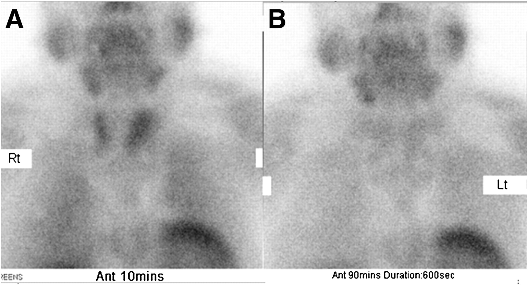
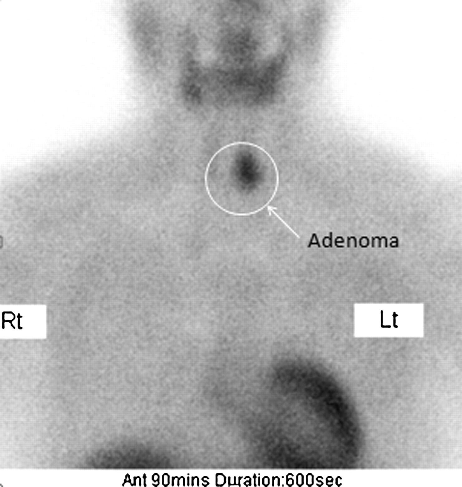
- FIGURE 3.
Adenoma in left inferior gland, visible in delayed phase of planar 99mTc-sestamibi scan.
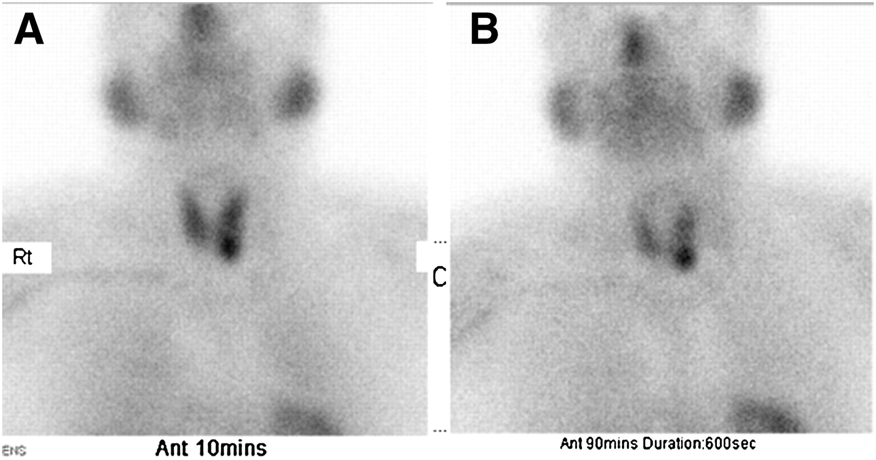
- FIGURE 4.
Early phase (A) and delayed phase (B) of planar 99mTc-sestamibi scan showing high uptake in parathyroid, consistent with adenoma.
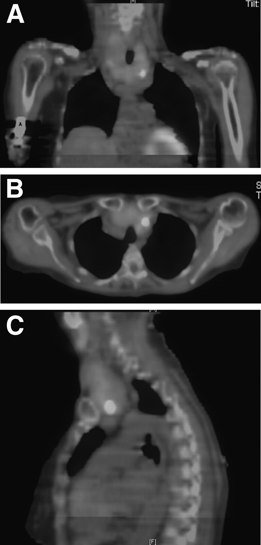
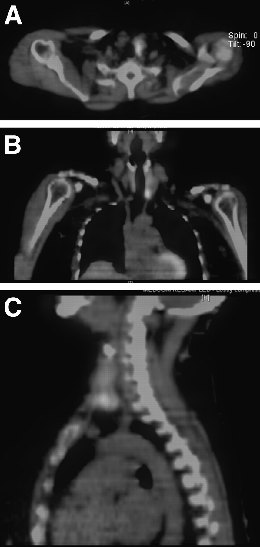
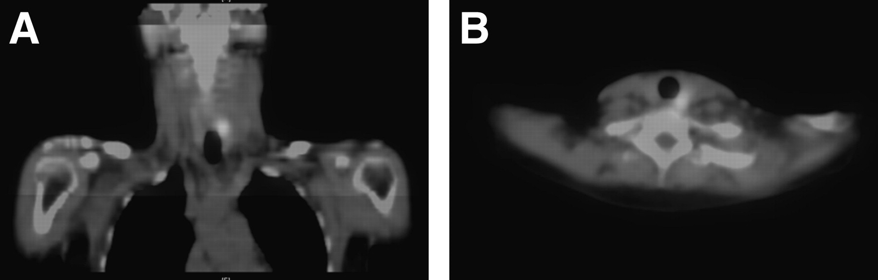
- FIGURE 5.
Transverse (A), fused coronal (B), and fused sagittal (C) SPECT/CT images showing higher uptake in neck, consistent with parathyroid adenoma, which helped to support information given by planar scan. A color version of this figure is available as a supplemental file at http://tech.snmjournals.org.
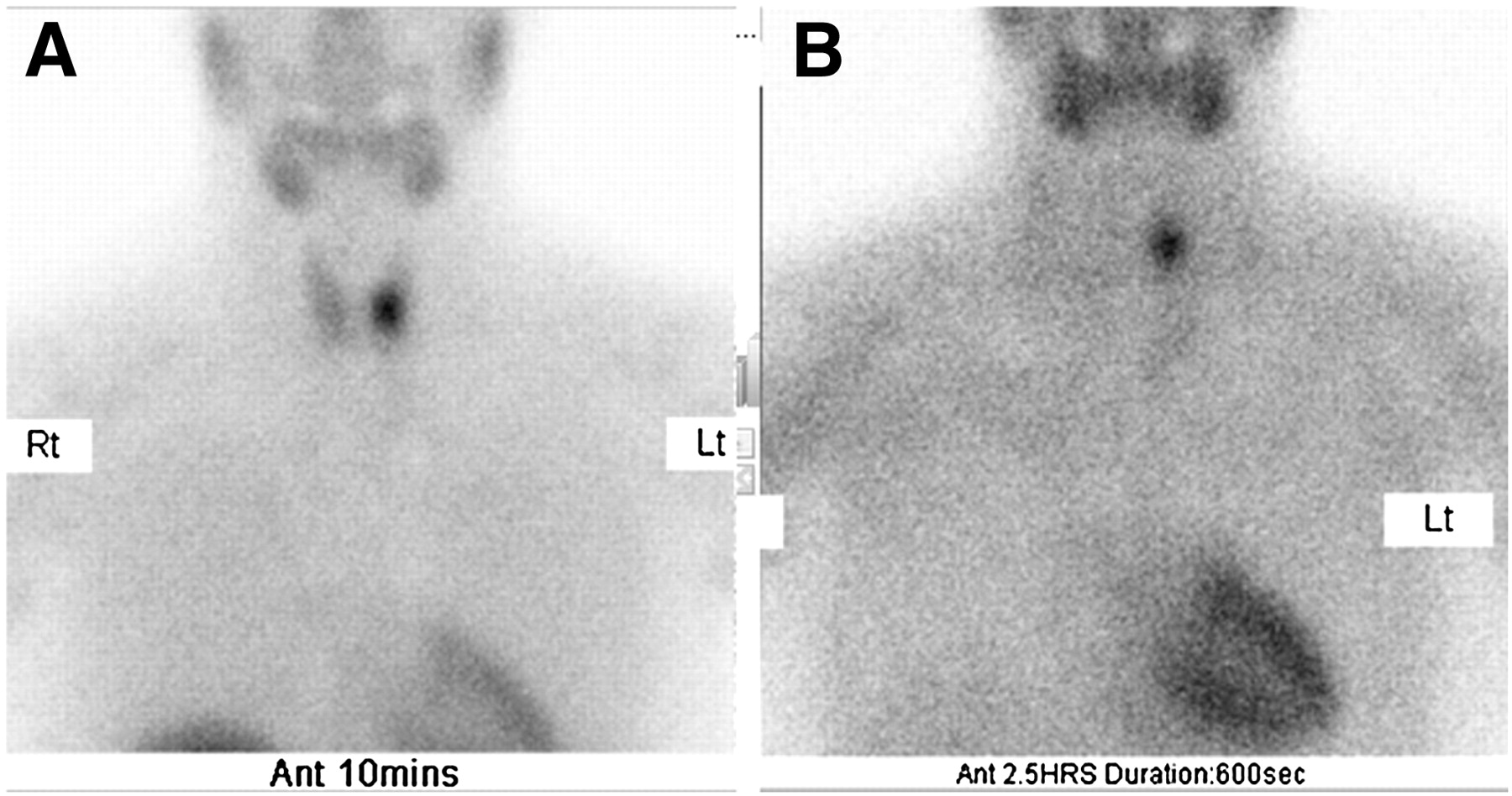
- FIGURE 6.
Early phase (A) and delayed phase (B) of planar 99mTc-sestamibi scan showing high-uptake area on left side of neck consistent with parathyroid adenoma.
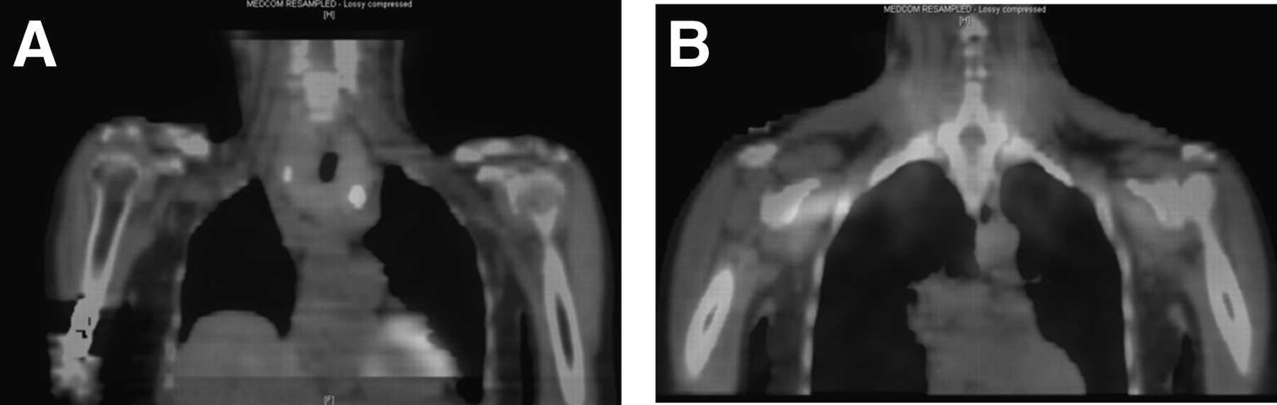
- FIGURE 7.
In same patient as in Figure 6, SPECT/CT coronal (A) and transverse (B) images that helped to confirm position of adenoma. Both images show high-uptake area consistent with parathyroid adenoma. A color version of this figure is available as a supplemental file at http://tech.snmjournals.org.
- FIGURE 8.
Early phase (A) and delayed phase (B) of planar 99mTc-sestamibi scan showing irregular uptake within thyroid gland.
- FIGURE 9.
Coronal (A), transverse (B), and sagittal (C) SPECT/CT images showing uptake within neck area. Even though fused images helped with anatomic localization, disease within parathyroid gland could not be excluded. A color version of this figure is available as a supplemental file at http://tech.snmjournals.org.
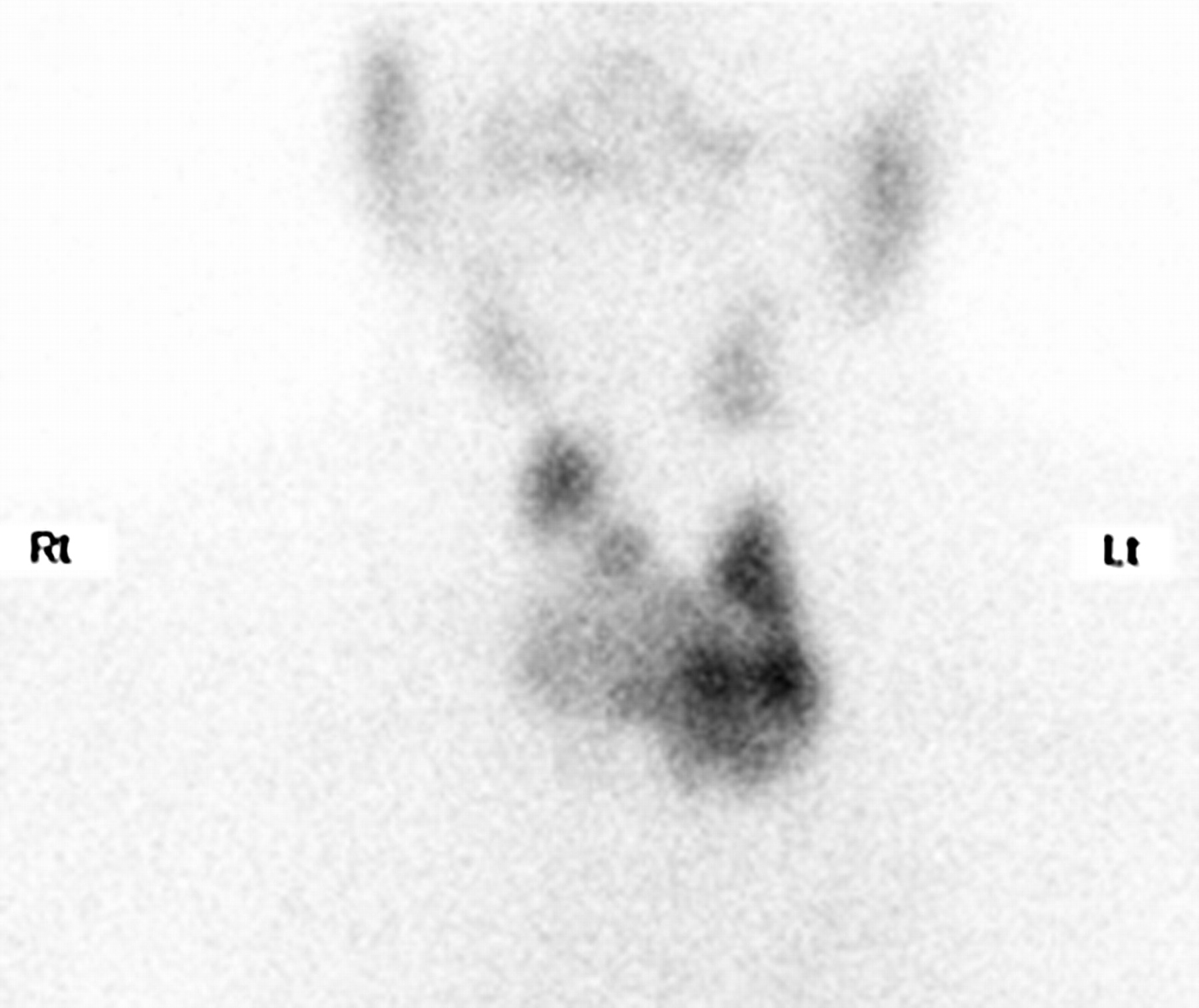
- FIGURE 10.
Planar 99mTc-sestamibi scan showing uneven thyroid uptake on 600-s anterior view. Scan was acquired 20 min after injection. Matrix is 128 × 128, and magnification is 2.0.


Tables
Sex Age (y) Reason for scan Activity administered (MBq) Results F 66 Preoperative localization 952 Parathyroid adenoma in left inferior gland M 65 Clinical signs of adenoma 955 No evidence of functioning parathyroid adenoma F 49 Clinical signs of adenoma 719 Parathyroid adenoma in left inferior gland F 53 Preoperative localization 899 No evidence of functioning parathyroid adenoma F 56 Clinical signs of adenoma 899 Parathyroid adenoma in left inferior gland F 60 Preoperative localization 880 Parathyroid adenoma in eccentric position F 79 Clinical signs of adenoma 770 Possible parathyroid adenoma in left inferior gland F 71 Clinical signs of adenoma 755 Possible parathyroid adenoma in left inferior gland F 50 Clinical signs of adenoma 742 Possible parathyroid adenoma in left inferior gland
Supplemental Data
Files in this Data Supplement:


