


Article Figures & Data
Figures
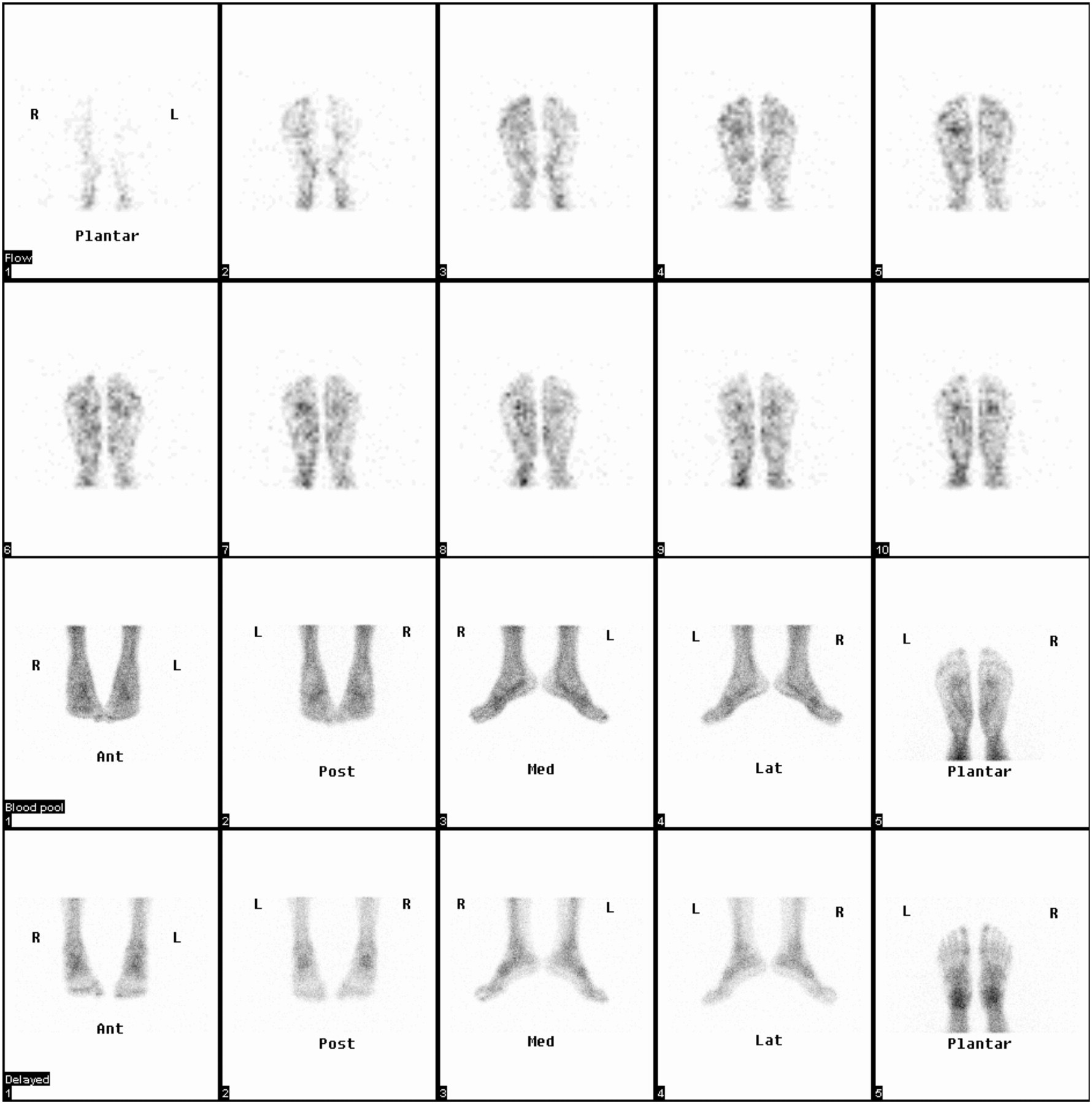
- FIGURE 1.
Three-phase bone study in control. Upper 2 rows display sequential 3-s/frame flow images acquired in plantar position. Third row shows blood-pool images, and in fourth row, corresponding delayed images in 5 positions are shown. Ant = anterior; Post = posterior; Med = medial; Lat = lateral.
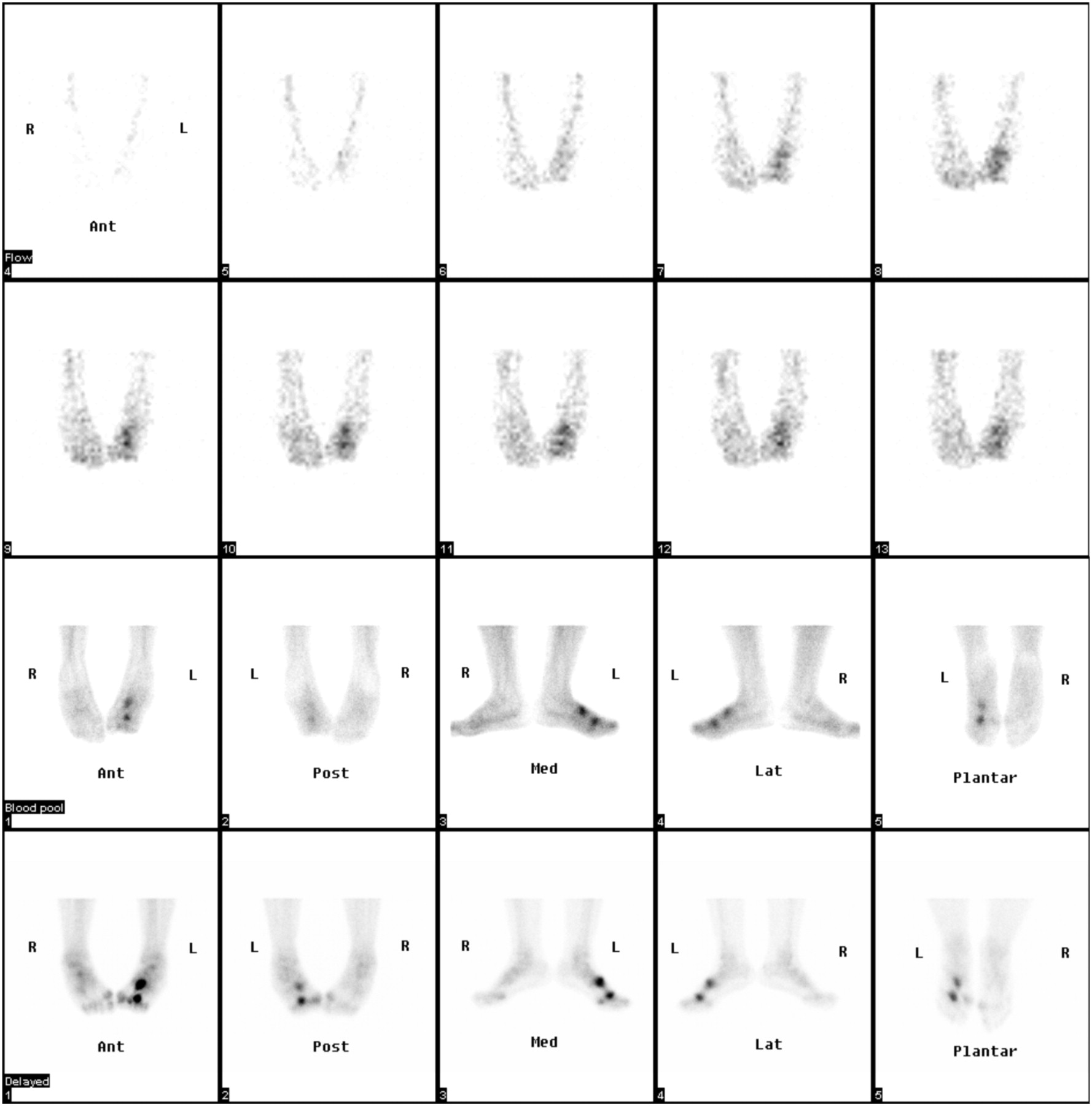
- FIGURE 2.
Three-phase bone scan in patient with trauma to left foot. Upper 2 rows display sequential 3-s/frame flow images acquired in anterior position, showing increased flow to mid left foot. Third row shows blood-pool images, and in fourth row, corresponding delayed images in 5 positions are shown. Anterior blood-pool and delayed views show clearly 2 abnormalities in third metatarsal bone, indicating fractures. Small tarsal bones are also better seen on these views. Ant = anterior; Post = posterior; Med = medial; Lat = lateral.
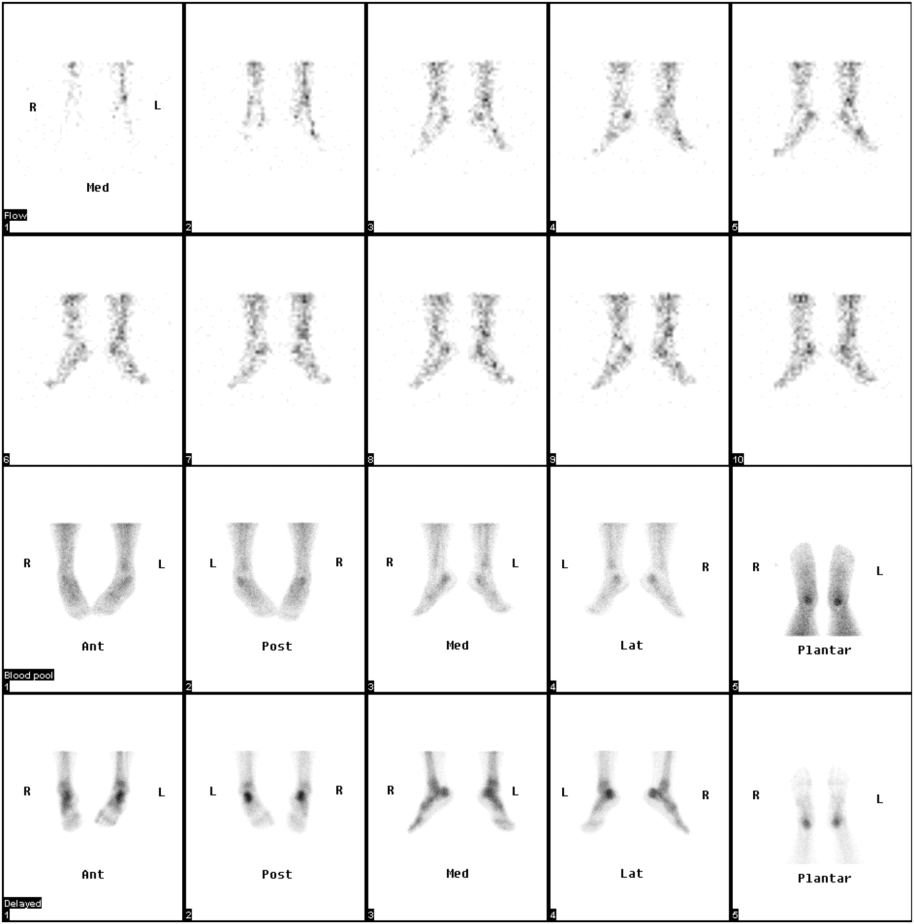
- FIGURE 3.
Three-phase bone scan in patient with bilateral heel pain. Upper 2 rows display sequential 3-s/frame flow images acquired in medial position, showing increased flow to hind-foot areas. Third row shows blood-pool images, and in fourth row, corresponding delayed images in 5 positions are shown. Medial–lateral blood-pool and delayed views show clearly increased uptake in calcaneus bilaterally. Positive findings are also seen on posterior and plantar views. Calcaneal spurs were found on radiographs. Ant = anterior; Post = posterior; Med = medial; Lat = lateral.


Tables
Demographic Controls Patients Average age ± SD (y) 49 ± 20 45 ± 17 Sex (n) Male 10 16 Female 16 11 Clinical condition (n) Diabetes — 14 Trauma — 5 Arthritis — 4 Foot pain — 4 Back pain 5 — Cancer (breast, prostate, or other) 21 — Phase of bone scan Flow Blood pool Delayed Region of foot Projection Score Best view Score Best view Score Forefoot Plantar 6 Phalanges Plantar 18 Plantar 15 Anterior 3 Anterior 4 Metatarsals Plantar 15 Plantar 14 Anterior 5 Anterior 5 MPJs Plantar 17 Plantar 15 Anterior 3 Anterior 3 Mid foot Anterior 10 Cuneiform bones Anterior 19 Anterior 18 Plantar 5 Plantar 4 Medial 1 Medial 1 Cuboid bone Anterior 22 Anterior 23 Plantar 1 Plantar 2 Medial 1 Medial 1 Navicular bone Anterior 22 Anterior 23 Plantar 2 Plantar 1 Medial 2 Medial 1 Hind foot Medial–lateral 10 Calcaneus Medial–lateral 17 Medial and lateral 20 Lateral 4 Lateral 4 Posterior 1 Posterior 2 Talus Medial–lateral 19 Medial and lateral 23 Lateral 1 Lateral 1 Ankle Posterior 10 Ankle joint Posterior 26 Posterior 26 Phase of bone scan Flow Blood pool Delayed images Region of foot Projection n Best view n Best view n Forefoot Phalanges Anterior 3 Anterior 6 Anterior 5 Posterior 1 Posterior 1 Plantar 3 Plantar 4 Metatarsals Anterior 1 Anterior 3 Anterior 5 Plantar 2 Plantar 8 MPJs Anterior 2 Anterior 8 Anterior 9 Posterior 2 Posterior 1 Plantar 5 Plantar 10 Mid foot Cuneiform bones Anterior 3 Anterior 2 Anterior 3 Plantar 2 Medial 1 Cuboid bone Anterior 3 Anterior 1 Anterior 3 Plantar 1 Plantar 3 Navicular bone Anterior 2 Anterior 1 Anterior 2 Posterior 1 Posterior 1 Hind foot Calcaneus Medial–lateral 2 Medial 3 Medial 4 Lateral 3 Lateral 3 Talus Medial 1 Medial 2 Lateral 3 Posterior 1 Ankle Medial–lateral 1 Medial 1 Medial 1 Ankle joint Lateral 2 Lateral 2 Posterior 2 Posterior 2 View Scintigraphic appearance Impact on interpretation Anterior–posterior (blood pool) Focal increased metatarsal uptake False-positive blood-pool image Medial or lateral Focal increased uptake in forefoot Difficulty in separating bones of forefoot Diffuse increased uptake in mid and hind foot Overlapping lesions in talus and navicular Posterior Diffuse increased uptake in mid foot Difficulty in separating bones of mid foot Diffuse increased uptake in hind foot Cannot differentiate talus and calcaneus Plantar Focal increased uptake in hind foot Cannot differentiate talus and calcaneus Foot region Best view Forefoot Plantar for flow, blood-pool, and delayed images; anterior will be useful to resolve overlap in some cases Mid foot Anterior for all phases; lateral or plantar will help in cases in which cuneiform bones are involved Hind foot Lateral and medial will be useful for all phases Ankle joint Posterior, medial, and lateral


Jump to section
Related Articles
Cited By...
- No citing articles found.