


Article Figures & Data
Figures
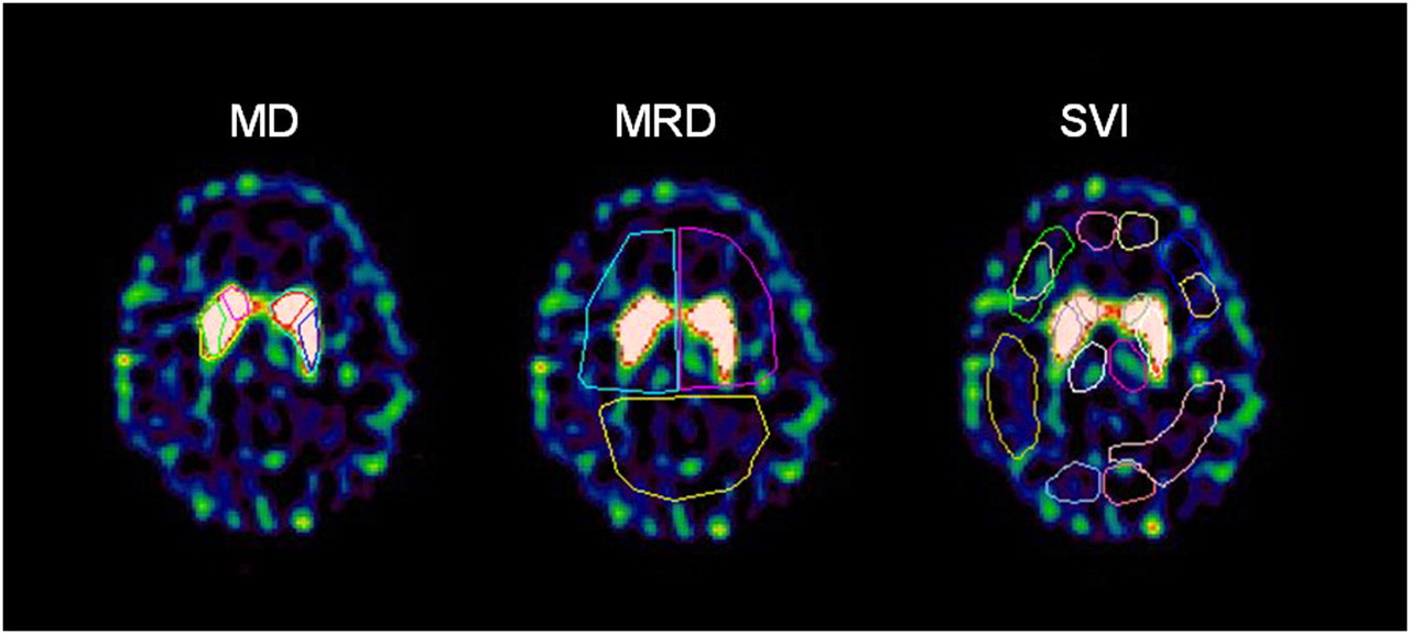
- FIGURE 1.
Three different methods of ROI delineation on SPECT images of same brain slice in same individual.
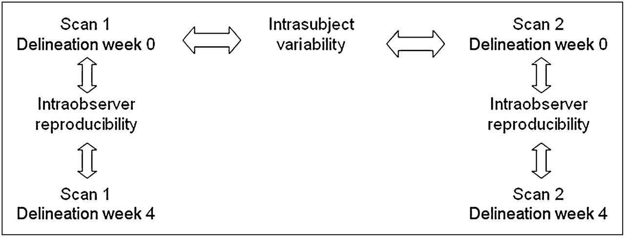
- FIGURE 2.
Flowchart of study. Eight individuals were scanned twice. Every SPECT (n = 16) image was delineated at week 0, and process was repeated 4 wk later. From these data, intraobserver reproducibility could be calculated according to Equation 2. For every individual, there were 14–21 d between first scan and rescan; from these data (n = 8), intrasubject variability was calculated according to Equation 3.
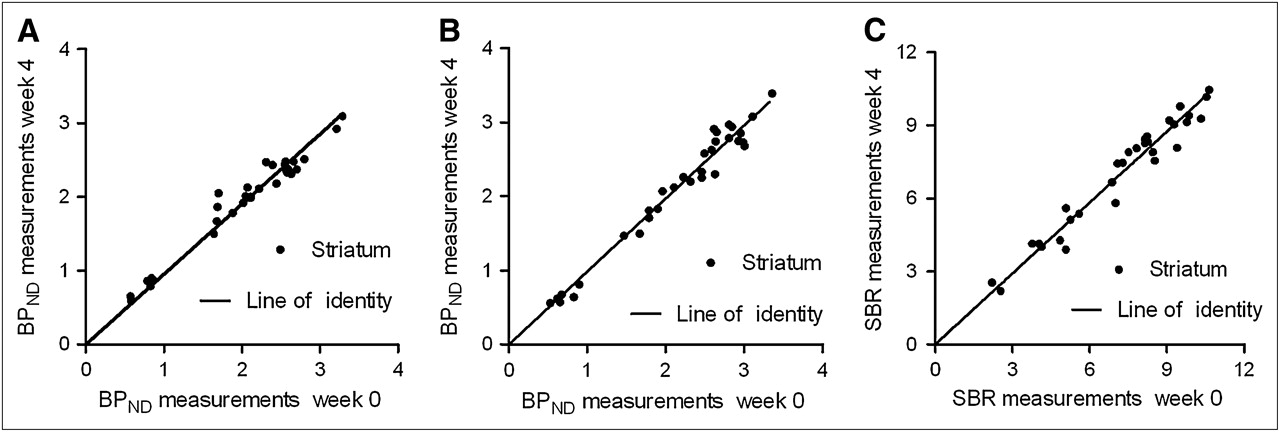
- FIGURE 3.
Intraobserver reproducibility of BPND in striatum for different delineation methods. MD (A), probability map–based delineation (B), and SVI (C). For all 3 methods, both test and retest scans were quantified (32 data points).
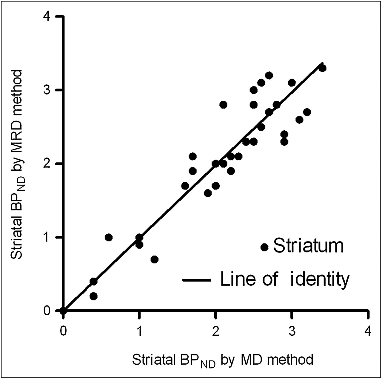
- FIGURE 4.
BPND values for caudate nucleus and putamen by ROI application with MD vs. probability map–based automatic delineation method. Linear regression analysis showed excellent correlation, R2 = 0.96.

Tables
Method Caudate nucleus Putamen Striatum ICC MD (BPND) 10.2% ± 9.2% 9.7% ± 5.4% 7.0% ± 4.1% 0.97 MRD (BPND) 14.2% ± 12.3% 8.1% ± 7.5% 5.7% ± 5.4%* 0.98 SVI (SBR) 6.7% ± 6.0% 0.98 ↵* Calculated striatum = volume-weighted (caudate nucleus+ putamen).
ICC = intraclass correlation coefficient.
No statistically significant better intraobserver reproducibility was observed for any method (MD vs. MRD, MD vs. SVI, MRD vs. SVI; P > 0.1), and all performed equally for intraclass correlation coefficient (n = 16).
Subject Method 1 2 3 4 5 6 7 8 Mean ± SD MD Striatal BPND test 2.06 0.87 2.07 3.21 2.11 0.57 2.58 1.64 1.9 Striatal BPND retest 1.88 0.87 2.55 2.66 2.63 0.58 2.56 2.05 2.0 Reproducibility (%) 9.3 0.1 20.5 18.8 21.6 1.8 0.5 22.2 11.9 ± 10.0 MRD Striatal* BPND test 1.90 0.67 2.46 2.64 2.99 0.65 2.46 1.47 1.9 Striatal* BPND retest 1.67 0.53 2.63 2.65 2.96 0.62 3.11 2.32 2.1 Reproducibility (%) 12.9 23.3 6.6 0.7 1.1 4.1 23.3 45.0 14.6 ± 15.3 SVI Striatal SBR test 7.51 2.21 8.46 10.33 8.23 4.13 8.53 5.08 6.8 Striatal SBR retest 7.08 2.54 8.15 9.85 9.09 4.03 9.78 7.01 7.2 Reproducibility (%) 5.8 14.0 3.7 4.8 9.9 2.5 13.7 32.0 10.8 ± 10.2 ↵* Calculated striatum = volume-weighted (caudate nucleus + putamen).
No significant difference in intrasubject variability was observed for any method (t test, P > 0.5). BPND outcome measurements were not significant different using MD vs. MRD method (t test, P > 0.5; n = 8).
Intrasubject variability* Study Ligand Total patients (n) Delineation Healthy volunteers Patients Reliability ICC Ziebell et al. (17) PE2I 7 MD HC 4.1 ± 3.2 0.96 Seibyl et al. (20) β-CIT 7 Template WS 12.8 ± 8.9† 0.82 Booij et al. (18) FP-CIT 6 Template HC 7.3 ± 3.2 0.92† Tsuchida et al. (21) FP-CIT 10 Template HC 11.1 ± 10.4 0.59 Pirker et al. (31) β-CIT 9 MD HC 8.2 ± 7.2 0.70 Ziebell et al., current study PE2I 8 MD HC 11.9 ± 10.0 0.88 Seibyl et al. (20) β-CIT 7 Template WS 16.8 ± 13.3† 0.82 Booij et al. (18) FP-CIT 6 Template HC 7.9 ± 6.9 0.72† Tsuchida et al. (21) FP-CIT 6 Template HC 7.8 ± 8.9 0.95 Hwang et al. (19) Trodat 20 MD HC 10.2 ± 6.2 0.95


