


Article Figures & Data
Figures
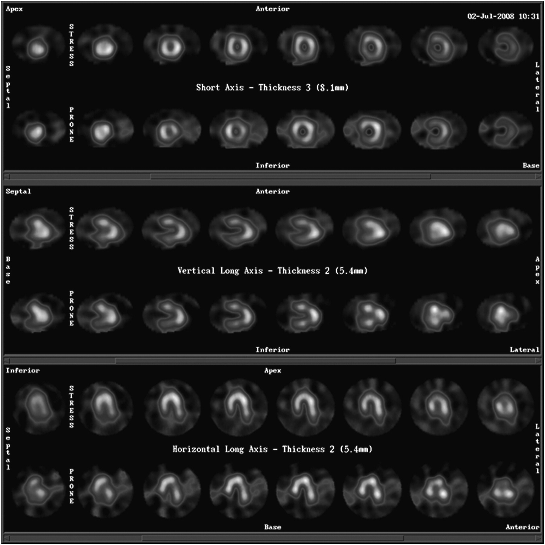
- FIGURE 1.
Supine (upper row) and prone (lower row) myocardial perfusion SPECT. Note irregularity of radiotracer uptake in myocardial walls on prone images.
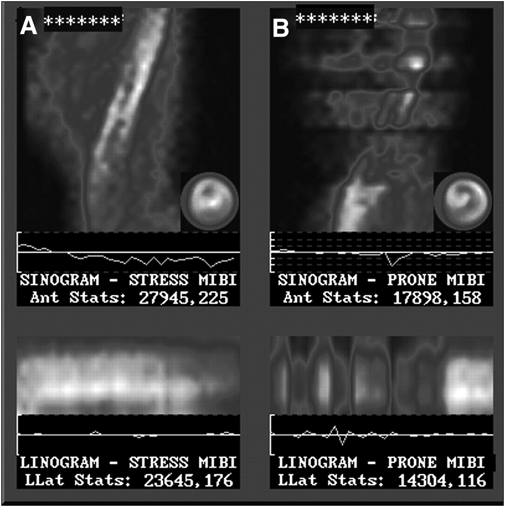
- FIGURE 2.
Linogram and sinogram of supine (A) and prone (B) myocardial perfusion SPECT. Note interruptions in linogram and sinogram on prone images.
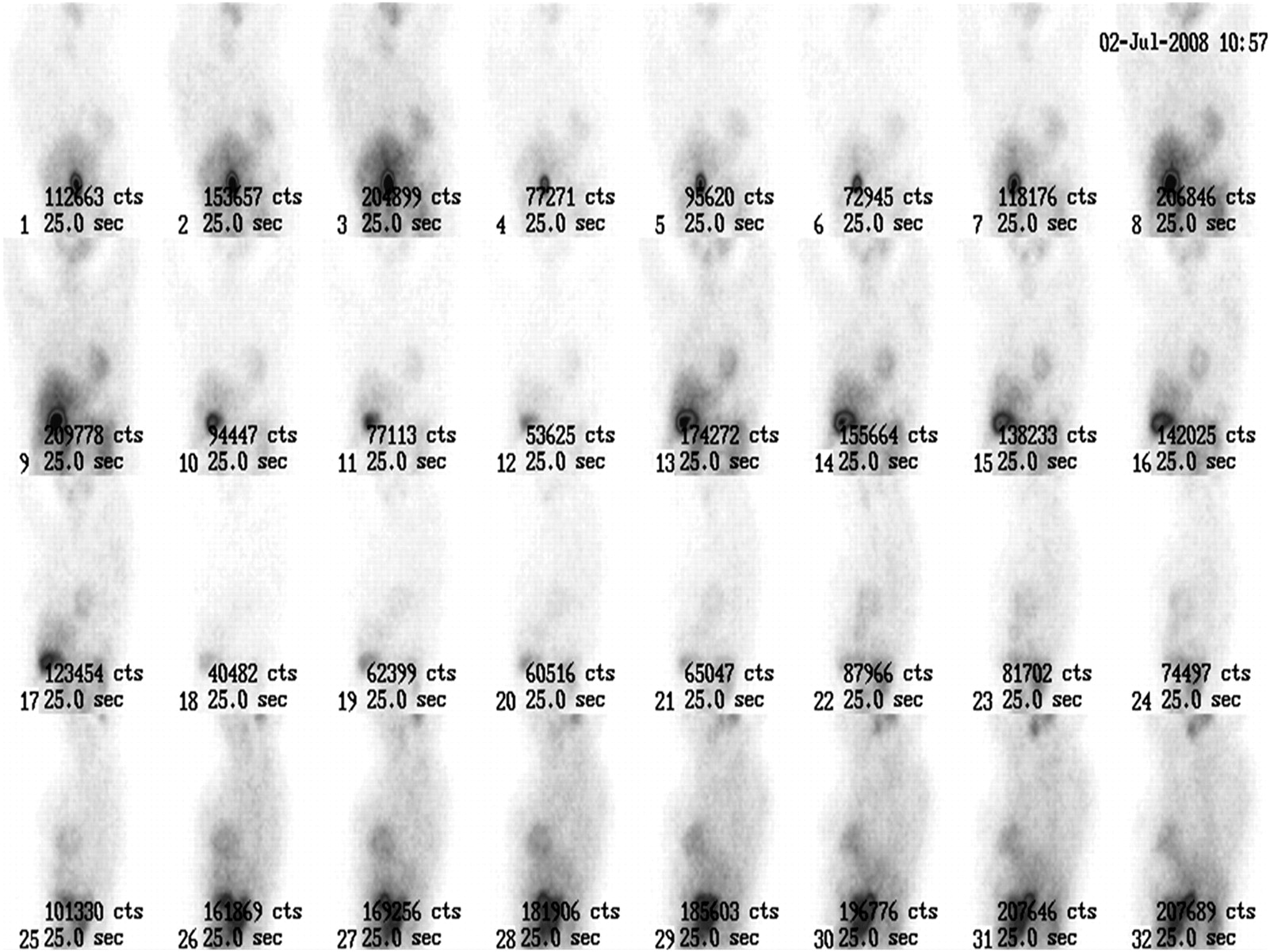
- FIGURE 3.
Thirty-two projections, 25 s each, obtained during myocardial perfusion SPECT. Total counts and intensity of images are significantly different among projections.


