


Article Figures & Data
Figures
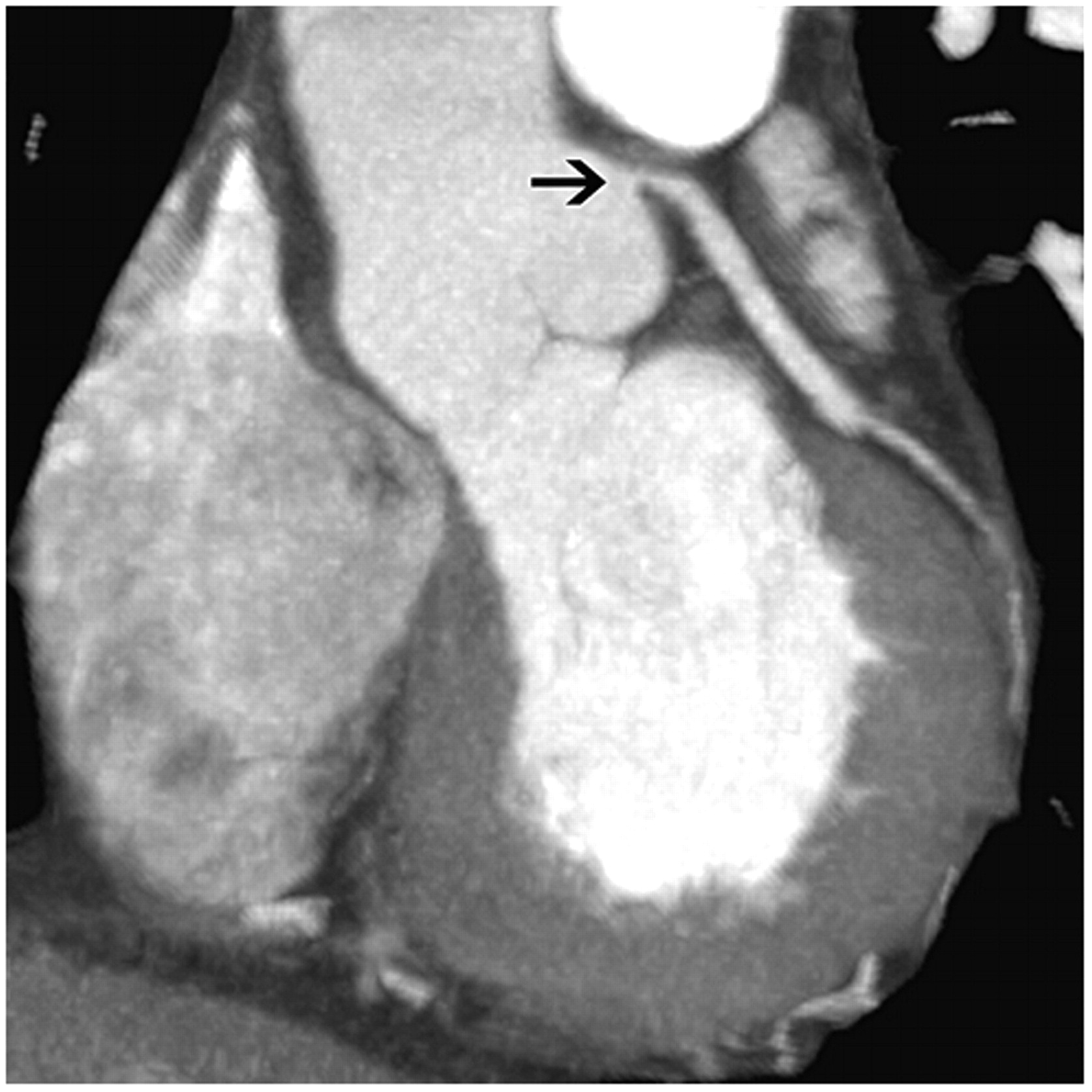
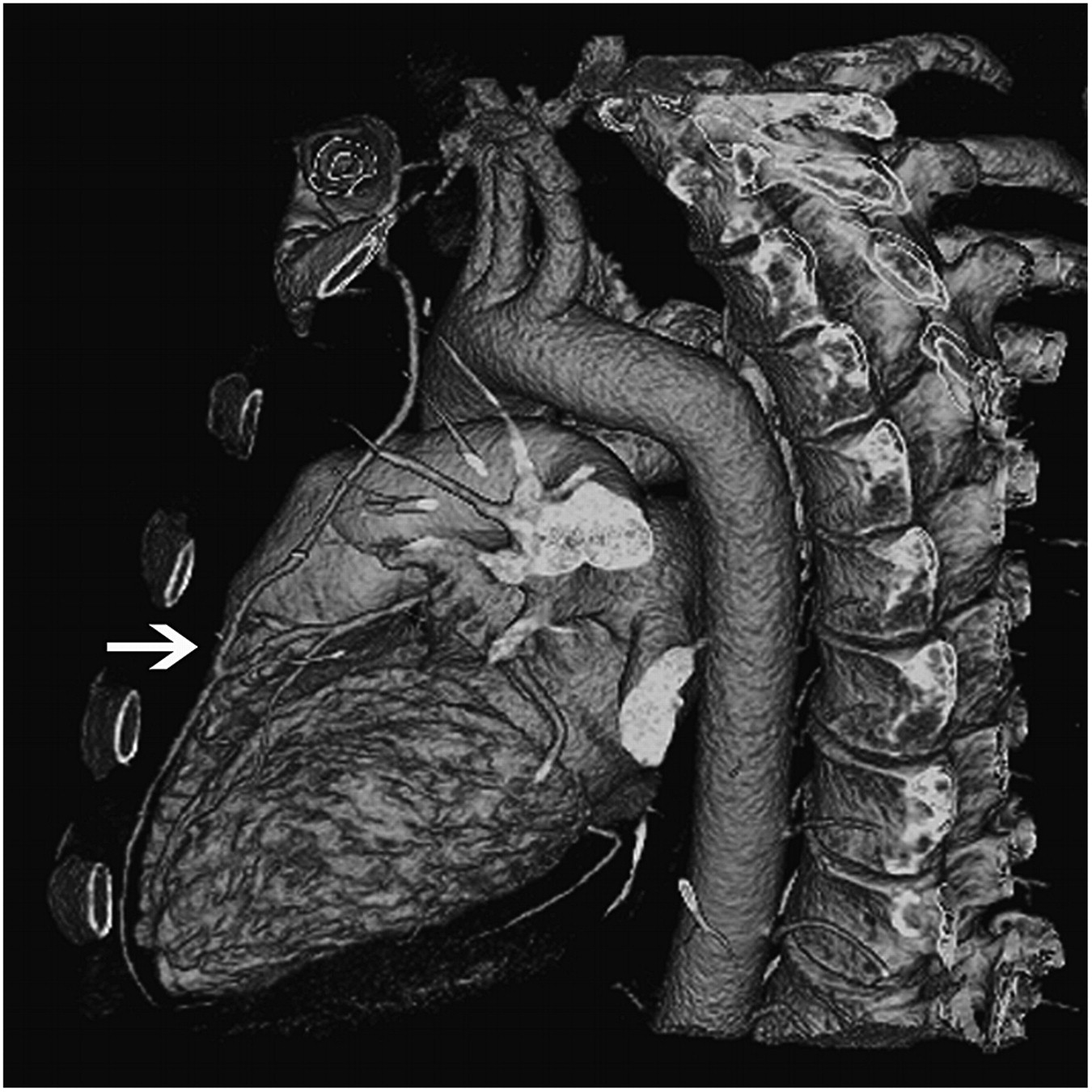
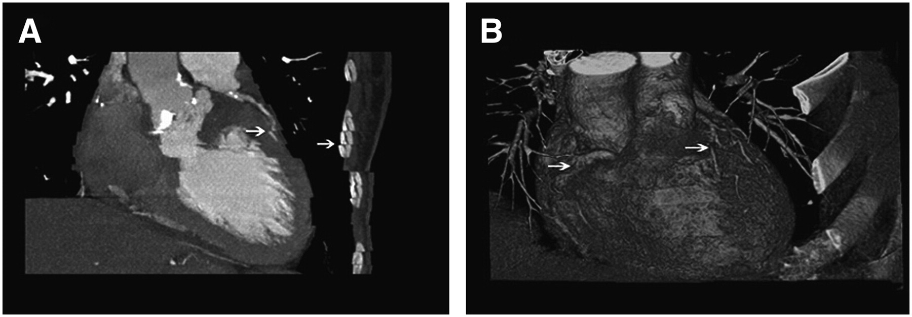
- FIGURE 1.
(A) CCT image obtained from patient who was breathing during image acquisition. Note “stair-step” artifacts, with displacement of trajectory of coronary vessels and chest wall (arrows). (B) 3-Dimensional volume-rendered CT image reconstruction of whole heart. Motion artifacts (arrows) are seen in patients who experience multiple extrasystolic beats during image acquisition.
- FIGURE 2.
(A) Axial image showing left main coronary ostium and its divisions into left anterior descending, ramus intermedius, and left circumflex arteries. (B) Multiplanar reconstructed image. (C) Anatomic 3-dimensional volume-rendered image showing relationships among left main artery, branches, and adjacent cardiac structures. (D) Curved multiplanar reconstruction of entire length of left circumflex artery. A = aorta; CX = left circumflex coronary artery; LA = left atrium; LAD = left anterior descending coronary artery; LM = left main artery.
- FIGURE 3.
Oblique coronal image obtained from patient with anginal symptoms and indeterminate stress test results, showing severe stenosis of ostium of left main coronary artery (arrow).
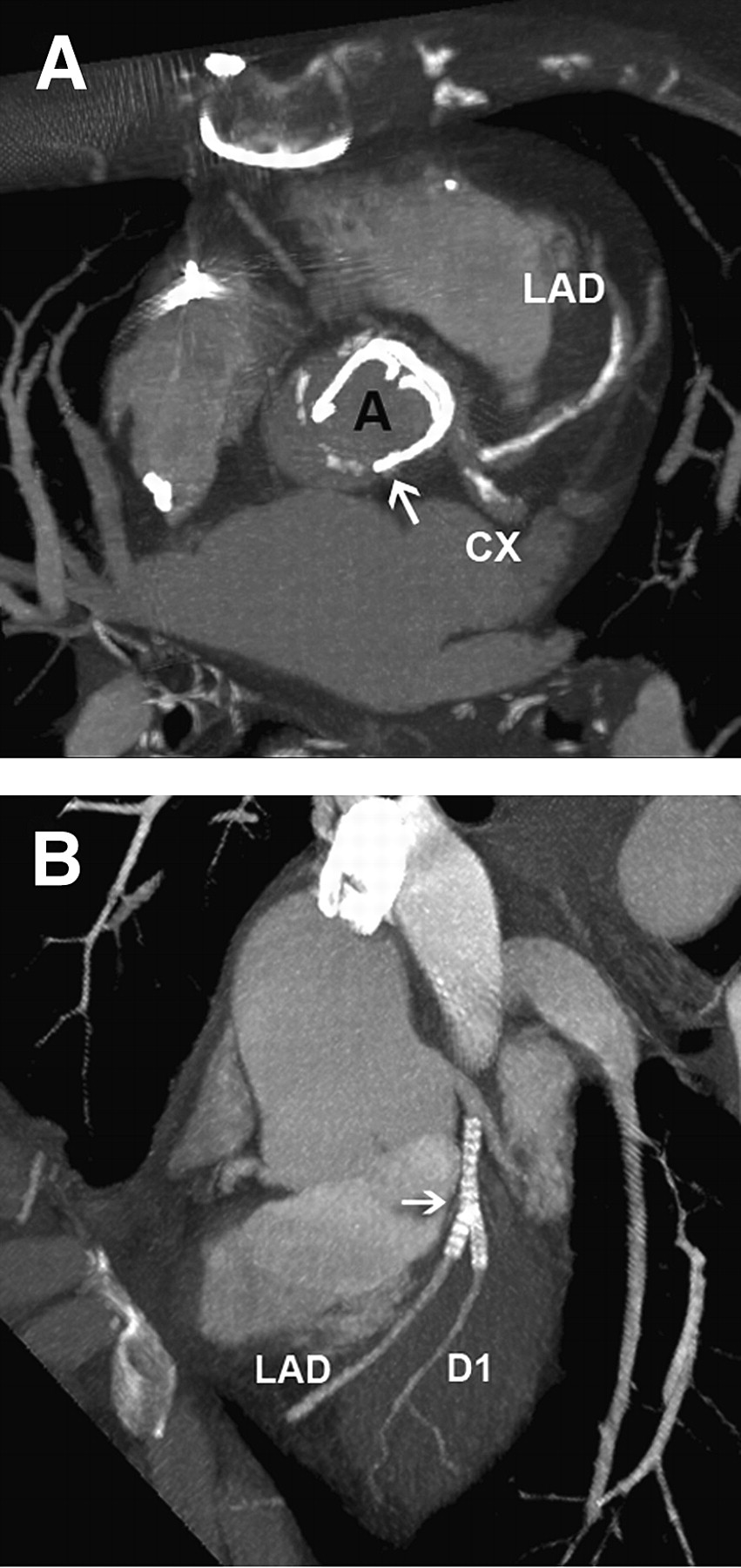
- FIGURE 4.
(A) Axial image obtained at level of origin of left main artery, showing extensive calcification in left anterior descending coronary artery. Aortic mechanical prosthetic valve is visualized (arrow). (B) Maximum-intensity-projection image obtained from patient with “kissing” stents in left anterior descending coronary artery and first diagonal branch. In this case, it is difficult to evaluate lumen because of metallic artifacts. Vessels distal to stents are widely patent. A = aorta; CX = left circumflex coronary artery; D1 = first diagonal branch; LAD = left anterior descending coronary artery.
- FIGURE 5.
Three-dimensional volume-rendered oblique sagittal view obtained from patient with previous bypass surgery. Arrow indicates distal anastomosis of aortocoronary bypass graft to left anterior descending artery.
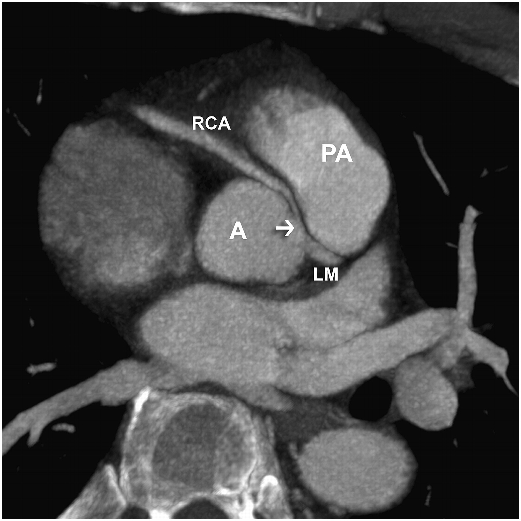
- FIGURE 6.
CCT image obtained for young patient with chest pain. Arrow indicates anomalous origin and course of right coronary artery between aorta and pulmonary arterial trunk. A = aorta; LM = left main artery; PA = pulmonary artery; RCA = right coronary artery.
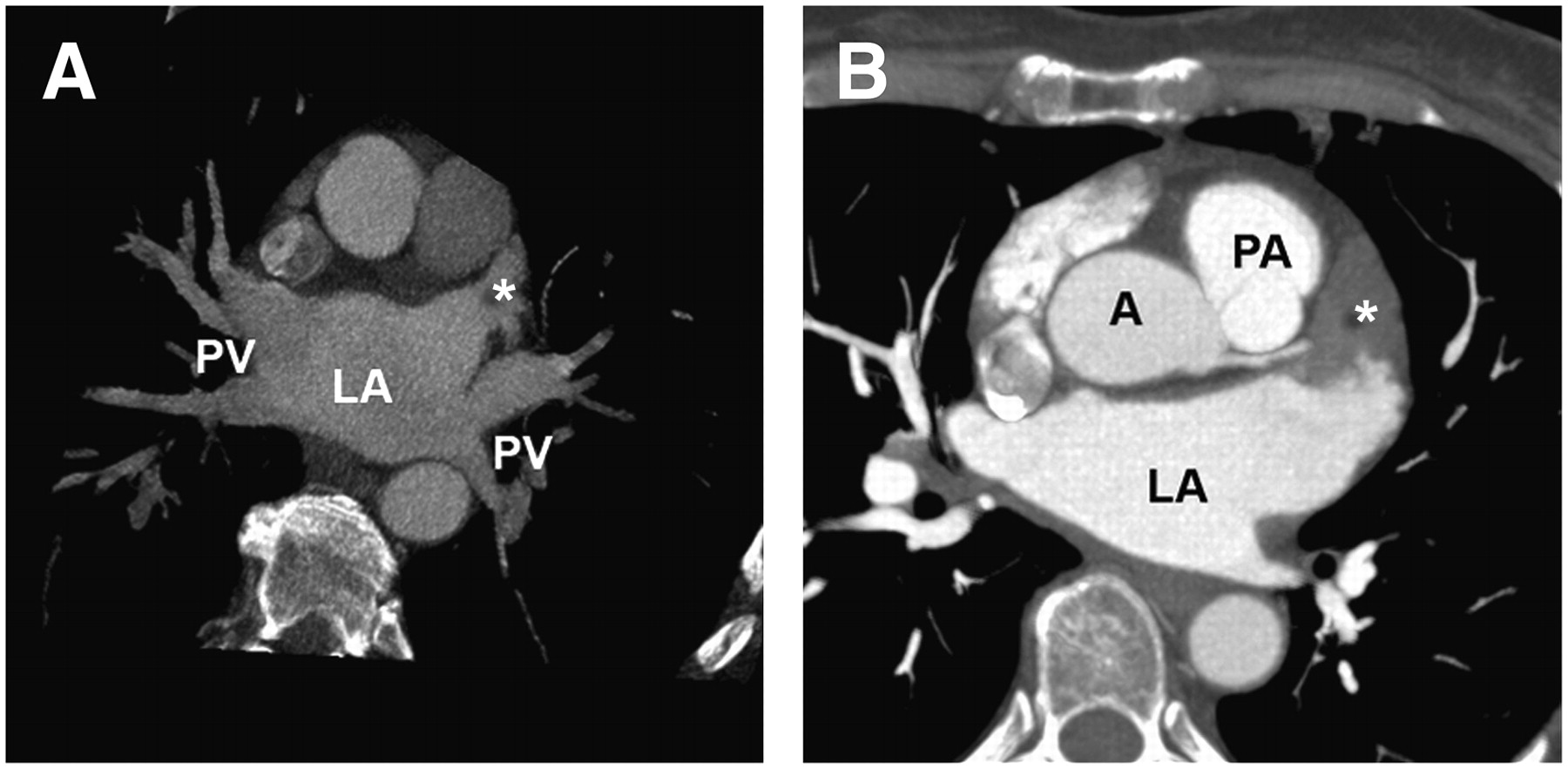
- FIGURE 7.
(A) Axial view showing normal anatomy of 4 pulmonary veins and left atrial appendage clear of thrombus (asterisk). (B) Axial view from another patient undergoing evaluation before radiofrequency ablation of atrial fibrillation. Thrombus (asterisk) is visible in left atrial appendage. A = aorta; LA = left atrium; PA = pulmonary artery; PV = pulmonary vein.


Jump to section
Related Articles
Cited By...
- No citing articles found.