
Main menu

  • Home
  • Content
    • Current
      • JNMT Supplement
    • Ahead of print
    • Past Issues
    • Continuing Education
    • JNMT Podcast
    • SNMMI Annual Meeting Abstracts
  • Subscriptions
    • Subscribers
    • Rates
    • Journal Claims
    • Institutional and Non-member
  • Authors
    • Submit to JNMT
    • Information for Authors
    • Assignment of Copyright
    • AQARA Requirements
  • Info
    • Reviewers
    • Permissions
    • Advertisers
    • Corporate & Special Sales
  • About
    • About Us
    • Editorial Board
    • Contact Information
  • More
    • Alerts
    • Feedback
    • Help
    • SNMMI Journals
  • SNMMI
    • JNMT
    • JNM
    • SNMMI Journals
    • SNMMI

User menu

  • Subscribe
  • My alerts
  • Log in
  • My Cart

Search

  • Advanced search
Journal of Nuclear Medicine Technology
  • SNMMI
    • JNMT
    • JNM
    • SNMMI Journals
    • SNMMI
  • Subscribe
  • My alerts
  • Log in
  • My Cart
Journal of Nuclear Medicine Technology

Advanced Search

  • Home
  • Content
    • Current
    • Ahead of print
    • Past Issues
    • Continuing Education
    • JNMT Podcast
    • SNMMI Annual Meeting Abstracts
  • Subscriptions
    • Subscribers
    • Rates
    • Journal Claims
    • Institutional and Non-member
  • Authors
    • Submit to JNMT
    • Information for Authors
    • Assignment of Copyright
    • AQARA Requirements
  • Info
    • Reviewers
    • Permissions
    • Advertisers
    • Corporate & Special Sales
  • About
    • About Us
    • Editorial Board
    • Contact Information
  • More
    • Alerts
    • Feedback
    • Help
    • SNMMI Journals
  • Watch or Listen to JNMT Podcast
  • Visit SNMMI on Facebook
  • Join SNMMI on LinkedIn
  • Follow SNMMI on Twitter
  • Subscribe to JNMT RSS feeds
OtherCONTINUING EDUCATION

Principles of CT: Radiation Dose and Image Quality

Lee W. Goldman
Journal of Nuclear Medicine Technology December 2007, 35 (4) 213-225; DOI: https://doi.org/10.2967/jnmt.106.037846
Lee W. Goldman
  • Find this author on Google Scholar
  • Find this author on PubMed
  • Search for this author on this site
  • Article
  • Figures & Data
  • Info & Metrics
  • PDF
Loading

Article Figures & Data

Figures

  • FIGURE 1. 
    • Download figure
    • Open in new tab
    • Download powerpoint
    FIGURE 1. 

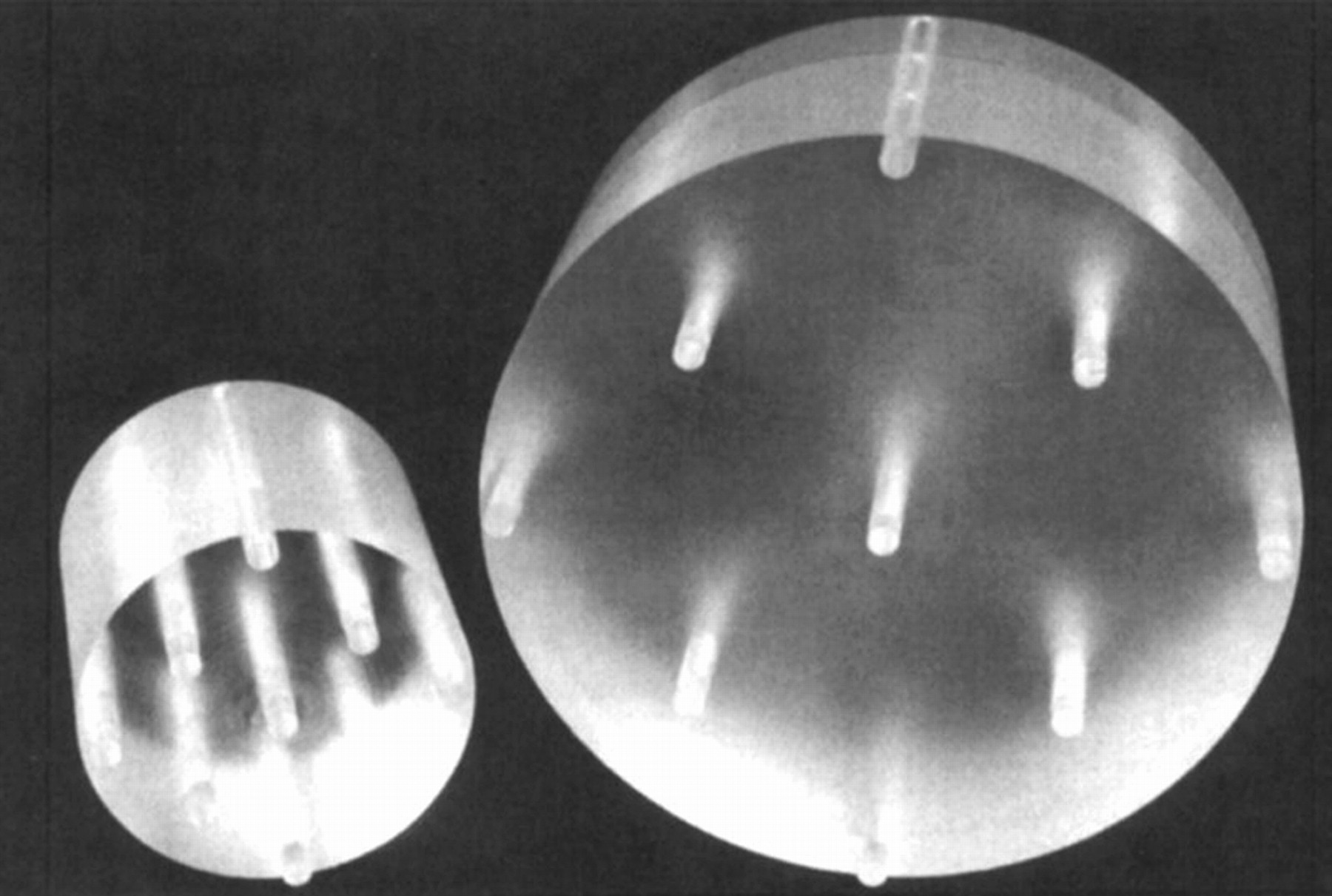
    Standard CT dosimetry phantoms consist of cylindric acrylic phantoms with holes for dosimeter insertion at various locations. The 2 sizes are 16 cm in diameter to represent heads and small pediatric bodies and 32 cm in diameter to represent adult bodies. (Courtesy of Lawrence Rothenberg.)

  • FIGURE 2. 
    • Download figure
    • Open in new tab
    • Download powerpoint
    FIGURE 2. 

    Phantom insert to hold TLDs used to measure dose profiles. TLDs are close together within x-ray primary beam and are farther apart to measure profile tails.

  • FIGURE 3. 
    • Download figure
    • Open in new tab
    • Download powerpoint
    FIGURE 3. 

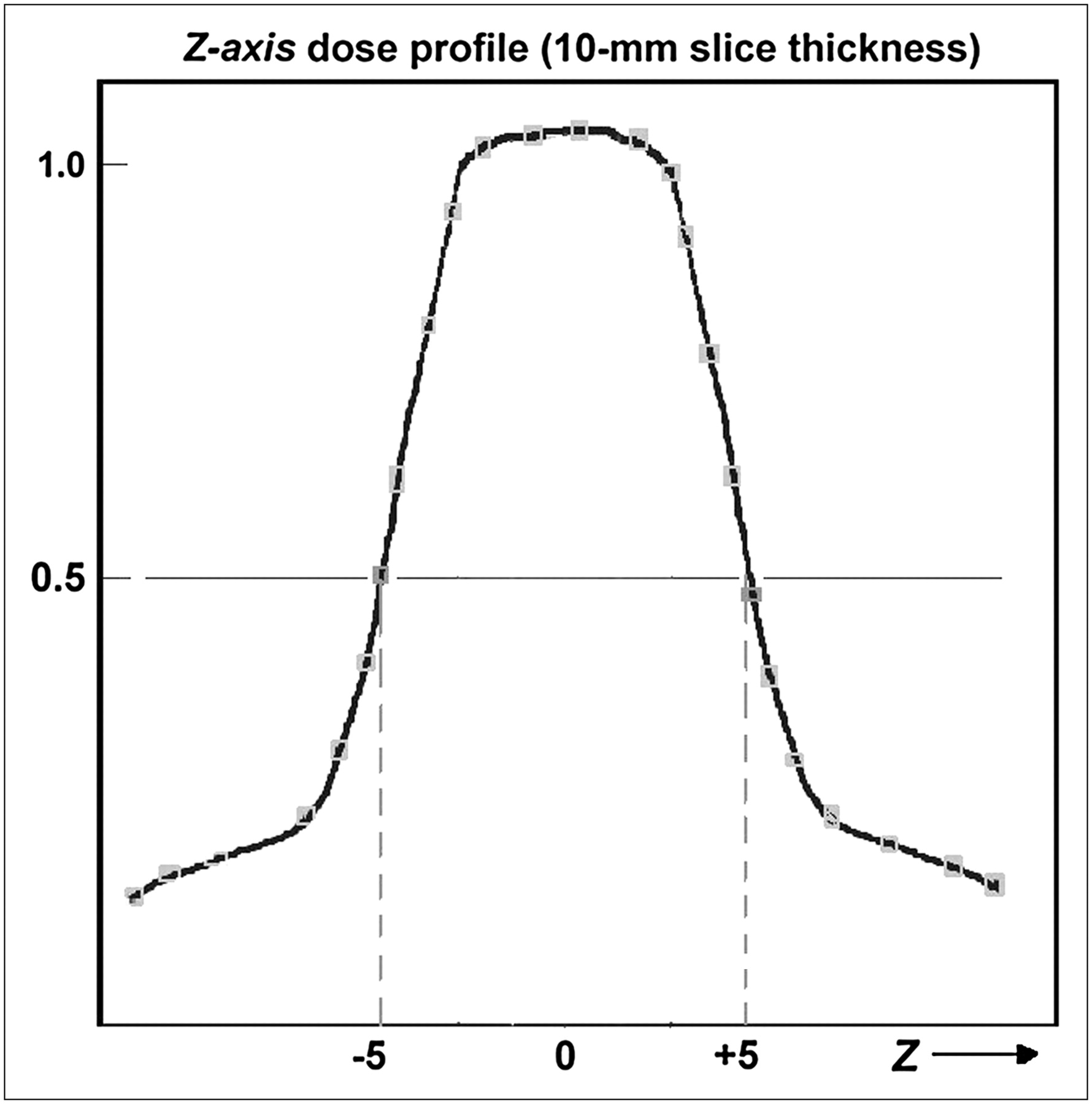
    Example of doses measured by TLDs inside phantom for a 10-mm slice thickness, plotted as function of TLD position along z-axis. This type of plot is referred to as a dose profile. Slice thickness for single-slice CT scanners is usually equivalent to full width at half maximum of dose profile.

  • FIGURE 4. 
    • Download figure
    • Open in new tab
    • Download powerpoint
    FIGURE 4. 

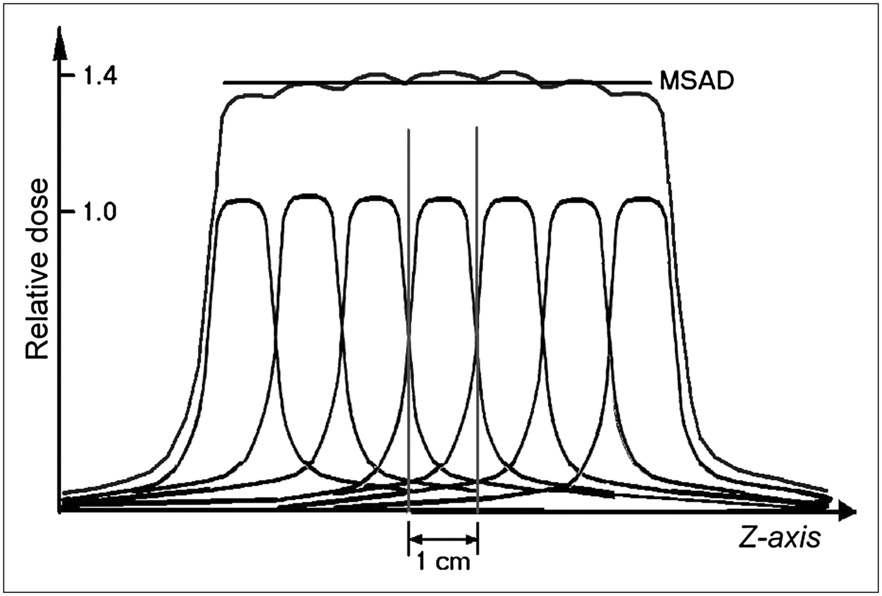
    Cumulative dose from a series of contiguous slices is known as multislice average dose (MSAD). Dose-profile tails extend quite far from center of x-ray beam and thus contribute radiation dose to nearby slices. Total dose for a procedure consists of several contiguous slices and is typically 25%–40% higher than single-slice dose.

  • FIGURE 5. 
    • Download figure
    • Open in new tab
    • Download powerpoint
    FIGURE 5. 

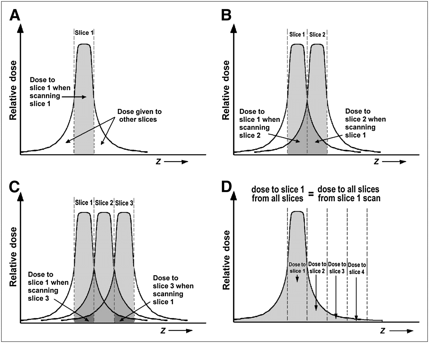
    (A) Dose to slice 1 from scanning of slice 1. (B) Dose to slice 1 from scanning of slice 2 equals dose to slice 2 from scanning of slice 1. (C) Dose to slice 1 from scanning of slice 3 equals dose to slice 3 from scanning of slice 1. (D) Dose to slice 1 from scanning of all slices equals dose to all slices from scanning of slice 1, or total area under dose profile, which is measured using long ionization chamber.
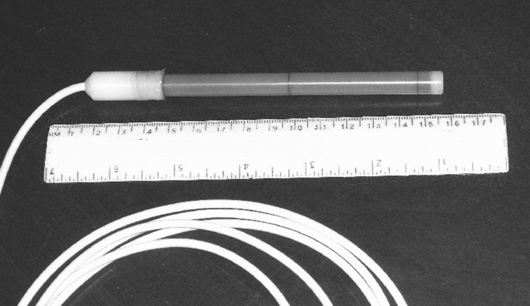
  • FIGURE 6. 
    • Download figure
    • Open in new tab
    • Download powerpoint
    FIGURE 6. 

    Example of 100-mm-long CT ionization chamber for measuring CTDI.

  • FIGURE 7. 
    • Download figure
    • Open in new tab
    • Download powerpoint
    FIGURE 7. 

    Typical central and peripheral doses (CTDI) in head and body phantoms. Central dose is about equal to peripheral dose in head phantom and is more than half the peripheral dose in body phantom. Weighted sum of central and peripheral doses, known as CTDIw, is single-number estimate of patient radiation dose to scanned volume.
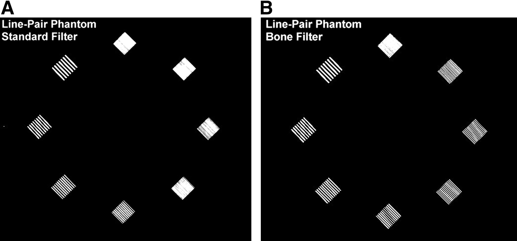
  • FIGURE 8. 
    • Download figure
    • Open in new tab
    • Download powerpoint
    FIGURE 8. 

    CT spatial resolution phantom, consisting of 4–12 line-pairs per centimeter (from American College of Radiology accreditation phantom), reconstructed using standard (A) and bone (B, high-resolution) filters.

  • FIGURE 9. 
    • Download figure
    • Open in new tab
    • Download powerpoint
    FIGURE 9. 

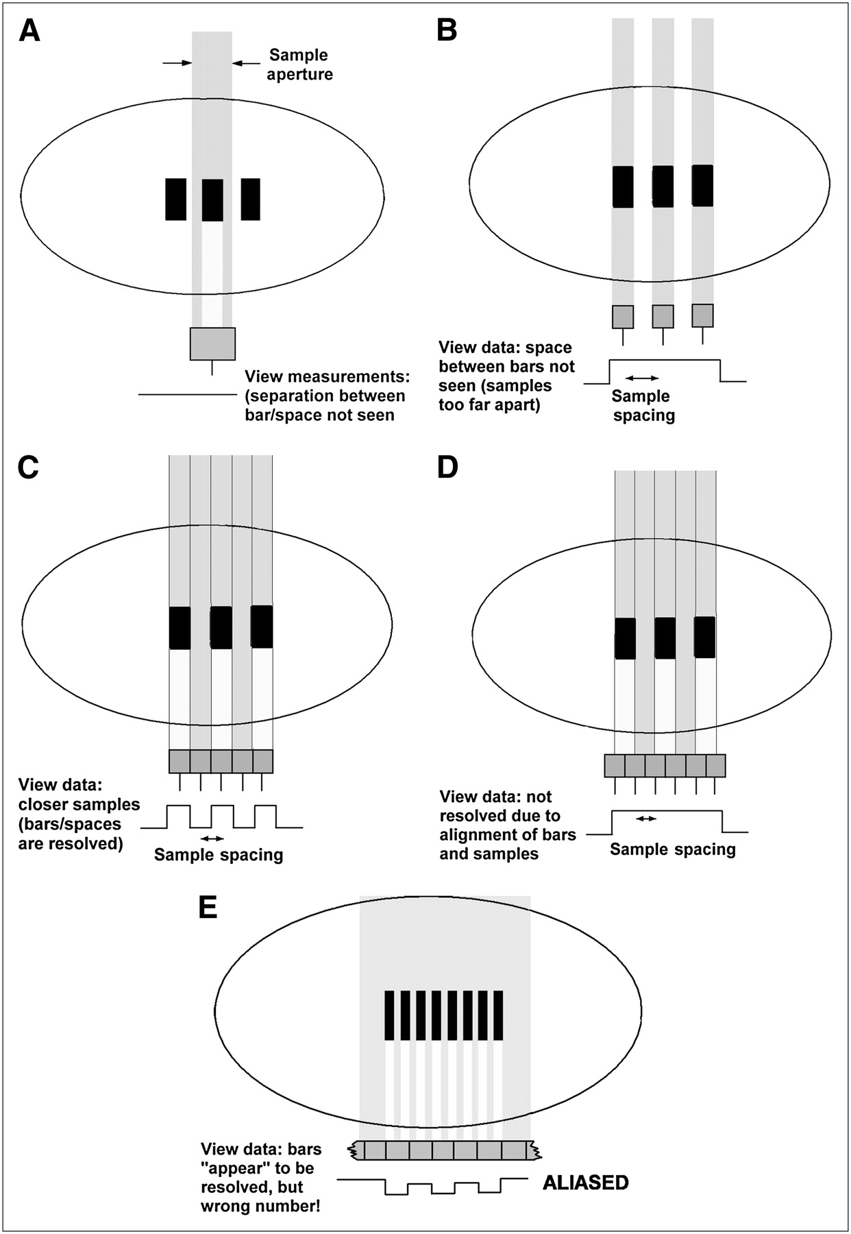
    CT resolution is limited by sampling—size and spacing of measurements (samples) used to form image. (A) Pattern is unresolved because sample size (aperture) is too large. (B) Pattern is unresolved because samples are too far apart. (C) Aperture size and sample spacing are adequate to resolve pattern. (D) “Effective” resolution may be lower than expected because of position of samples relative to pattern. (E) In aliasing, pattern seems to be resolved but with incorrect number of bars.

  • FIGURE 10. 
    • Download figure
    • Open in new tab
    • Download powerpoint
    FIGURE 10. 

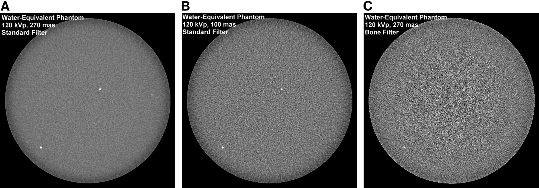
    CT image noise depends on number of x-ray photons contributing to image. (A and B) Comparison of noise from scans using 270 mAs (typical clinical value) and 100 mAs. (C) Appearance of image noise is strongly affected by reconstruction filter; sharp filter such as bone also sharpens (enhances) appearance of noise.

  • FIGURE 11. 
    • Download figure
    • Open in new tab
    • Download powerpoint
    FIGURE 11. 

    Low-contrast phantom to test CT performance in presence of typical image noise levels (from American College of Radiology accreditation phantom). Five-millimeter rods are visible, but smaller ones are obscured by noise.

  • FIGURE 12. 
    • Download figure
    • Open in new tab
    • Download powerpoint
    FIGURE 12. 

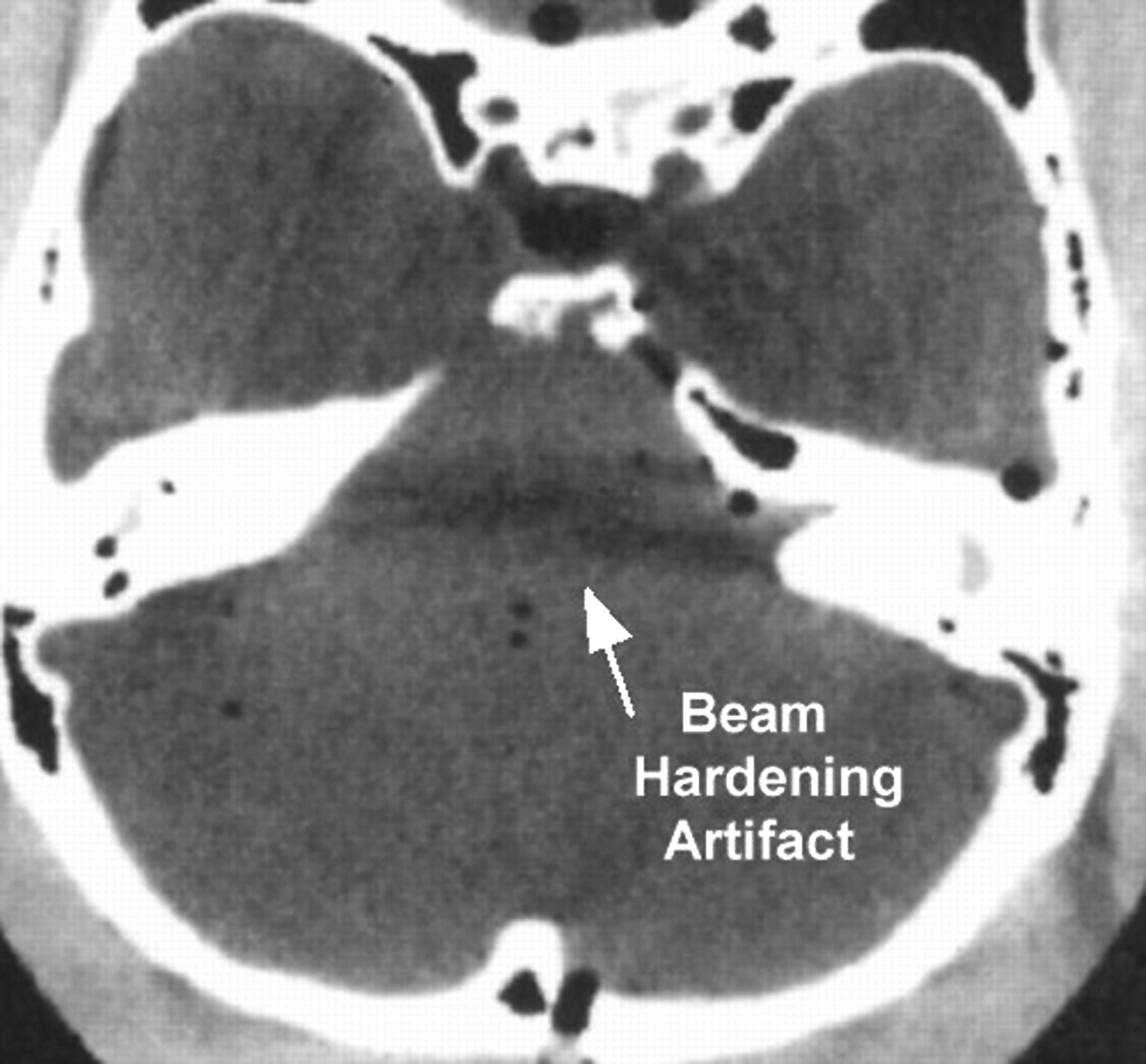
    Beam-hardening artifact caused by unusually severe hardening of x-rays passing though thick bone.

  • FIGURE 13. 
    • Download figure
    • Open in new tab
    • Download powerpoint
    FIGURE 13. 

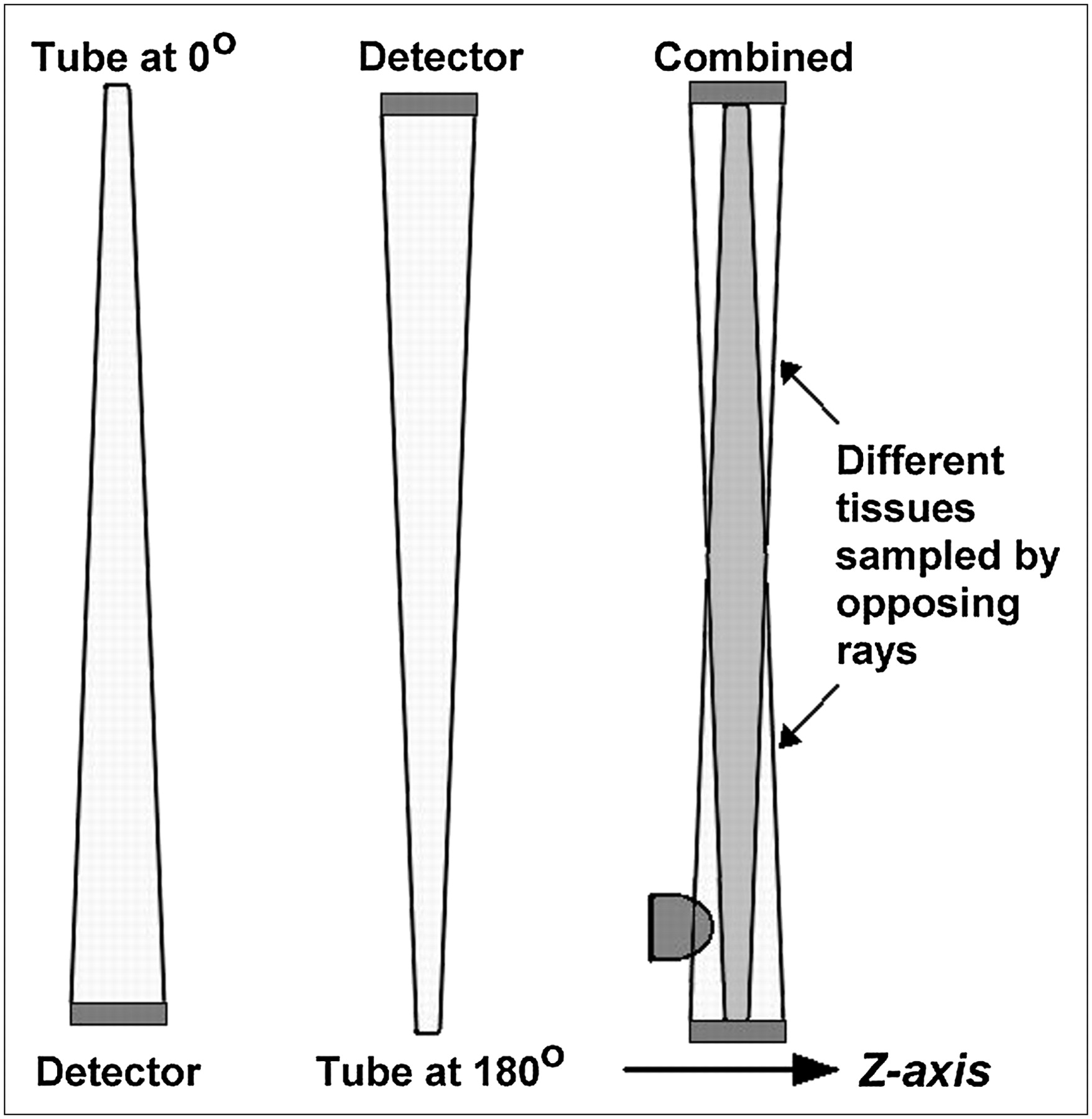
    Partial-volume streaks are caused by opposing x-ray beams, which nominally pass through the same voxels but actually sample slightly different cone-shaped tissue volumes as a result of beam divergence. Small structure, such as piece of bone, is detected by beam from one direction but is missed by opposing beam. Resulting inconsistency leads to streak artifact.

PreviousNext
Back to top

In this issue

Journal of Nuclear Medicine Technology: 35 (4)
Journal of Nuclear Medicine Technology
Vol. 35, Issue 4
December 2007
  • Table of Contents
  • About the Cover
  • Index by author
Print
Download PDF
Article Alerts
Sign In to Email Alerts with your Email Address
Email Article

Thank you for your interest in spreading the word on Journal of Nuclear Medicine Technology.

NOTE: We only request your email address so that the person you are recommending the page to knows that you wanted them to see it, and that it is not junk mail. We do not capture any email address.

Enter multiple addresses on separate lines or separate them with commas.
Principles of CT: Radiation Dose and Image Quality
(Your Name) has sent you a message from Journal of Nuclear Medicine Technology
(Your Name) thought you would like to see the Journal of Nuclear Medicine Technology web site.
Citation Tools
Principles of CT: Radiation Dose and Image Quality
Lee W. Goldman
Journal of Nuclear Medicine Technology Dec 2007, 35 (4) 213-225; DOI: 10.2967/jnmt.106.037846

Citation Manager Formats

  • BibTeX
  • Bookends
  • EasyBib
  • EndNote (tagged)
  • EndNote 8 (xml)
  • Medlars
  • Mendeley
  • Papers
  • RefWorks Tagged
  • Ref Manager
  • RIS
  • Zotero
Share
Principles of CT: Radiation Dose and Image Quality
Lee W. Goldman
Journal of Nuclear Medicine Technology Dec 2007, 35 (4) 213-225; DOI: 10.2967/jnmt.106.037846
Twitter logo Facebook logo LinkedIn logo Mendeley logo
  • Tweet Widget
  • Facebook Like
  • Google Plus One
Bookmark this article

Jump to section

  • Article
    • Abstract
    • CT DOSIMETRY AND RADIATION DOSE
    • CT IMAGE QUALITY
    • CONCLUSION
    • APPENDIX A
    • APPENDIX B
    • Footnotes
    • References
  • Figures & Data
  • Info & Metrics
  • PDF

Related Articles

  • No related articles found.
  • PubMed
  • Google Scholar

Cited By...

  • Optimizing Contrast Resolution in Digital Chest Radiography by Varying Copper Filtration and kVp
  • Assessment of Image Quality in Chest CT With Precision Matrix and Increased Framing Rate Using Single Source CT: A Phantom Study
  • Micro-CT analyses of the lung in mice: Parameters influencing the radiation dose and acquisition quality
  • Radiation Protection in Computed Tomography
  • A comparative evaluation of cone beam CT and micro-CT on trabecular bone structures in the human mandible
  • Influence of cone beam CT scanning parameters on grey value measurements at an implant site
  • Influence of scan setting selections on root canal visibility with cone beam CT
  • Adult patient radiation doses from non-cardiac CT examinations: a review of published results
  • Assessment of Patient Exposure to X-Radiation from SPECT/CT Scanners
  • Principles of CT: Multislice CT
  • Google Scholar

More in this TOC Section

  • Illuminating the Hidden: Standardizing Cardiac MIBG Imaging for Sympathetic Dysfunction
  • PET/CT Case Series: Unmasking the Mystery of Cardiac Sarcoidosis
  • Delivery Methods of Radiopharmaceuticals: Exploring Global Strategies to Minimize Occupational Radiation Exposure
Show more Continuing Education

Similar Articles

SNMMI

© 2025 SNMMI

Powered by HighWire