


Article Figures & Data
Figures
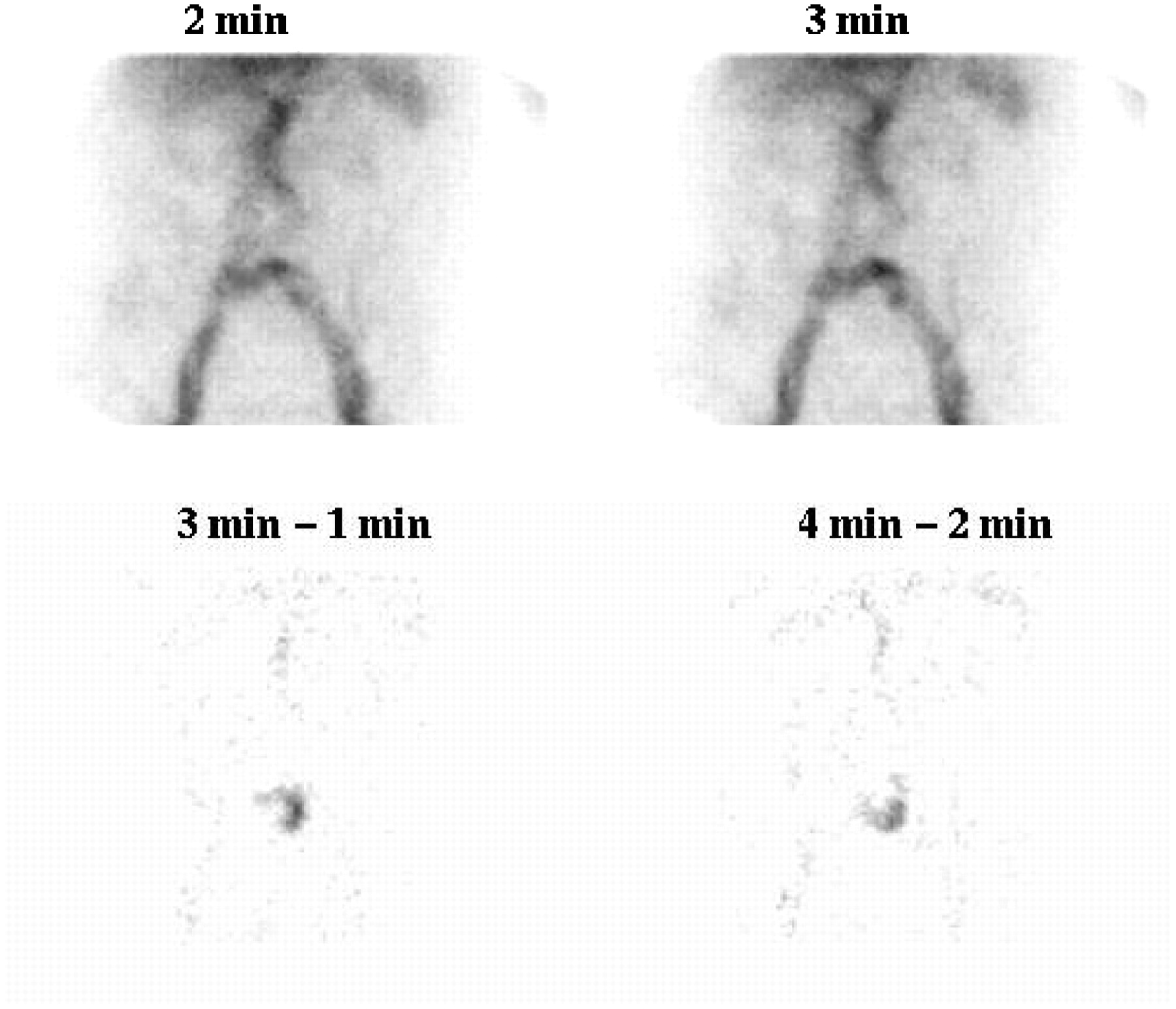
- FIGURE 1.
(Top) Study showing accumulation of activity that 3 observers identified on conventional scintigraphy as a bleed. One observer thought the bleed to originate in small bowel; another observer, in ascending colon; and a third observer, in cecum. (Bottom) Alternate sequential subtraction scintigraphy demonstrates that activity remains constant over time (indicated by absence of activity), neither increasing in intensity (no additional bleeding) nor moving within bowel. Activity most likely represents a vessel consistent with prominent superficial vasculature.
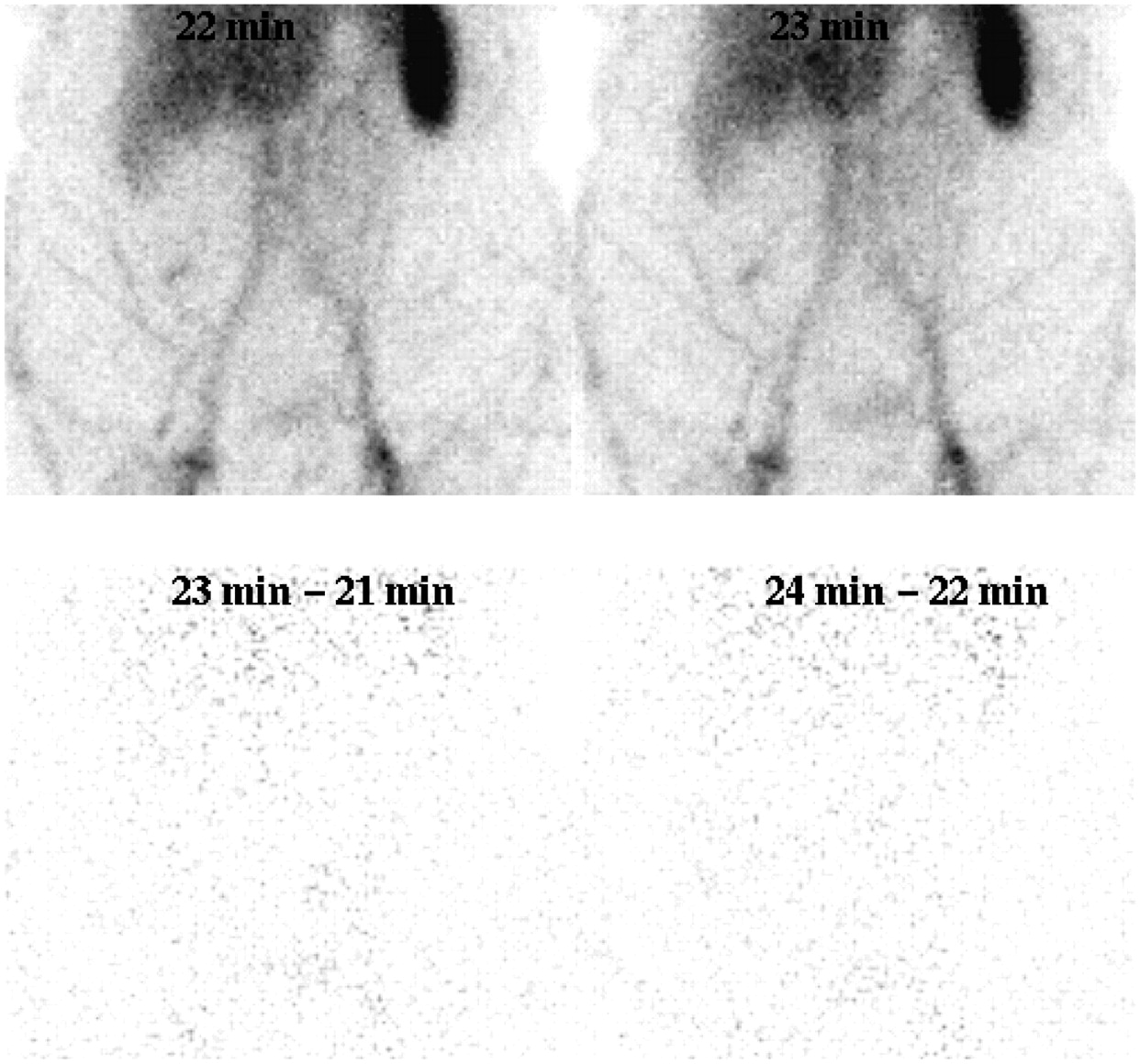
- FIGURE 2.
Study showing accumulation of activity superimposed over blood vessels on early conventional scintigrams (top). This finding may be viewed as a normal variant or tortuosity. Alternate sequential subtraction scintigraphy (bottom) clearly shows presence and location of bleed site.

Tables
- TABLE 1
Diagnostic Outcomes for 4 Interpreting Physicians for Each of 2 Interpretive Rounds
Processing method Outcome Observer 1 Observer 2 Observer 3 Observer 4 Conventional alone False-positive 0% (0) 23.8% (10) 7.1% (3) 11.9% (5) False-negative 14.3% (1) 14.3% (1) 0% (0) 0% (0) True-positive 85.7% (6) 85.7% (6) 100% (7) 100% (7) True-negative 100% (42) 76.2% (32) 92.9% (39) 88.1% (37) All 3 methods False-positive 0% (0) 14.3% (6) 0% (0) 2.4% (1) False-negative 14.3% (1) 14.3% (1) 14.3% (1) 0% (0) True-positive 85.7% (6) 85.7% (6) 85.7% (6) 100% (7) True-negative 100% (42) 85.7% (36) 100% (42) 97.6% (41) - TABLE 2
Interobserver Agreement and Agreement with Actual Diagnostic Outcome for Interpretive Rounds 1 and 2
Versus observer Conventional method alone All 3 processing methods Observer R2 κ R2 κ One Two 0.437 — 0.512 — Three 0.578 — 1.0 — Four 0.512 — 0.793 — Two Three 0.439 0.436 0.512 — Four 0.441 0.367 0.800 0.574 Three Four 0.632 0.673 0.793 — Cohen's κ-coefficient could not be calculated when no false-positives and false-negatives existed for an observer.
- TABLE 3
Contingency Analysis of Confidence of Bleed Detection (or Absence) Between Interpretive Rounds 1 and 2

Processing method n Mean contribution (%) 95% CI of mean (%) P against conventional P against reference subtraction P against alternate sequential subtraction Conventional 116 84.8 81.3–88.3 <0.001 <0.001 Reference subtraction 116 3.6 2.1–5.1 <0.001 <0.001 Alternate sequential subtraction 116 11.6 8.5–14.7 <0.001 <0.001 Statistically significant differences between means are supported by lack of overlap of 95% CIs.
Processing method n Mean contribution (%) 95% CI of mean (%) P against conventional P against reference subtraction P against alternate sequential subtraction Conventional 30 87.3 81.1–93.6 <0.001 <0.001 Reference subtraction 30 10.0 4.2–15.8 <0.001 0.015 Alternate sequential subtraction 30 2.7 1.1–4.2 <0.001 0.015 Statistically significant differences between means are supported by lack of overlap of 95% CIs.
Processing method n Mean contribution (%) 95% CI of mean (%) P against conventional P against reference subtraction P against alternate sequential subtraction Conventional 158 83.8 81.0–86.6 <0.001 <0.001 Reference subtraction 158 5.0 3.7–6.4 <0.001 <0.001 Alternate sequential subtraction 158 11.2 8.7–13.7 <0.001 <0.001 Statistically significant differences between means are supported by lack of overlap of 95% CIs.
Processing method Sensitivity Specificity Accuracy Area under ROC curve Conventional 100% 61.9% 67.3% 0.973 Reference subtraction 100% 81.0% 83.7% 0.917 Alternate sequential subtraction 100% 100% 100% 1.0


Jump to section
Related Articles
Cited By...
- No citing articles found.