
Main menu

  • Home
  • Content
    • Current
      • JNMT Supplement
    • Ahead of print
    • Past Issues
    • Continuing Education
    • JNMT Podcast
    • SNMMI Annual Meeting Abstracts
  • Subscriptions
    • Subscribers
    • Rates
    • Journal Claims
    • Institutional and Non-member
  • Authors
    • Submit to JNMT
    • Information for Authors
    • Assignment of Copyright
    • AQARA Requirements
  • Info
    • Reviewers
    • Permissions
    • Advertisers
    • Corporate & Special Sales
  • About
    • About Us
    • Editorial Board
    • Contact Information
  • More
    • Alerts
    • Feedback
    • Help
    • SNMMI Journals
  • SNMMI
    • JNMT
    • JNM
    • SNMMI Journals
    • SNMMI

User menu

  • Subscribe
  • My alerts
  • Log in
  • My Cart

Search

  • Advanced search
Journal of Nuclear Medicine Technology
  • SNMMI
    • JNMT
    • JNM
    • SNMMI Journals
    • SNMMI
  • Subscribe
  • My alerts
  • Log in
  • My Cart
Journal of Nuclear Medicine Technology

Advanced Search

  • Home
  • Content
    • Current
    • Ahead of print
    • Past Issues
    • Continuing Education
    • JNMT Podcast
    • SNMMI Annual Meeting Abstracts
  • Subscriptions
    • Subscribers
    • Rates
    • Journal Claims
    • Institutional and Non-member
  • Authors
    • Submit to JNMT
    • Information for Authors
    • Assignment of Copyright
    • AQARA Requirements
  • Info
    • Reviewers
    • Permissions
    • Advertisers
    • Corporate & Special Sales
  • About
    • About Us
    • Editorial Board
    • Contact Information
  • More
    • Alerts
    • Feedback
    • Help
    • SNMMI Journals
  • Watch or Listen to JNMT Podcast
  • Visit SNMMI on Facebook
  • Join SNMMI on LinkedIn
  • Follow SNMMI on Twitter
  • Subscribe to JNMT RSS feeds
OtherIMAGING

Improved Detection and Localization of Lower Gastrointestinal Tract Hemorrhage by Subtraction Scintigraphy: Phantom Analysis

Geoffrey M. Currie, Philip A. Towers and Janelle M. Wheat
Journal of Nuclear Medicine Technology September 2006, 34 (3) 160-168;
Geoffrey M. Currie
  • Find this author on Google Scholar
  • Find this author on PubMed
  • Search for this author on this site
Philip A. Towers
  • Find this author on Google Scholar
  • Find this author on PubMed
  • Search for this author on this site
Janelle M. Wheat
  • Find this author on Google Scholar
  • Find this author on PubMed
  • Search for this author on this site
  • Article
  • Figures & Data
  • Info & Metrics
  • PDF
Loading

Article Figures & Data

Figures

  • Tables
  • FIGURE 1. 
    • Download figure
    • Open in new tab
    • Download powerpoint
    FIGURE 1. 

    Sequential subtraction images of normal study. Liver (top arrow) and large vessel (bottom arrow) activities can be faintly visualized.
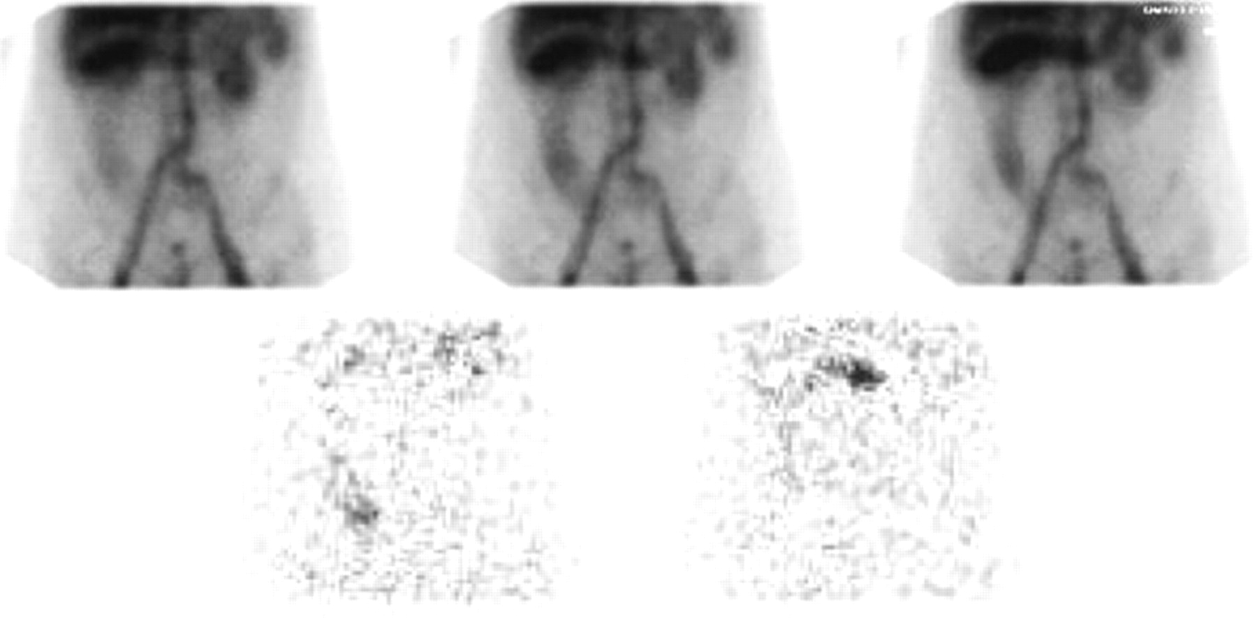
  • FIGURE 2. 
    • Download figure
    • Open in new tab
    • Download powerpoint
    FIGURE 2. 

    (Top) Conventional 99mTc-labeled RBC scintigrams demonstrating diffuse ascending colon accumulation of radiopharmaceutical without identification of specific bleeding site. Focal accumulation of radiopharmaceutical in transverse colon may suggest bleeding origin with rapid retrograde movement of blood into ascending colon. (Bottom) SSS demonstrates proximal ascending colon bleeding source that precedes the transverse colon accumulation.
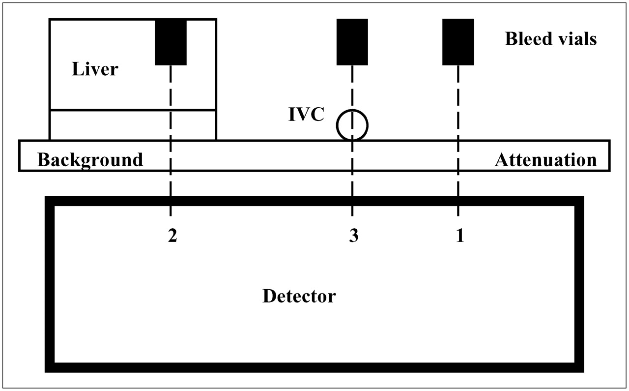
  • FIGURE 3. 
    • Download figure
    • Open in new tab
    • Download powerpoint
    FIGURE 3. 

    Cross section through phantom apparatus simulating bleed locations in background (1), superimposed on liver (2), and superimposed on IVC (3).
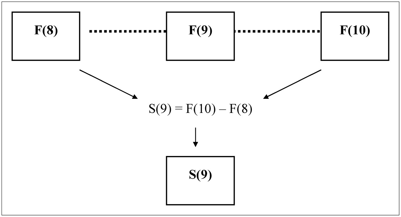
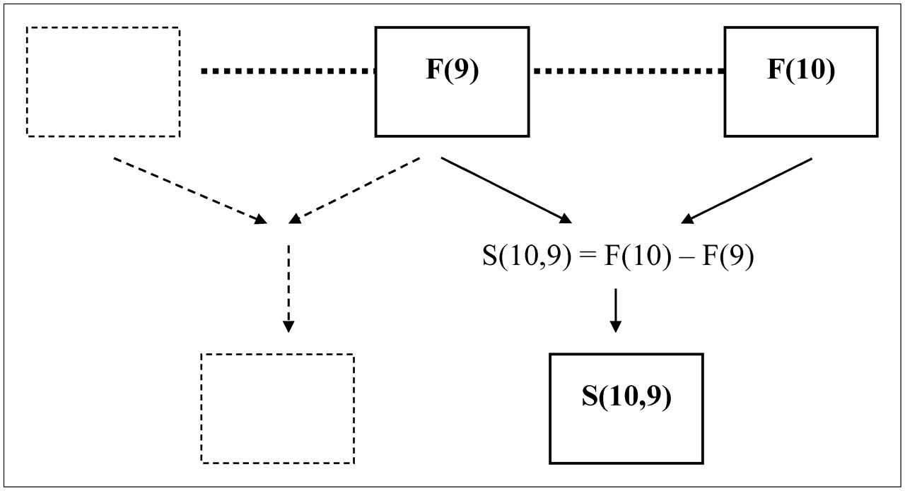
  • FIGURE 4. 
    • Download figure
    • Open in new tab
    • Download powerpoint
    FIGURE 4. 

    Schematic representation of RSS. Each individual frame has subtracted from it the initial reference frame.
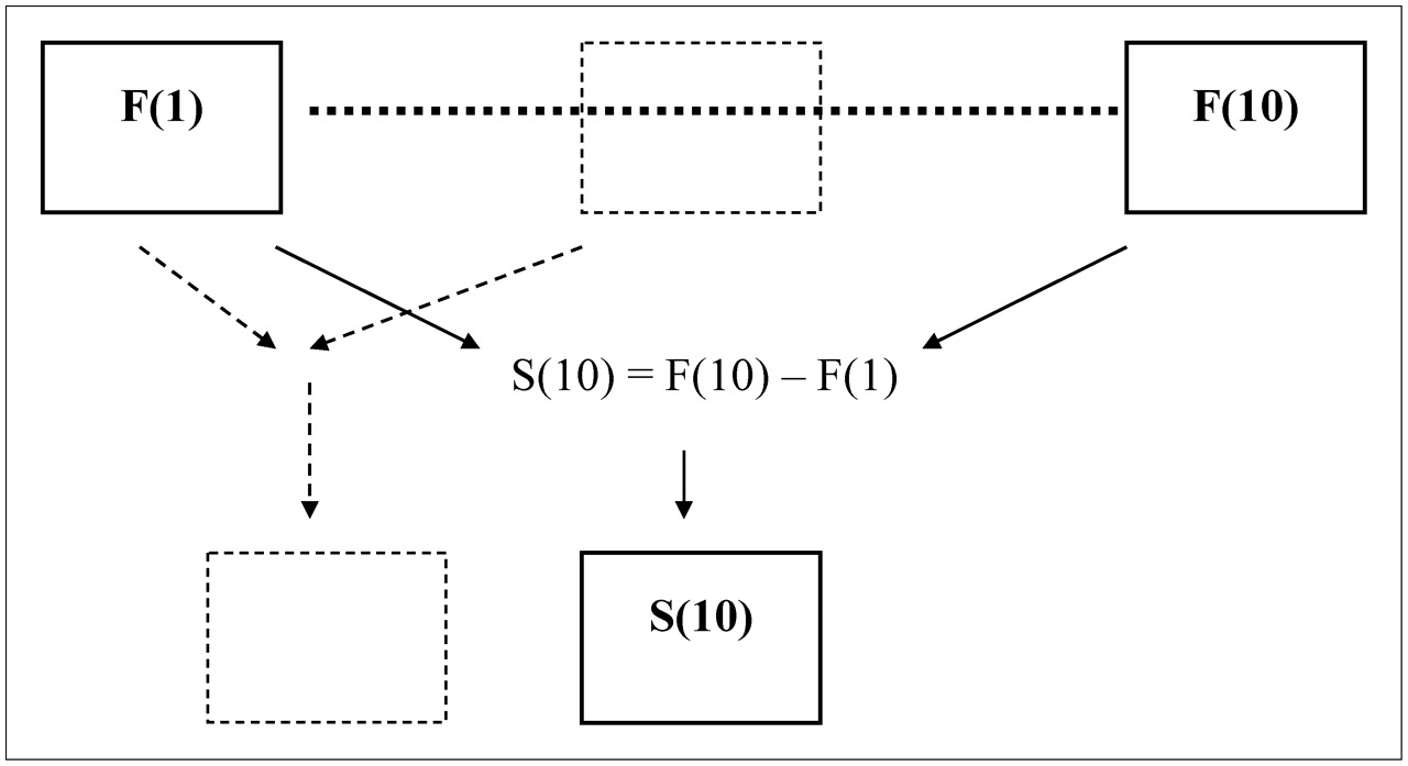
  • FIGURE 5. 
    • Download figure
    • Open in new tab
    • Download powerpoint
    FIGURE 5. 

    Schematic representation of SSS. Each individual frame has subtracted from it the preceding frame.

  • FIGURE 6. 
    • Download figure
    • Open in new tab
    • Download powerpoint
    FIGURE 6. 

    Schematic representation of ASSS. Each new frame is produced by subtraction of the preceding frame from the subsequent frame.
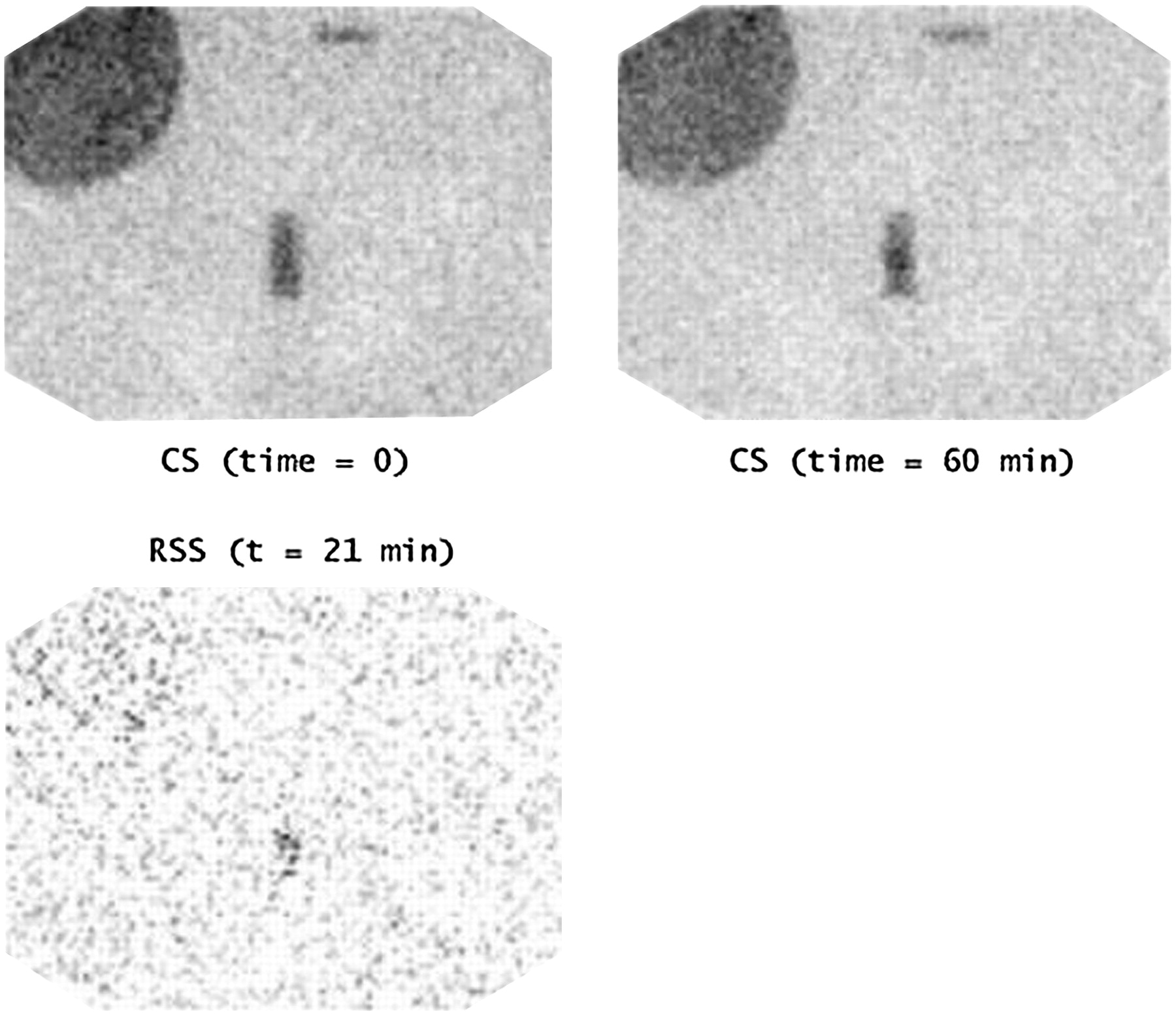
  • FIGURE 7. 
    • Download figure
    • Open in new tab
    • Download powerpoint
    FIGURE 7. 

    Bleed rate of 0.5 mL·min−1 with 10-s sampling interval. Interpretation with CS at 60 min is probably equivocal, especially if one were not aware of precisely where bleed was expected. However, RSS at just 21 min provides “probably present” outcome for IVC bleed position.
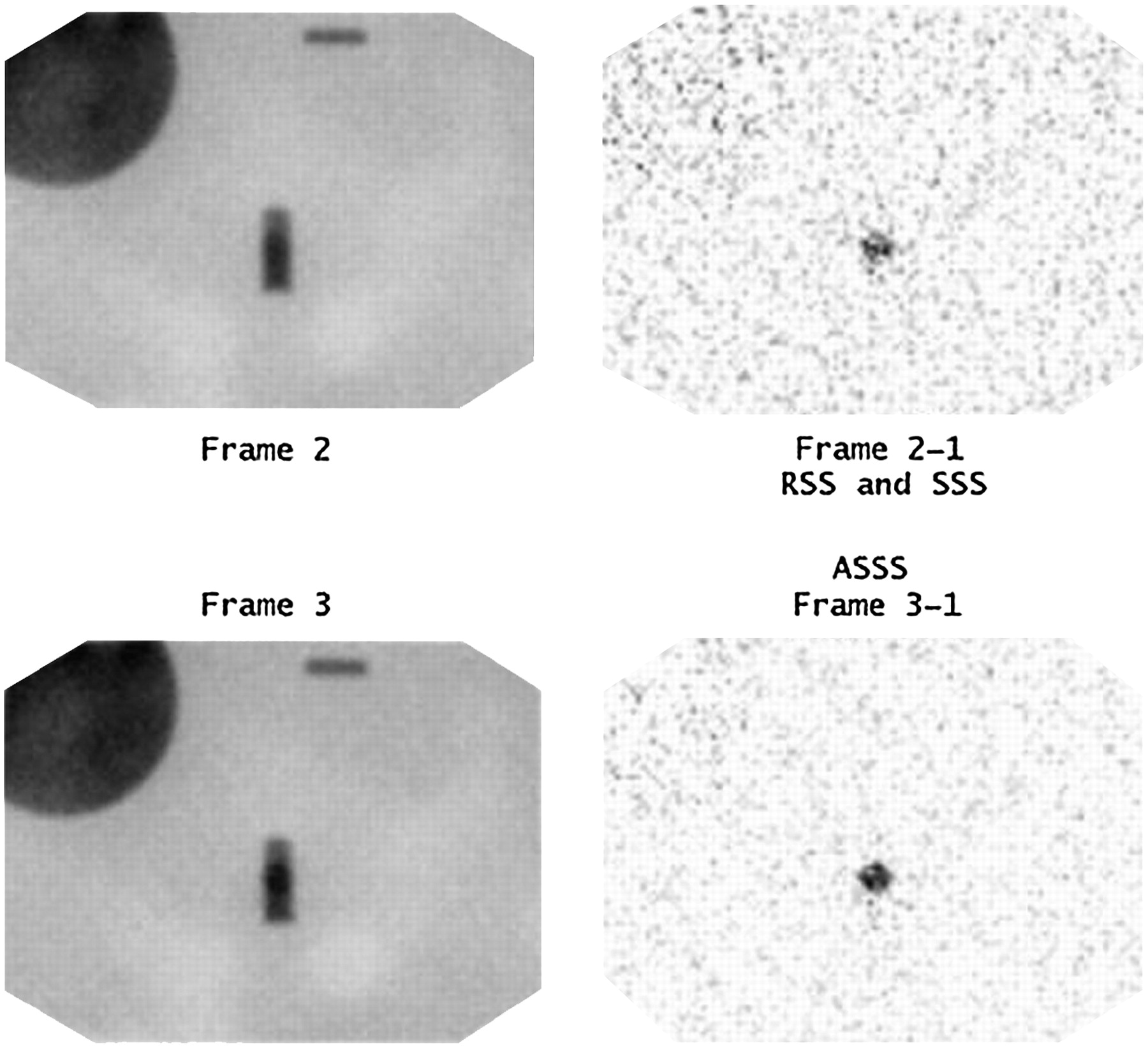
  • FIGURE 8. 
    • Download figure
    • Open in new tab
    • Download powerpoint
    FIGURE 8. 

    Bleed rate of 1.0 mL·min−1 with 5-min sampling interval. Lack of certainty exists for this IVC bleed position in CS frames 2 and 3 (left images). However, RSS (frame 2 – frame 1) and ASSS (frame 3 – frame 1) provide “definitely present” outcome. In this example, RSS and SSS produce the same subtraction image.
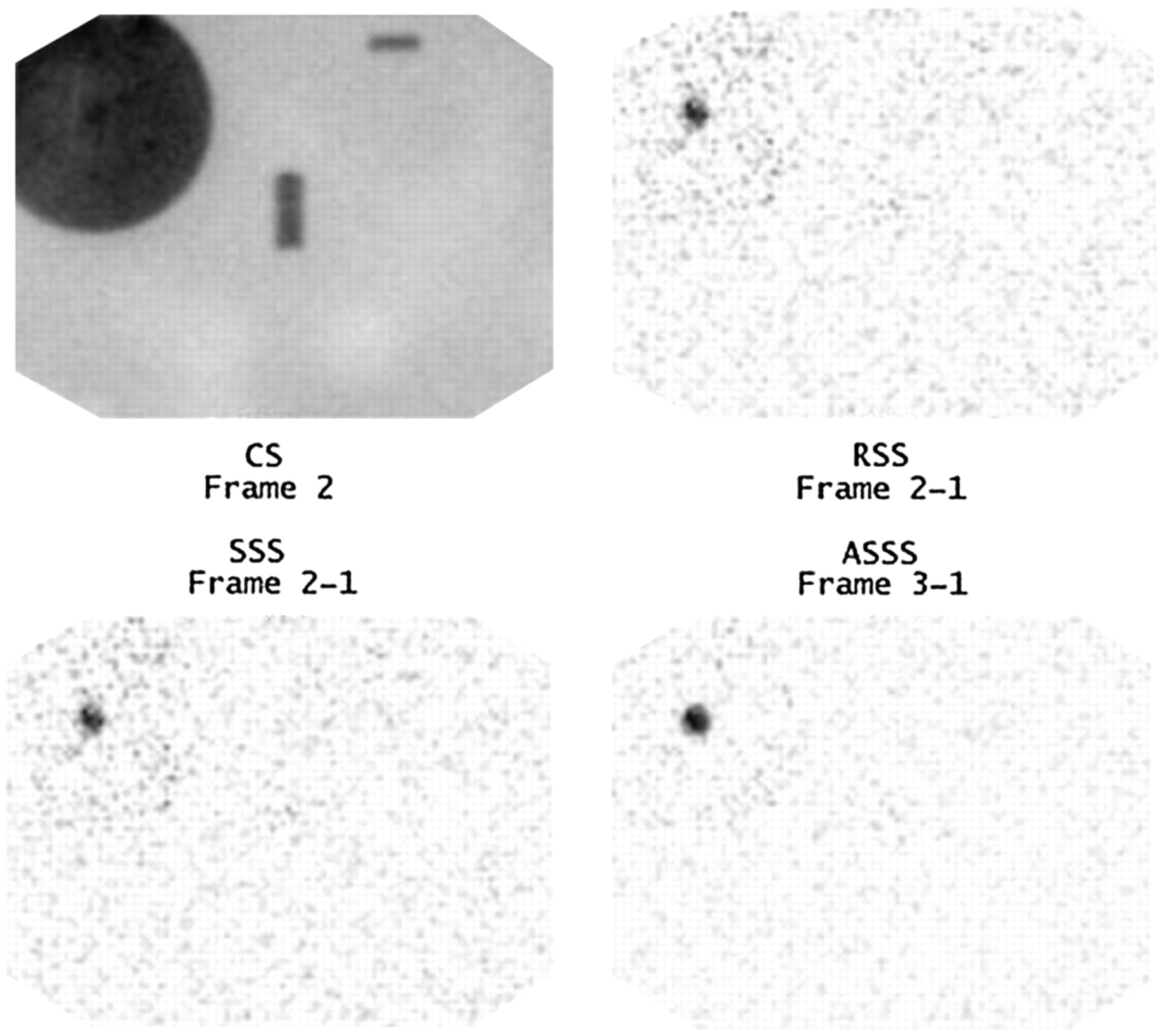
  • FIGURE 9. 
    • Download figure
    • Open in new tab
    • Download powerpoint
    FIGURE 9. 

    Bleed rate of 1.0 mL·min−1 with 5-min sampling interval. Lack of certainty exists for this liver bleed position in CS frame 2 (top left image). However, RSS (frame 2 – frame 1), SSS (frame 2 – frame 1), and ASSS (frame 3 – frame 1) provide “definitely present” outcome.

Tables

  • Figures
    • View popup
    TABLE 1

    Comparison of Maximum and Minimum Counts and Ratios for Structures from Clinical Database with Those from Phantom*

    ParameterClinical minimum–maximum (phantom)Clinical mean (95% CI)P (mean vs. phantom)
    Liver counts/pixel135.6−365.6 (212.8)230.3 (112.7−348.0)0.70
    IVC counts/pixel118.8−247.6 (167.2)164.7 (96.4−233.0)0.92
    Background counts/pixel39.8−122.0 (70.0)72.8 (25.9−119.8)0.87
    Liver-to-IVC ratio1.0−1.9 (1.3)1.40 (0.95−1.86)0.56
    Liver-to-background ratio2.6−4.8 (3.0)3.38 (2.26−4.51)0.40
    IVC-to-background ratio1.6−3.1 (2.4)2.47 (1.78−3.16)0.80
    • ↵* All phantom parameters fell within clinical range.

PreviousNext
Back to top

In this issue

Journal of Nuclear Medicine Technology: 34 (3)
Journal of Nuclear Medicine Technology
Vol. 34, Issue 3
September 2006
  • Table of Contents
  • About the Cover
  • Index by author
Print
Download PDF
Article Alerts
Sign In to Email Alerts with your Email Address
Email Article

Thank you for your interest in spreading the word on Journal of Nuclear Medicine Technology.

NOTE: We only request your email address so that the person you are recommending the page to knows that you wanted them to see it, and that it is not junk mail. We do not capture any email address.

Enter multiple addresses on separate lines or separate them with commas.
Improved Detection and Localization of Lower Gastrointestinal Tract Hemorrhage by Subtraction Scintigraphy: Phantom Analysis
(Your Name) has sent you a message from Journal of Nuclear Medicine Technology
(Your Name) thought you would like to see the Journal of Nuclear Medicine Technology web site.
Citation Tools
Improved Detection and Localization of Lower Gastrointestinal Tract Hemorrhage by Subtraction Scintigraphy: Phantom Analysis
Geoffrey M. Currie, Philip A. Towers, Janelle M. Wheat
Journal of Nuclear Medicine Technology Sep 2006, 34 (3) 160-168;

Citation Manager Formats

  • BibTeX
  • Bookends
  • EasyBib
  • EndNote (tagged)
  • EndNote 8 (xml)
  • Medlars
  • Mendeley
  • Papers
  • RefWorks Tagged
  • Ref Manager
  • RIS
  • Zotero
Share
Improved Detection and Localization of Lower Gastrointestinal Tract Hemorrhage by Subtraction Scintigraphy: Phantom Analysis
Geoffrey M. Currie, Philip A. Towers, Janelle M. Wheat
Journal of Nuclear Medicine Technology Sep 2006, 34 (3) 160-168;
Twitter logo Facebook logo LinkedIn logo Mendeley logo
  • Tweet Widget
  • Facebook Like
  • Google Plus One
Bookmark this article

Jump to section

  • Article
    • Abstract
    • MATERIALS AND METHODS
    • RESULTS
    • DISCUSSION
    • CONCLUSION
    • Acknowledgments
    • Footnotes
    • References
  • Figures & Data
  • Info & Metrics
  • PDF

Related Articles

  • No related articles found.
  • PubMed
  • Google Scholar

Cited By...

  • The SNMMI Procedure Standard/ACNM Practice Guideline for Gastrointestinal Bleeding Scintigraphy 3.0
  • The SNMMI Procedure Standard/EANM Practice Guideline for Gastrointestinal Bleeding Scintigraphy 2.0
  • Cost-Effectiveness Analysis of Subtraction Scintigraphy in Patients with Acute Lower Gastrointestinal Tract Hemorrhage
  • Improved Detection and Localization of Lower Gastrointestinal Hemorrhage Using Subtraction Scintigraphy: Clinical Evaluation
  • Google Scholar

More in this TOC Section

  • Early 10-Minute Postinjection [18F]F-FAPI-42 uEXPLORER Total-Body PET/CT Scanning Protocol for Staging Lung Cancer Using HYPER Iterative Reconstruction
  • Single- Versus Dual-Time-Point Imaging for Transthyretin Cardiac Amyloid Using 99mTc-Pyrophosphate
  • Does Arthrography Improve Accuracy of SPECT/CT for Diagnosis of Aseptic Loosening in Patients with Painful Knee Arthroplasty: A Systematic Review and Metaanalysis
Show more Imaging

Similar Articles

SNMMI

© 2025 SNMMI

Powered by HighWire