


Article Figures & Data
Figures
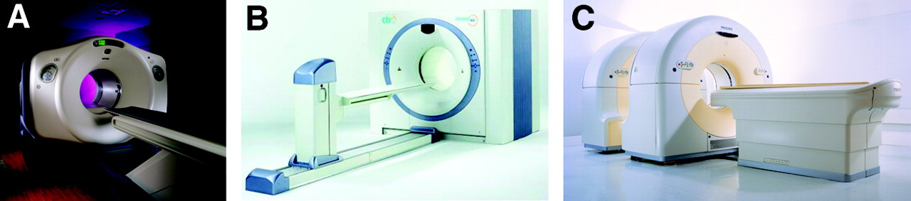
- FIGURE 1.
Current commercial PET/CT scanners from 3 vendors: Discovery ST (GE Healthcare) (A), Biograph Sensation 16 (Siemens Medical Systems) or Reveal XVI (CTI, Inc.) (B), and Gemini (Philips Medical) (C).
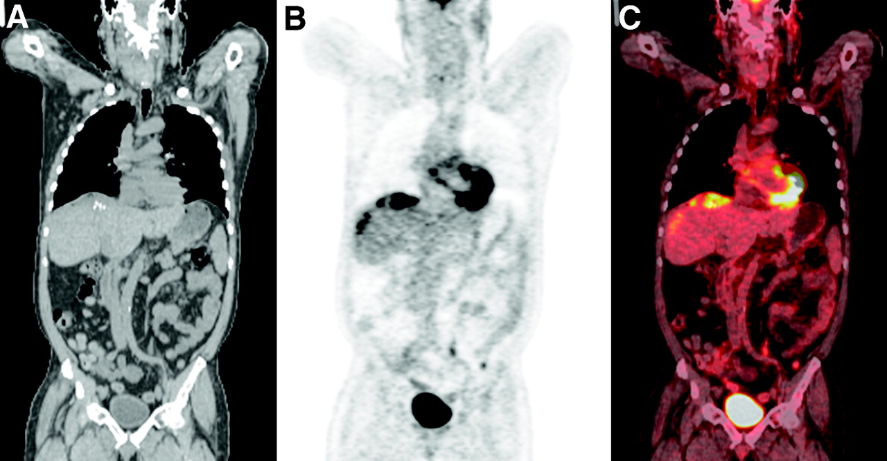
- FIGURE 2.
PET/CT image consisting of coronal whole-body CT image (A), PET image with CT attenuation correction (B), and fused image (C).
- FIGURE 3.
(A) High-density metallic implants generate streaking artifacts and high CT numbers (arrow) on CT image. (B) High CT numbers will then be mapped to high PET attenuation coefficients, leading to overestimation of activity concentration. (C) PET images without attenuation correction help to rule out metal-induced artifacts.
- FIGURE 4.
Metal ring on left breast of patient (arrow) produces streaking artifacts and high CT numbers (A), resulting in falsely increased radiotracer uptake on PET images with CT attenuation correction (B), whereas PET image without attenuation correction shows only background activity at level of metal ring (C).
- FIGURE 5.
(A) Hip prosthesis produces photopenic area (arrow) on PET image without attenuation correction because of photon absorption. (B) No radiotracer uptake is seen on PET image with attenuation correction.
- FIGURE 6.
Curvilinear cold artifact (arrow) is commonly seen on dome of diaphragm/liver or at lung base because of respiration mismatch on PET images with CT attenuation correction.
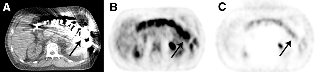
- FIGURE 7.
(A) 58-y-old man with colon cancer. Lesion at dome of liver is mislocalized to right lung (arrow) because of respiratory motion. (B) Image without attenuation correction shows that all lesions are confined to liver.
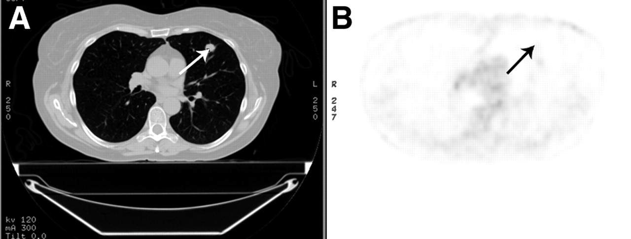
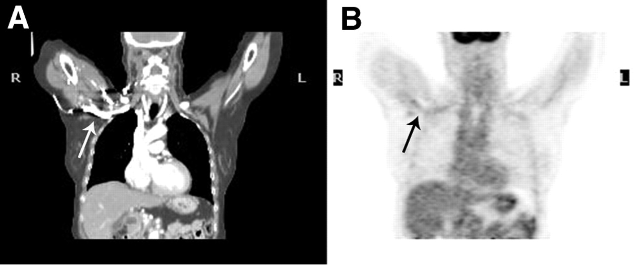
- FIGURE 8.
Mismatch of lesion localization between CT and PET scans. Lesion (arrow) seen on upper left lung of CT transaxial image (A) is not present on corresponding PET image (B), creating inaccurate attenuation correction values for that lesion.
- FIGURE 9.
(A) 61-y-old patient with lung cancer who ingested barium for an esophagogram 1 d before PET/CT scan. Concentration of contrast medium in colon (arrow) increased because of significant water reabsorption, shown on CT image. (B) High CT numbers of residual barium overcorrect attenuation of PET emission data and mimic increased 18F-FDG uptake on PET image with CT attenuation correction. (C) No increased 18F-FDG uptake is seen on image without attenuation correction.
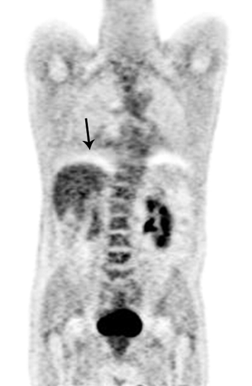
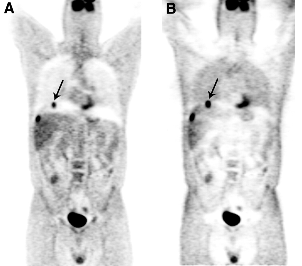
- FIGURE 10.
(A) CT scan shows intravenous contrast medium in right subclavian vein (arrow). (B) Radiotracer uptake is increased in corresponding region of PET image but does not change clinical diagnosis.
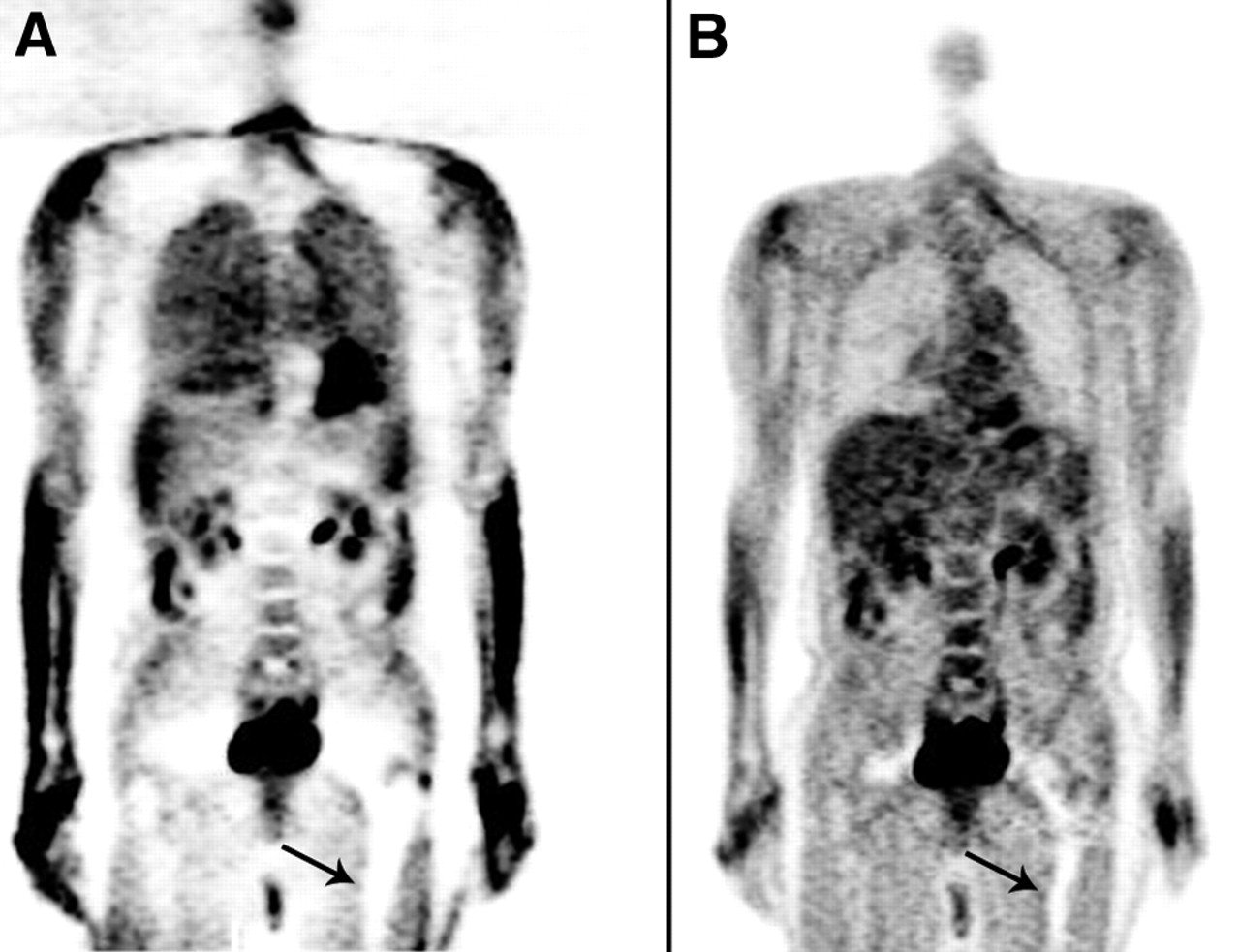
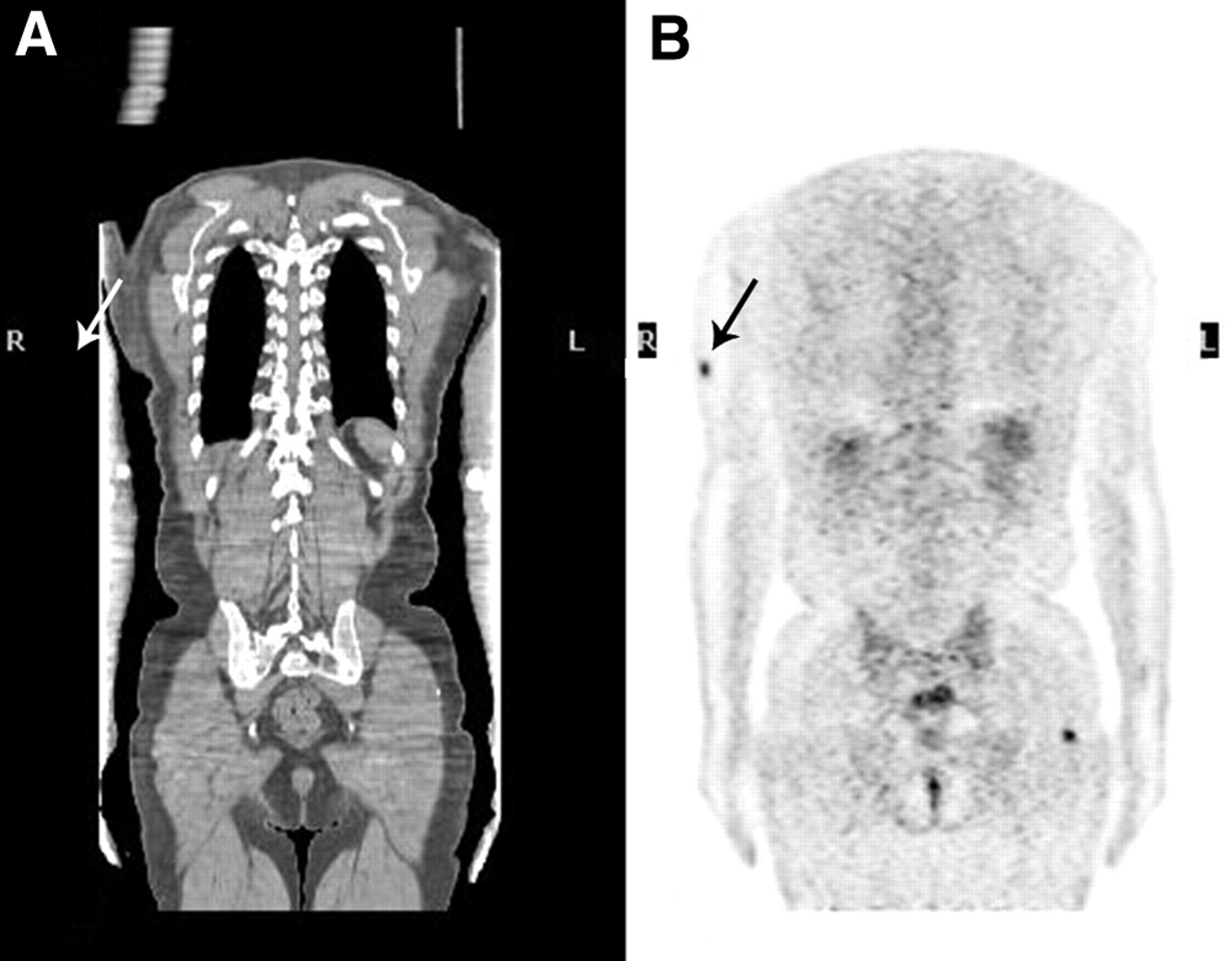
- FIGURE 11.
54-y-old man with history of metastatic melanoma (arrow). CT image appears truncated at sides (A) and biases PET attenuation-corrected image (B).


Jump to section
Related Articles
Cited By...
- Diagnostic Accuracy of FDG PET/CT in Suspected LVAD Infections: A Case Series, Systematic Review, and Meta-Analysis
- Measuring PET Spatial Resolution Using a Cylinder Phantom Positioned at an Oblique Angle
- Clinical Impact of Respiratory Motion Correction in Simultaneous PET/MR, Using a Joint PET/MR Predictive Motion Model
- Pitfalls and Pearls of Wisdom in 18F-FDG PET Imaging of Tumors
- PET artefact masquerading as a PET positive lung mass
- Technical Considerations in Brain Amyloid PET Imaging with 18F-Florbetapir
- Whole-Body 18F-FDG PET/CT: The Need for a Standardized Field of View--A Referring-Physician Aid