


Article Figures & Data
Figures
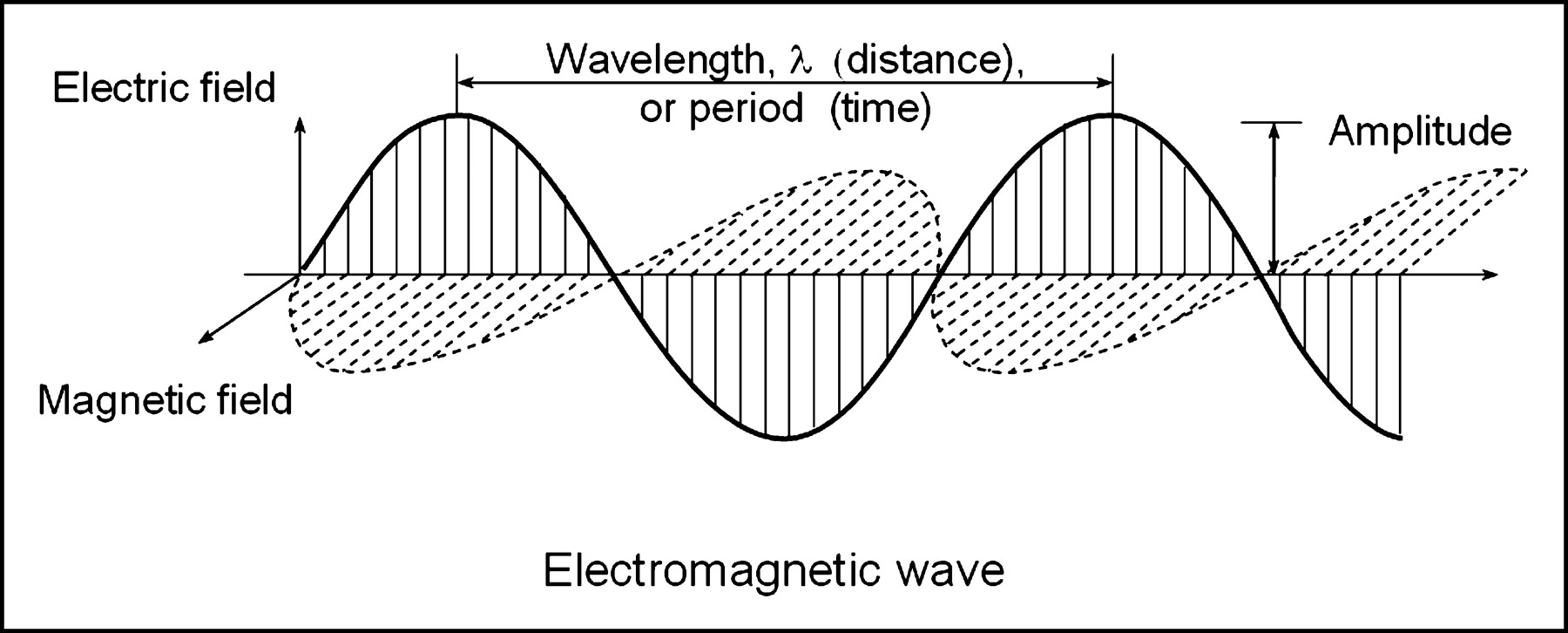
- FIGURE 1.
Electromagnetic radiation is described as a cyclic repeating wave having electrical and magnetic fields with amplitude (peak value from the average) and period (time between repeating portions of the wave). Frequency equals the number of cycles per second, and the wavelength is the distance between repeating points as determined from the frequency and velocity (see text for relationship between velocity, wavelength, and frequency).
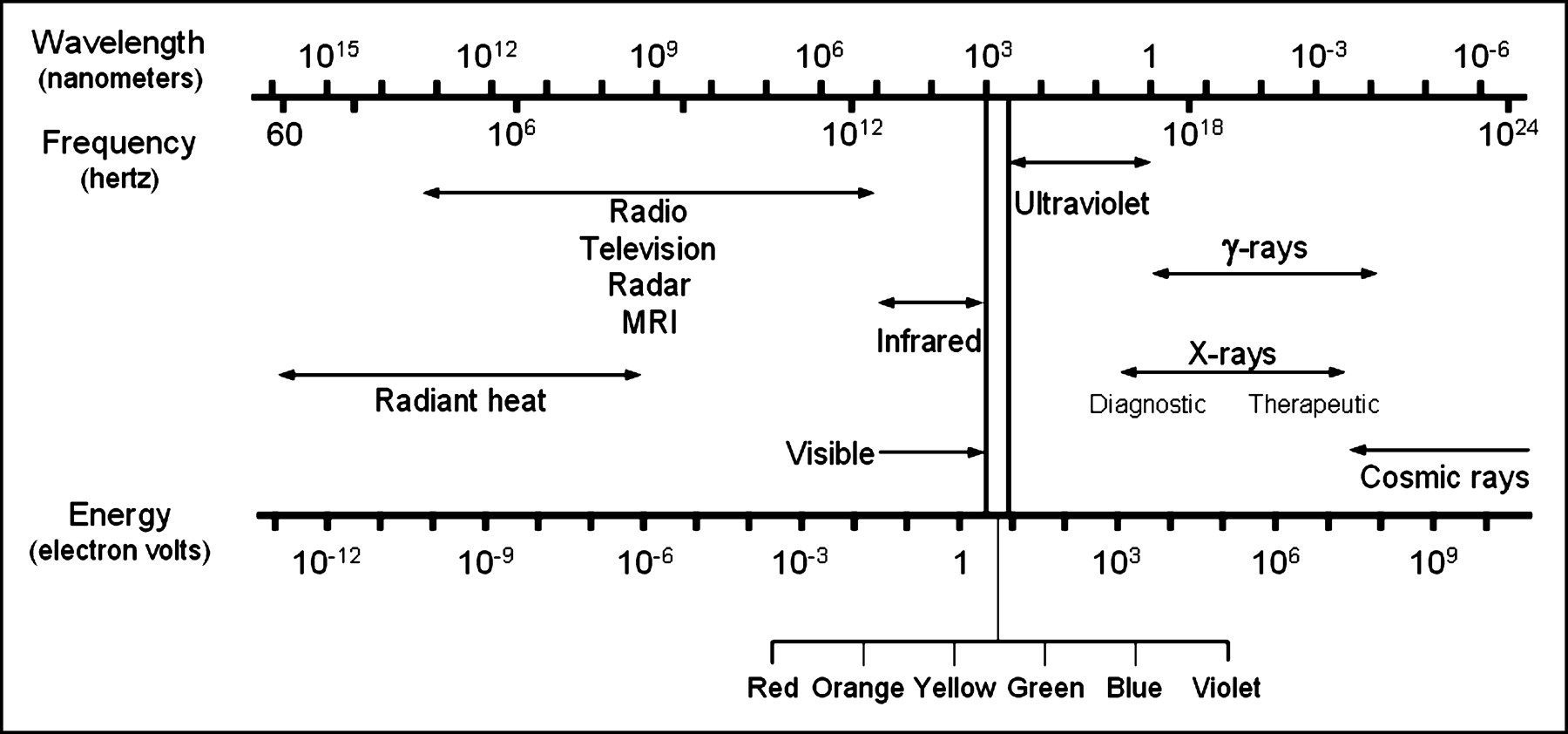
- FIGURE 2.
The electromagnetic spectrum, presented as a function of wavelength, frequency, and energy. X-rays and γ-rays comprise the high-energy portion of the electromagnetic spectrum.
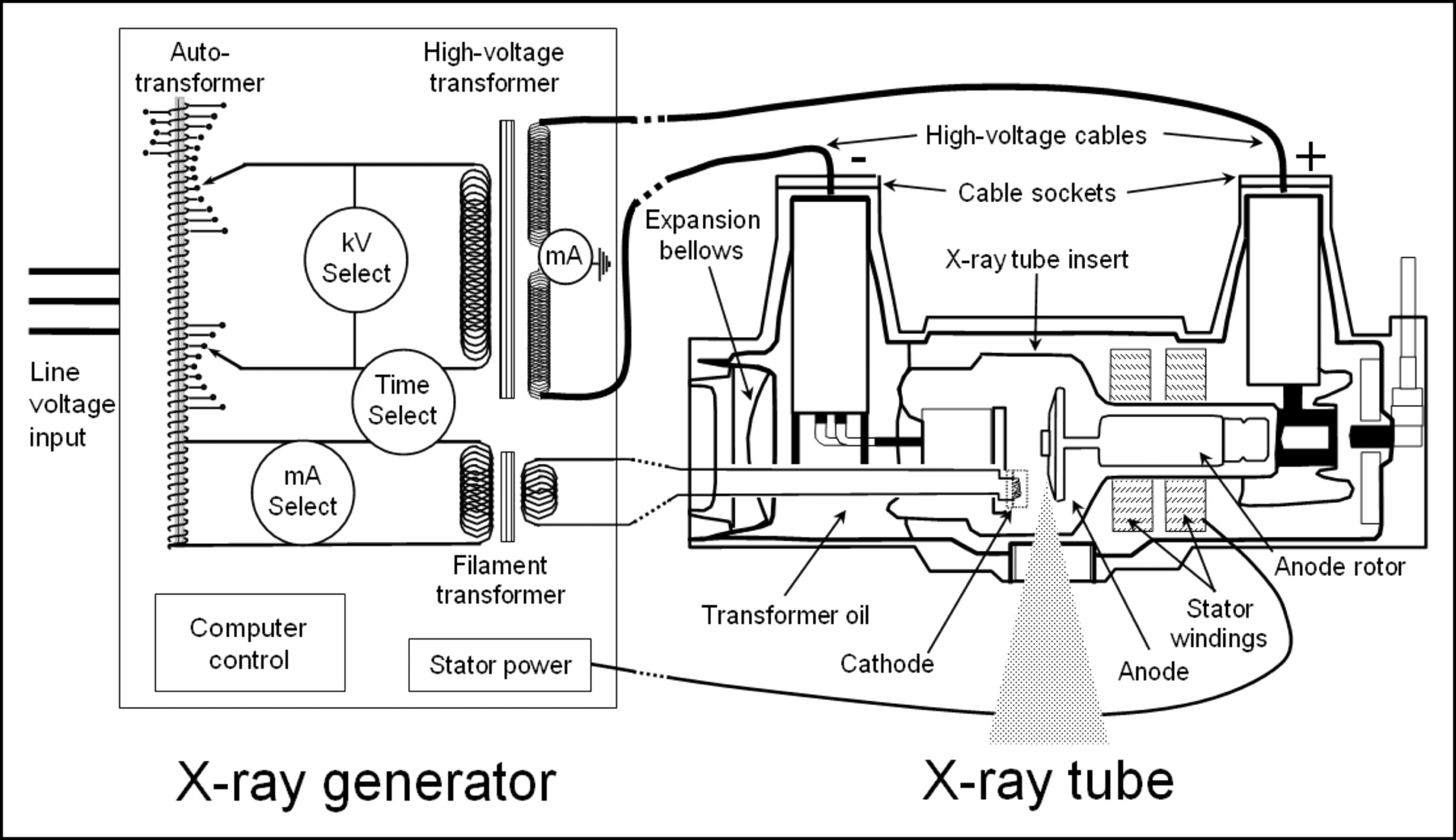
- FIGURE 3.
X-ray generator and x-ray tube components are illustrated. The x-ray generator provides operator control of the radiographic techniques, including tube voltage (kVp), tube current (mA), and exposure duration, and delivers power to the x-ray tube. The x-ray tube provides the environment (evacuated x-ray tube insert and high-voltage cable sockets), source of electrons (cathode), source of x-rays (anode), induction motor to rotate the anode (rotor/stator), transformer oil and expansion bellows to provide electrical and heat build-up protection, and the tube housing to support the insert and provide protection from leakage radiation.
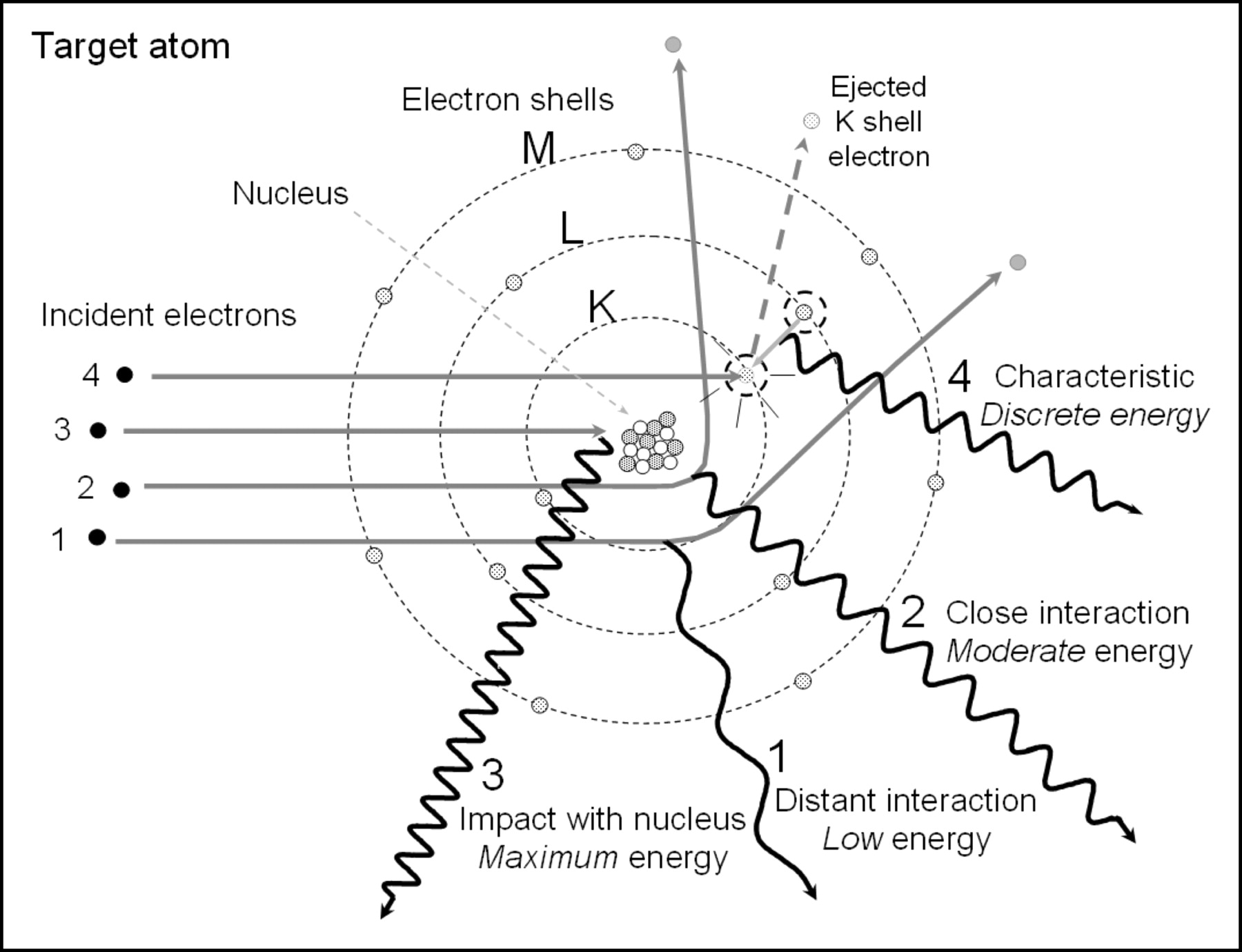
- FIGURE 4.
X-ray production by energy conversion. Events 1, 2, and 3 depict incident electrons interacting in the vicinity of the target nucleus, resulting in bremsstrahlung production caused by the deceleration and change of momentum, with the emission of a continuous energy spectrum of x-ray photons. Event 4 demonstrates characteristic radiation emission, where an incident electron with energy greater than the K-shell binding energy collides with and ejects the inner electron creating an unstable vacancy. An outer shell electron transitions to the inner shell and emits an x-ray with energy equal to the difference in binding energies of the outer electron shell and K shell that are “characteristic” of tungsten.
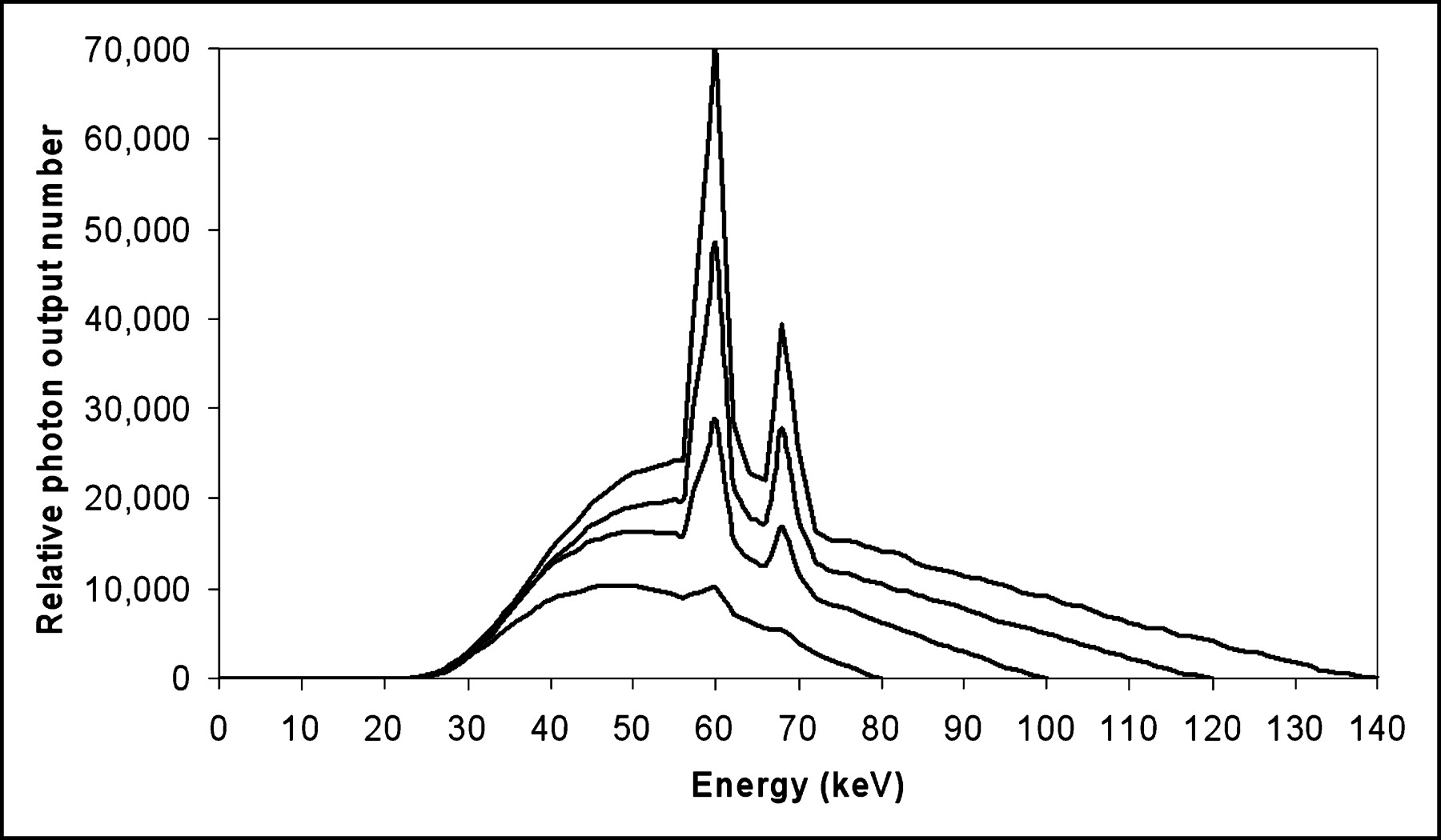
- FIGURE 5.
Bremsstrahlung and characteristic radiation spectra are shown for a tungsten anode with x-ray tube operation at 80, 100, 120, and 140 kVp and equal tube current.


