
Main menu

  • Home
  • Content
    • Current
      • JNMT Supplement
    • Ahead of print
    • Past Issues
    • Continuing Education
    • JNMT Podcast
    • SNMMI Annual Meeting Abstracts
  • Subscriptions
    • Subscribers
    • Rates
    • Journal Claims
    • Institutional and Non-member
  • Authors
    • Submit to JNMT
    • Information for Authors
    • Assignment of Copyright
    • AQARA Requirements
  • Info
    • Reviewers
    • Permissions
    • Advertisers
    • Corporate & Special Sales
  • About
    • About Us
    • Editorial Board
    • Contact Information
  • More
    • Alerts
    • Feedback
    • Help
    • SNMMI Journals
  • SNMMI
    • JNMT
    • JNM
    • SNMMI Journals
    • SNMMI

User menu

  • Subscribe
  • My alerts
  • Log in
  • My Cart

Search

  • Advanced search
Journal of Nuclear Medicine Technology
  • SNMMI
    • JNMT
    • JNM
    • SNMMI Journals
    • SNMMI
  • Subscribe
  • My alerts
  • Log in
  • My Cart
Journal of Nuclear Medicine Technology

Advanced Search

  • Home
  • Content
    • Current
    • Ahead of print
    • Past Issues
    • Continuing Education
    • JNMT Podcast
    • SNMMI Annual Meeting Abstracts
  • Subscriptions
    • Subscribers
    • Rates
    • Journal Claims
    • Institutional and Non-member
  • Authors
    • Submit to JNMT
    • Information for Authors
    • Assignment of Copyright
    • AQARA Requirements
  • Info
    • Reviewers
    • Permissions
    • Advertisers
    • Corporate & Special Sales
  • About
    • About Us
    • Editorial Board
    • Contact Information
  • More
    • Alerts
    • Feedback
    • Help
    • SNMMI Journals
  • Watch or Listen to JNMT Podcast
  • Visit SNMMI on Facebook
  • Join SNMMI on LinkedIn
  • Follow SNMMI on Twitter
  • Subscribe to JNMT RSS feeds
OtherCONTINUING EDUCATION

Techniques for Using Bexxar for the Treatment of Non-Hodgkin’s Lymphoma*

David W. Seldin
Journal of Nuclear Medicine Technology September 2002, 30 (3) 109-114;
David W. Seldin
  • Find this author on Google Scholar
  • Find this author on PubMed
  • Search for this author on this site
  • Article
  • Figures & Data
  • Info & Metrics
  • PDF
Loading

Article Figures & Data

Figures

  • Tables
  • FIGURE 1.
    • Download figure
    • Open in new tab
    • Download powerpoint
    FIGURE 1.

    Graphic representation of developmental stages of B-lymphocytes from undifferentiated stem cells to plasma cells. Predominant location of these cell types, degree of expression of CD20 antigen, and associated malignant transformations are shown. CML = chronic myelogenous leukemia; CLL = chronic lymphocytic leukemia.

  • FIGURE 2.
    • Download figure
    • Open in new tab
    • Download powerpoint
    FIGURE 2.

    Anterior whole-body images from same patient taken 1 h after injection of 131I-tositumomab. (Left) Image shows biodistribution without predose of unlabeled tositumomab, with most activity in spleen. (Right) Image, for which patient had received unlabeled predose, shows markedly less splenic uptake and better visualization of rest of body.

  • FIGURE 3.
    • Download figure
    • Open in new tab
    • Download powerpoint
    FIGURE 3.

    Total radiation dose from 131I-tositumomab is dependent on rate of biologic clearance of radiopharmaceutical. Patients with rapid clearance require higher treatment dose than those with slow clearance to deliver same absorbed dose. Total administered dose is proportional to area under curve.

  • FIGURE 4.
    • Download figure
    • Open in new tab
    • Download powerpoint
    FIGURE 4.

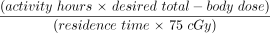
    Distribution of administered doses of 131I-tositumomab required to deliver total administered dose of 75 cGy in group of 201 patients enrolled in clinical trials.

  • FIGURE 5.
    • Download figure
    • Open in new tab
    • Download powerpoint
    FIGURE 5.

    Images (scan speed, 100 cm/min) obtained as part of dosimetric study of patient enrolled in clinical trial. Background and standard images are similar at all time points; changes in distribution and clearance of activity are seen on patient images taken at 3 time points.

Tables

  • Figures
    • View popup
    TABLE 1

    Sample Data and Calculations from Patient Dosimetric Study

    ParameterDay 0Day 4Day 6
    Dose calibrator measurements
     Time of measurement13:2012:5111:30
     131I standard activity (kBq [μCi])9,694 (262)6,993 (189)5,846 (158)
    Gamma-camera counts (whole-body mode)
     131I source
      Time started13:1112:5211:19
      Anterior counts35,92524,30920,411
     Background
      Time started13:0512:4211:14
      Anterior counts2,5442,5102,345
     Patient
      Time started14:1313:0111:27
      Anterior counts327,581125,24874,381
    Quality control calculation
     Time from initial count (h)095.7142.1
     Background-corrected source counts33,38121,79918,066
     % initial count1006554
     Counts per 37 kBq (μCi) in standard127.4115.3114.3
    Residence time calculation
     Background-corrected patient counts325,037122,73872,036
     % initial count1003822
     Residence time (h) from graph9896
    Therapy dose calculation
     Administered 131I dose
      Embedded Image75.476.9
    • Whole-body scan speed, 100 cm/min; collimator, medium energy; patient platelet count, 133,000; patient height, 165 cm; maximum effective mass (from table), 78.7 kg; camera height above table, 36 cm; whole-body scan field of view, 179 cm; desired total-body dose, 65 cGy; patient weight, 82.5 kg; activity hours (from table), 8,523.

    • View popup
    TABLE 2

    Calculations for Determining Release Criteria and Length of Time for Observing Precautions After Therapy Dose Administration

    Measured residence time96 h
    Measured dose rate at 1 m after therapy0.1 mSv/h (10 mrem/h)
    Releasable dose rate (from table)0.2 mSv/h (20 mrem/h)
    Calculated exposure to others (0.25 × 0.1 × [7.15 + 0.99 × 96])2.56 mSv (255.5 mrem)
    Duration of precautions from table provided by manufacturer
     Sleep at least 1.83 m from others (0.098 × 96)9.4 d
     Do not take long trip (>4 h) near others (0.019 × 96)1.8 d
     Stay at least 1.83 m from pregnant women and children (<1 mSv [<100 mrem] exposure) (0.135 × 96)13 d
PreviousNext
Back to top

In this issue

Journal of Nuclear Medicine Technology: 30 (3)
Journal of Nuclear Medicine Technology
Vol. 30, Issue 3
September 1, 2002
  • Table of Contents
  • About the Cover
  • Index by author
Print
Download PDF
Article Alerts
Sign In to Email Alerts with your Email Address
Email Article

Thank you for your interest in spreading the word on Journal of Nuclear Medicine Technology.

NOTE: We only request your email address so that the person you are recommending the page to knows that you wanted them to see it, and that it is not junk mail. We do not capture any email address.

Enter multiple addresses on separate lines or separate them with commas.
Techniques for Using Bexxar for the Treatment of Non-Hodgkin’s Lymphoma*
(Your Name) has sent you a message from Journal of Nuclear Medicine Technology
(Your Name) thought you would like to see the Journal of Nuclear Medicine Technology web site.
Citation Tools
Techniques for Using Bexxar for the Treatment of Non-Hodgkin’s Lymphoma*
David W. Seldin
Journal of Nuclear Medicine Technology Sep 2002, 30 (3) 109-114;

Citation Manager Formats

  • BibTeX
  • Bookends
  • EasyBib
  • EndNote (tagged)
  • EndNote 8 (xml)
  • Medlars
  • Mendeley
  • Papers
  • RefWorks Tagged
  • Ref Manager
  • RIS
  • Zotero
Share
Techniques for Using Bexxar for the Treatment of Non-Hodgkin’s Lymphoma*
David W. Seldin
Journal of Nuclear Medicine Technology Sep 2002, 30 (3) 109-114;
Twitter logo Facebook logo LinkedIn logo Mendeley logo
  • Tweet Widget
  • Facebook Like
  • Google Plus One
Bookmark this article

Jump to section

  • Article
    • Abstract
    • NHL
    • THERAPEUTIC USE OF ANTIBODIES
    • TOSITUMOMAB PROTOCOL
    • THERAPEUTIC DOSE DETERMINATION
    • TOSITUMOMAB ADMINISTRATION
    • PATIENT RELEASE CRITERIA
    • INITIAL CLINICAL RESULTS
    • CONCLUSION
    • Footnotes
    • REFERENCES
  • Figures & Data
  • Info & Metrics
  • PDF

Related Articles

  • No related articles found.
  • PubMed
  • Google Scholar

Cited By...

  • Proceedings of the Second NCI-SNMMI Workshop on Targeted Radionuclide Therapy
  • Targeted Radionuclide Therapy: Proceedings of a Joint Workshop Hosted by the National Cancer Institute and the Society of Nuclear Medicine and Molecular Imaging
  • Collimator Selection, Acquisition Speed, and Visual Assessment of 131I-Tositumomab Biodistribution in a Phantom Model
  • 89Zr as a PET Surrogate Radioisotope for Scouting Biodistribution of the Therapeutic Radiometals 90Y and 177Lu in Tumor-Bearing Nude Mice After Coupling to the Internalizing Antibody Cetuximab
  • Google Scholar

More in this TOC Section

  • Illuminating the Hidden: Standardizing Cardiac MIBG Imaging for Sympathetic Dysfunction
  • PET/CT Case Series: Unmasking the Mystery of Cardiac Sarcoidosis
  • Delivery Methods of Radiopharmaceuticals: Exploring Global Strategies to Minimize Occupational Radiation Exposure
Show more Continuing Education

Similar Articles

SNMMI

© 2025 SNMMI

Powered by HighWire