


Abstract
Objective: The process of making slides for presentation of nuclear medicine data has historically used methods that require some photographic expertise. This paper describes the use of readily available software and hardware to achieve the same result. Surface images generated for SPECT images of a normal patient and a patient with Alzheimer’s dementia were used to develop the technique described. Images were scanned and processed using commercially available software and a personal computer. Slides were generated by a slide printer and compared with electronic presentations of the same images. The method described is easier than the photographic method used in the past, and the quality of the presentation material (either slide or electronic) is readily predictable.
The recent development of computer tools allows us to obtain digital information and present research results in a filmless or slideless presentation. Despite these developments, slides are still frequently used to present research results or teaching material. The ability to transfer images from hard copy to file types compatible with commercial software programs would be helpful, whether slides or computer presentations of data are the desired result. A personal computer and common software applications have been used to create a digital radiographic teaching file and database (1). The addition of a slide printer or high-quality color printer will result in the ability to produce either slides or prints of nuclear medicine scan data for presentation. It is expected that slides or prints made using the methods and tools presented in this paper will be high-quality and representative of the outcome of any slide or print made using this technique.
MATERIALS AND METHODS
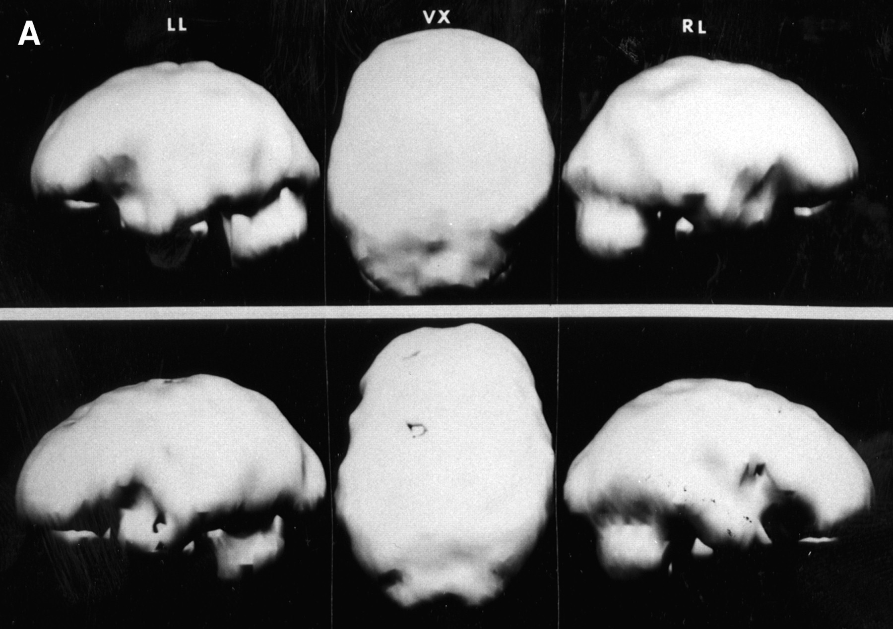
Nine composite photo-plates were used for this process. Each composite photo consists of 3 to 6 Polaroid (Polaroid Corp., Cambridge, MA) photos of the brain of a normal patient (Fig. 1A) and a patient with Alzheimer’s dementia (Fig. 1B). To illustrate the degree of the abnormalities by visual evaluation, the extent of the perfusion defects in the cerebral cortex has been semiquantitatively graded: 0, normal; 2, mild; 4, moderate; 6, severe; and 8, profound (2). SPECT brain images were obtained using a 3-head gamma camera (Prism 3000s; Marconi Medical Systems, Cleveland, OH). The data were reconstructed and three-dimensional surface rendered images were generated. The Polaroid photos were taken from the screen view of 3 different perspectives of the surface image. The 2 datasets demonstrate marked differences in brain perfusion between the 2 patients.

Photoplate composite of 6 Polaroid images of a normal patient. Images were obtained from the computer screen of a 3-head camera. Left lateral (LL), right lateral (RL), and vertex (VX) views of 2 different normal patients were obtained.

The photoplate from a patient with autopsy-proven Alzheimer’s dementia imaged over a 3-mo interval. Images were graded as severe (grade 6, top) from the initial scan and profound (grade 8, bottom) 3 mo later. Left lateral (LL), right lateral (RL) and vertex (VX) views were obtained.
The images were scanned using a flatbed scanner (Astra 12203; UMax Technologies, Inc., Freemont, CA) interfaced to a personal computer (PC). The PC contained software for the manipulation of the images (PhotoShop; Adobe Systems Inc., San Jose, CA) and incorporation into slide presentations (PowerPoint; Microsoft Corp., Redmond, WA). The PC was interfaced to a slide printer (Laser Graphics, Oakland, CA) for the production of the slides. Digital images were obtained using the scanner configured for 1-sided photos (this scanner can also be configured for transparency film). These images were scanned at 300 dots/inch (dpi); each scan took 30 s. The digital data were saved as tagged image file format (TIFF) and imported into Adobe PhotoShop, which was used to manipulate the images to obtain optimal contrast and brightness. In addition, pertinent information such as the interval between the SPECT studies, image orientation (anterior (ANT); posterior (POST); right lateral (RL); and left lateral (LL)) was added to each image. The images were then copied to PowerPoint, which was used to produce the slides in conjunction with the slide printer. The slides were photographed using a laser printer with Eastman Kodak EPP (Rochester, NY) 35-mm slide film. The slide film was processed in the departmental film processor, and the slides were then mounted and compared with the digital presentation for evaluation of quality.
RESULTS
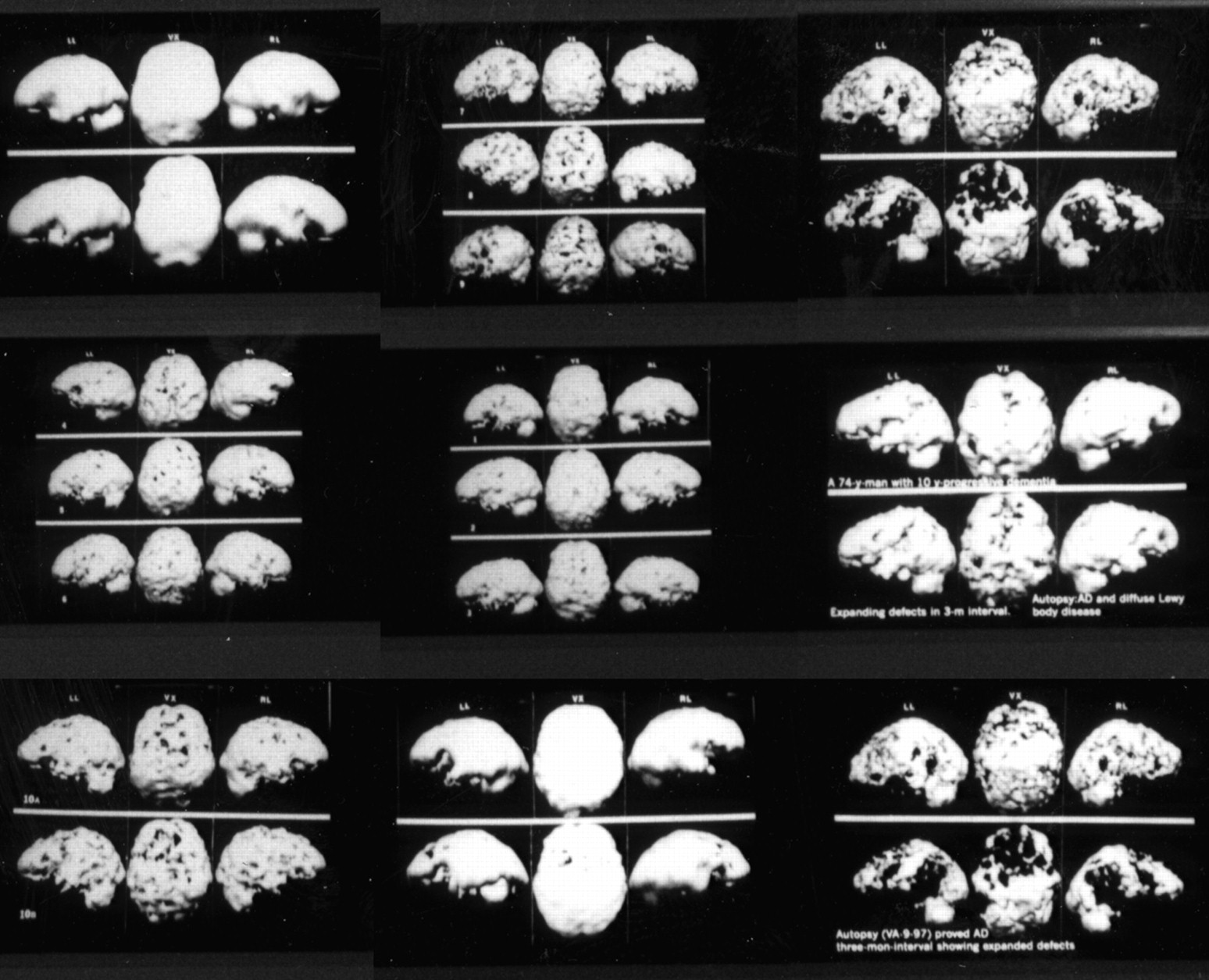
Figure 2 shows the slides produced using this method. The slides were used for presentation and were well-received. The quality of the slides was found to be at least comparable to that of the digital presentation. Some individuals thought that the three-dimensional effect was better with the slides than the electronic presentation.

Final products of the slides made with the Laser Graphic slide printer.
DISCUSSION
Making slides from nuclear medicine transparency film and paper print has been common practice for many years. It does require, however, photographic expertise and special equipment, such as a 35-mm camera, light boxes, and photographic lights. Adjustments must be made to the lights, focus, and exposure depending on the quality of the original film. Although experience improves efficiency, the first version of the slides or photographs may not be successful and it may be necessary to repeat the process. The advantage of the method we have described is the minimal operator experience required for a high-quality slide or photographic output. Utilization of a scanner interfaced to a PC with commercially available software, such as PhotoShop or PowerPoint, offers a quick and easy way to scan and edit images for presentation. Relevant information, such as image orientation, dose and scan time, is easily added to the images.
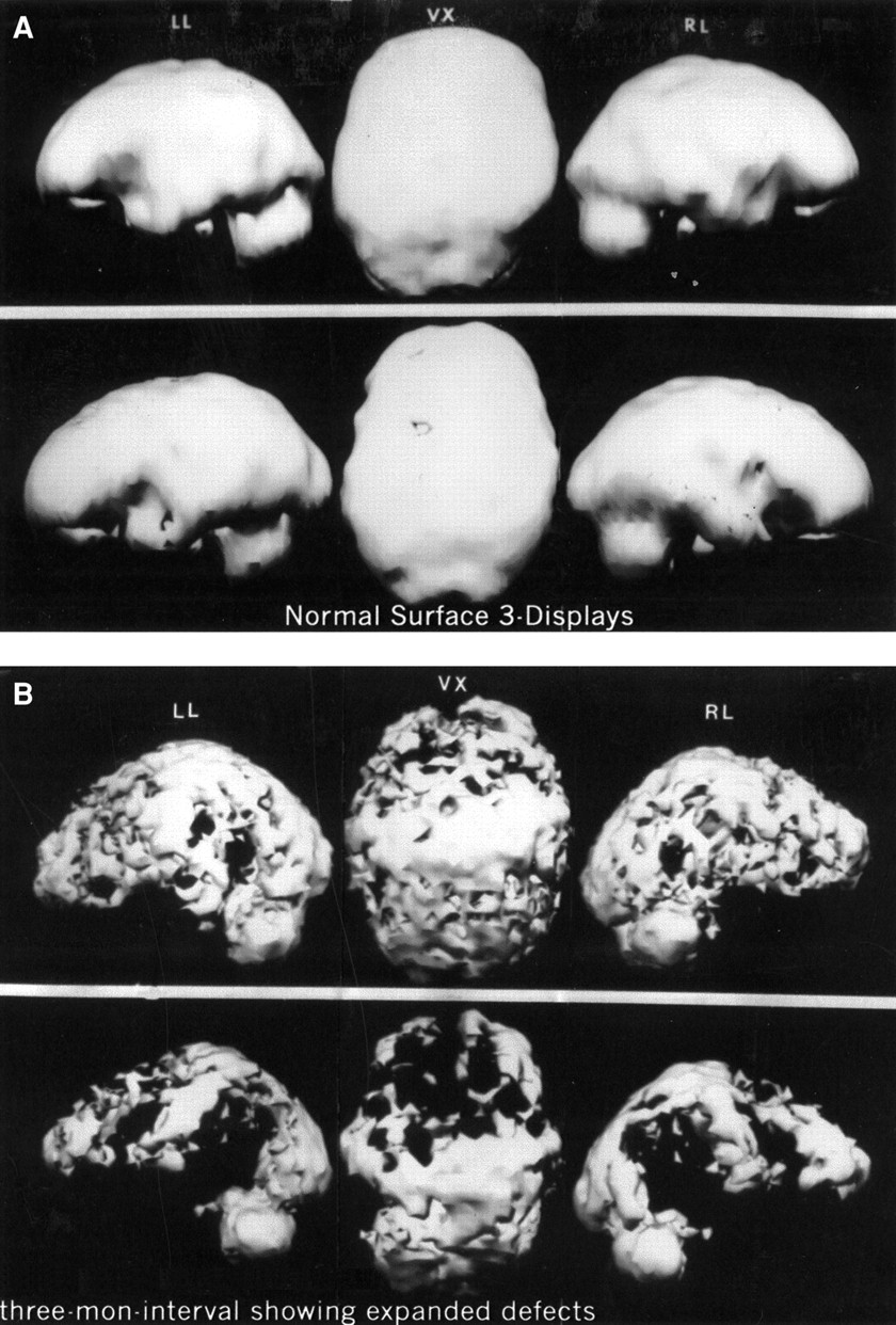
We have discussed the process for color slide production with Kodak EPP slide film. Replacing the color film with RPC B/W (Eastman Kodak, Rochester, NY) film can produce black and white prints. In addition to generating slides for projection, the digital image can also be printed out on photographic paper and then used for poster presentations (Fig. 3). In all instances, the digital image is readily accessible electronically and can be copied exactly to the desired medium (either slide or paper).

(A) Normal surface three-dimensional displays were printed using commercially available software and printer. This image corresponds to Figure 1A. These quality prints can be used for poster presentations. (B) This three-dimensional display of an Alzheimer’s patient demonstrates progression of perfusion defects, from severe to profound, that were printed using commercially available software and printer. This image corresponds to Figure 1B. Note that the patient’s clinical information can be labeled on the bottom using this method.
It was observed that the quality of the three-dimensional characteristic is better in the slides than in the electronic presentation. Although this finding could be attributed to the higher resolution capacity of the slide printer (4000–5000 dpi), it is unlikely because the electronic image at 100 dpi was the basis for the slide image.
CONCLUSION
A camera, scanner, and PC can be used to produce images for presentation. Judicious choice of film will allow for processing of the resultant slides in the nuclear medicine department. Eventually, it is likely that meeting presentations will be made via computer instead of with slides or film. The method described in this paper will allow individuals to transition slides produced using older, less reliable methods to electronic presentations.
Acknowledgments
The authors would like to thank Jason McDonald, MS, manager of the Instructional Technology Center, University of Kentucky Medical Center, and Robert P. Fons, MS, Manager of Instructional Technology Center, College of Medicine, University of Kentucky Medical Center, Lexington, KY, for their technical advice.
Footnotes
For correspondence or reprints contact: Wei-Jen Shih, MD, Dept. of Diagnostic Radiology, College of Medicine, University of Kentucky 40536.


Jump to section
Related Articles
Cited By...
- No citing articles found.