


Abstract
In vitro gastrointestinal (GI) bleed testing such as the UltraTag kits has been successfully used for a number of years. None of the research studies identified reported any significant bone marrow and liver uptake. An 80-y-old male patient was admitted to the hospital for rectal bleeding. An initial GI bleed study using the UltraTag kit was normal. Two days later, a second GI bleed scan was performed while the patient was undergoing blood transfusion with packed red blood cells. The technologist withdrew a blood sample from the blood transfusion bag. The second GI bleed test demonstrated unusual liver and bone marrow uptake. We believe that this liver uptake might have been caused by the labeling of fragmented red blood cells or oxidation of pertechnetate in the form of technetium dioxide.
Many different nuclear medicine gastrointestinal (GI) bleed techniques have been used to assess intermittent bleeding or correctly localize the unknown site of bleeding (1–3). A review of the literature revealed studies that have evaluated the effectiveness of these methodologies (2,3). Most gastrointestinal hemorrhages, including those that are clinically massive, are intermittent. Thus, to detect bleeding, it is preferable to use an indicator that remains in the intravascular space for the duration of the bleeding episode. Autologous human red blood cells can be readily labeled with 99mTc and remain intact in the vasculature for many hours. Numerous studies have compared the effectiveness and efficiency of a variety of in vitro and in vivo red blood cell labeling techniques for nuclear medicine GI bleeding studies (1–7). Other studies have specifically examined the UltraTag red blood cell labeling kit (Mallinckrodt) preparation technique (1–3). In vitro labeling (such as the UltraTag kit) has been shown to result in higher binding efficiency (1–3). Red blood cell imaging is also reported to be more sensitive than angiography and could be performed for up to 24 h after the administration of the tracer (2,8–10).
MATERIALS AND METHODS
An 80-y-old male inpatient arrived in the Nuclear Medicine Department presenting with lower GI bleeding and a history of colon diverticuli. Three milliliters of the patient’s blood were drawn using a heparinized syringe. The patient’s blood was then injected into the UltraTag vial. After a 5-min incubation period, 0.6 mg sodium hypochlorite in sterile water (in a syringe) and 8.7 mg citric acid, 32.5 mg sodium citrate, and 12 mg dextrose (in another syringe) were added to the UltraTag vial. 99mTc (762.2 MBq [20.6 mCi]) was then injected into the UltraTag vial. The sample was incubated for 20 min, allowing the 99mTc to properly tag the red blood cells. An Elscint 609 large-field-of-view single-head camera with a low-energy, all-purpose collimator was then programmed to perform a dynamic study at a rate of 4 s per frame for the first minute and then 1 min per frame for 90 min using a 128 × 128 matrix. The patient was positioned under the camera supine. A technetium marker was used to properly position the patient. The xiphoid process was marked and positioned at the top of the field of view. The camera was started just before the contents of the vial were reinjected via the antecubital vein. After 2 d, the patient returned to the Nuclear Medicine Department for a second GI bleed study. The technologist was unable to locate a suitable vein to draw the needed blood sample. The patient was currently undergoing blood transfusion with packed red blood cells. The technologist withdrew the 3-mL blood sample from the blood transfusion bag using a heparinized syringe. The content of the syringe was then injected into the UltraTag vial. After the 5-min incubation period, 0.6 mg sodium hypochlorite in sterile water (in a syringe) and 8.7 mg citric acid and 12 mg of dextrose (in another syringe) were added to the UltraTag vial. 99mTc (810.3 MBq [21.9 mCi]) was then added to the UltraTag vial. The sample was incubated for 20 min, allowing the 99mTc to properly tag the red blood cells. No quality control was performed to assess radiochemical purity. An Elscint 609 large-field-of-view single-head camera with a low-energy, all-purpose collimator was then programmed to perform a dynamic study at a rate of 4 s per frame for the first minute and then 1 min per frame for 90 min using a 128 × 128 matrix. The patient was positioned under the camera supine. A technetium marker was used to properly position the patient. The xiphoid process was marked and positioned at the top of the field. The camera was started just before the contents of the vial were injected into the patient through an intravenous line.
DISCUSSION
After drawing blood from a patient’s blood transfusion bag for a GI bleed study with UltraTag, unusual liver and bone marrow uptake was visualized on the nuclear medicine scan. The first examination using the in vitro UltraTag kit was negative (Fig. 1). After 2 d (Fig. 2), another UltraTag GI bleed study was performed on the same patient while the patient was receiving a blood transfusion with packed red blood cells. Both liver and bone marrow uptake were observed (Fig. 3), after the technologist used the packed red blood cells instead of the patient’s blood for labeling. The fact that the technologist was unable to locate a vein and, instead, used the blood sample from the blood transfusion bag may have resulted in the unusual liver and bone marrow uptake seen on the second study. The nuclear medicine physician who interpreted this study thought that labeling of red blood cell fragments might have caused the unusual liver and bone marrow uptake. The red blood cell fragments may have been present in the bag of packed red blood cells. Donated red blood cells are more fragile so they could have fragmented during labeling as well. The red blood cell fragments could have behaved as colloid particles, accumulating in the bone marrow and liver. A second possible, although unlikely, scenario could be the presence of excessive colloidal impurities, such as technetium dioxide or hydroxides. Another possible cause of this unusual uptake may have been a preparation error such as inadvertent addition of 99mTc sulfur colloid rather than 99mTc pertechnetate to the UltraTag kit. Also, the possibility of some interacting effect with anticoagulants or preservatives cannot be ruled out. The causation in this case is unknown but might have been avoided if radiochemical quality testing had been performed.

First GI bleed UltraTag red blood cell: normal study.

Second GI bleed study performed using UltraTag kit (2 d later). Anterior view: abnormal liver uptake is visualized.
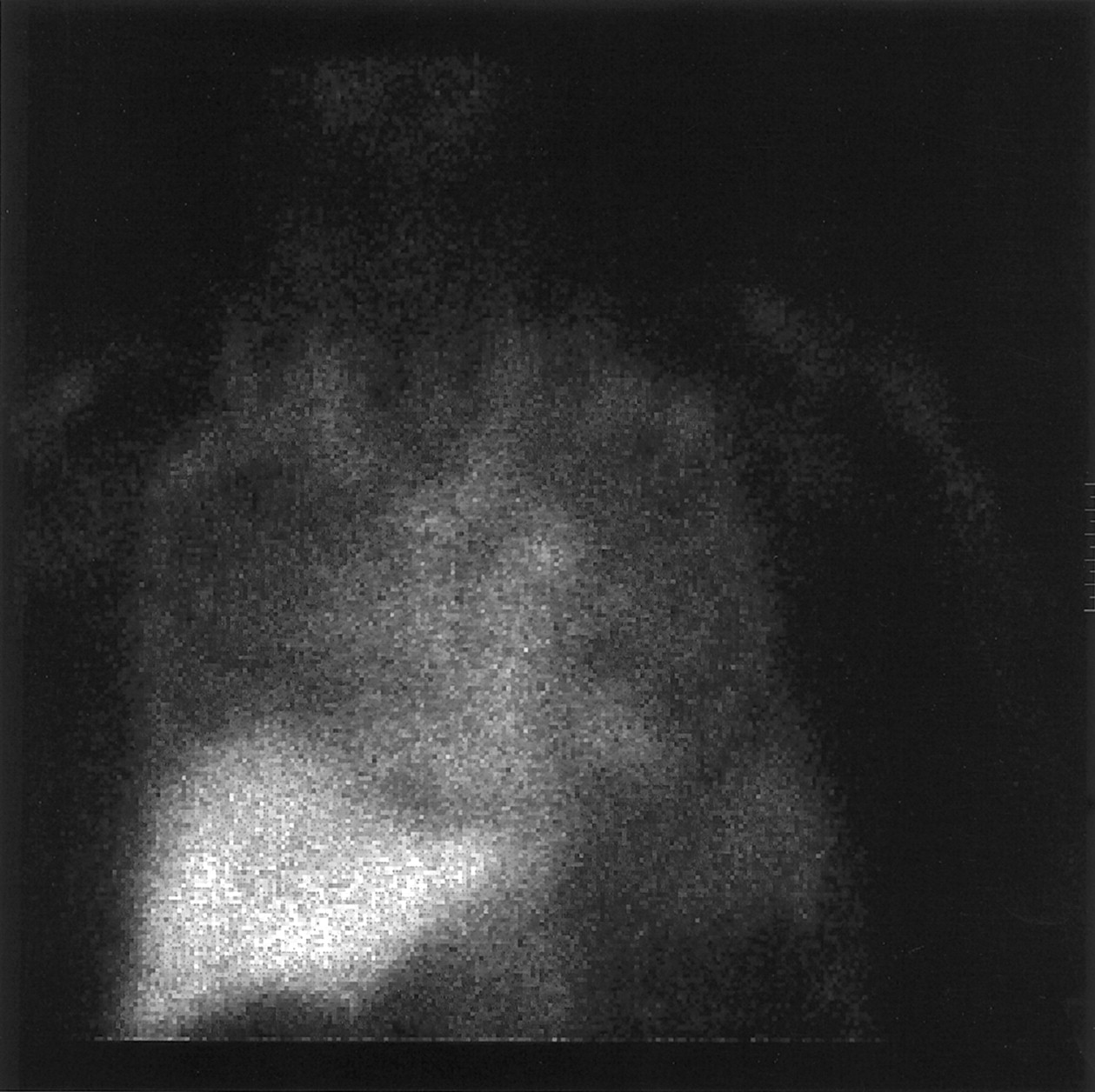
Second GI bleed study performed using UltraTag kit (2 d later) Anterior view: abnormal liver and bone marrow uptake is visualized.
CONCLUSION
Cases such as this underscore the recommendation that a routine radiopharmaceutical quality control program should be developed and implemented to detect unexpected radiopharmaceutical problems, especially when deviating from the standard preparation procedure, before administration to the patient.
Footnotes
For correspondence or reprints contact: Art Meyers, EdD, NMTCB, ARRT (N), Department of Health Physics, Nuclear Medicine Program, University of Nevada, 4505 Maryland Pky., Las Vegas, NV 89154.
E-mail: ameyers{at}ccmail.nevada.edu


Jump to section
Related Articles
Cited By...
- No citing articles found.