
Main menu

  • Home
  • Content
    • Current
      • JNMT Supplement
    • Ahead of print
    • Past Issues
    • Continuing Education
    • JNMT Podcast
    • SNMMI Annual Meeting Abstracts
  • Subscriptions
    • Subscribers
    • Rates
    • Journal Claims
    • Institutional and Non-member
  • Authors
    • Submit to JNMT
    • Information for Authors
    • Assignment of Copyright
    • AQARA Requirements
  • Info
    • Reviewers
    • Permissions
    • Advertisers
    • Corporate & Special Sales
  • About
    • About Us
    • Editorial Board
    • Contact Information
  • More
    • Alerts
    • Feedback
    • Help
    • SNMMI Journals
  • SNMMI
    • JNMT
    • JNM
    • SNMMI Journals
    • SNMMI

User menu

  • Subscribe
  • My alerts
  • Log in
  • My Cart

Search

  • Advanced search
Journal of Nuclear Medicine Technology
  • SNMMI
    • JNMT
    • JNM
    • SNMMI Journals
    • SNMMI
  • Subscribe
  • My alerts
  • Log in
  • My Cart
Journal of Nuclear Medicine Technology

Advanced Search

  • Home
  • Content
    • Current
    • Ahead of print
    • Past Issues
    • Continuing Education
    • JNMT Podcast
    • SNMMI Annual Meeting Abstracts
  • Subscriptions
    • Subscribers
    • Rates
    • Journal Claims
    • Institutional and Non-member
  • Authors
    • Submit to JNMT
    • Information for Authors
    • Assignment of Copyright
    • AQARA Requirements
  • Info
    • Reviewers
    • Permissions
    • Advertisers
    • Corporate & Special Sales
  • About
    • About Us
    • Editorial Board
    • Contact Information
  • More
    • Alerts
    • Feedback
    • Help
    • SNMMI Journals
  • Watch or Listen to JNMT Podcast
  • Visit SNMMI on Facebook
  • Join SNMMI on LinkedIn
  • Follow SNMMI on Twitter
  • Subscribe to JNMT RSS feeds
Review ArticleTEACHING FILE

Scintigraphic Evaluation of a Suspected Cavernous Hemangioma

Prashant Jolepalem, Catalin Grigore and Jane C. Palka
Journal of Nuclear Medicine Technology December 2012, 40 (4) 290-292; DOI: https://doi.org/10.2967/jnmt.112.114280
Prashant Jolepalem
Department of Radiology and Molecular Imaging, Oakland University William Beaumont Health Systems, Royal Oak, Michigan
MD
  • Find this author on Google Scholar
  • Find this author on PubMed
  • Search for this author on this site
Catalin Grigore
Department of Radiology and Molecular Imaging, Oakland University William Beaumont Health Systems, Royal Oak, Michigan
CNMT
  • Find this author on Google Scholar
  • Find this author on PubMed
  • Search for this author on this site
Jane C. Palka
Department of Radiology and Molecular Imaging, Oakland University William Beaumont Health Systems, Royal Oak, Michigan
DO
  • Find this author on Google Scholar
  • Find this author on PubMed
  • Search for this author on this site
  • Article
  • Figures & Data
  • Info & Metrics
  • PDF
Loading

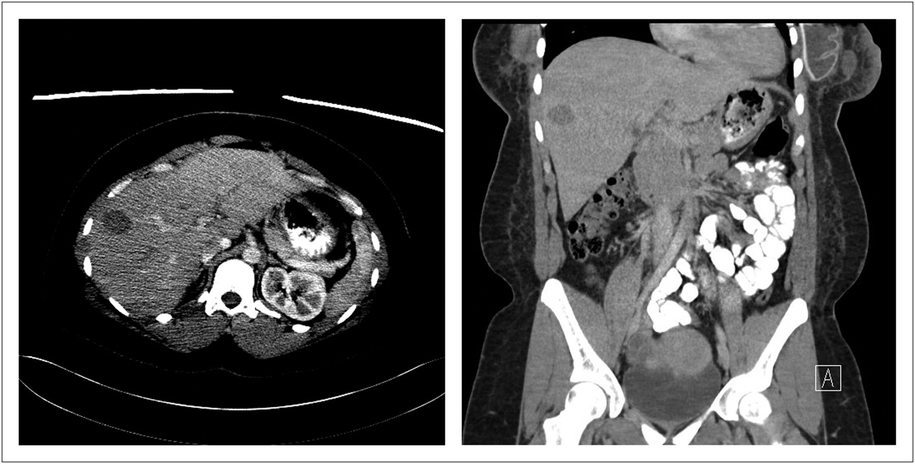
The patient was a 31-y-old woman with invasive ductal carcinoma of the right breast without evidence of metastases to the axillary lymph nodes. She had undergone bilateral mastectomy and was currently undergoing radiation therapy. A CT scan performed 10 d previously demonstrated “a 3 cm hypodensity within the anterior right hepatic lobe that is suspicious for a metastatic focus although benign etiology such as hemangioma cannot be excluded” (Fig. 1). A 99mTc-red blood cell (RBC) scan was ordered to exclude malignancy.

FIGURE 1.
  • Download figure
  • Open in new tab
  • Download powerpoint
FIGURE 1.

Transverse (liver window; left) and coronal (soft-tissue window; right) CT scans of abdomen and pelvis.

The patient was injected with 1,106.3 MBq (29.9 mCi) of 99mTc in vitro–labeled RBCs. Early and 2-h delayed planar images were taken in the anterior and posterior projections of the abdomen (heart through aortic bifurcation), with SPECT images of the early phase and SPECT/CT images of the 2-h delayed phase. Limited-field-of-view, low-dose CT was performed for anatomic localization and attenuation correction of the SPECT data.

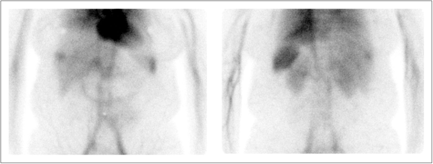
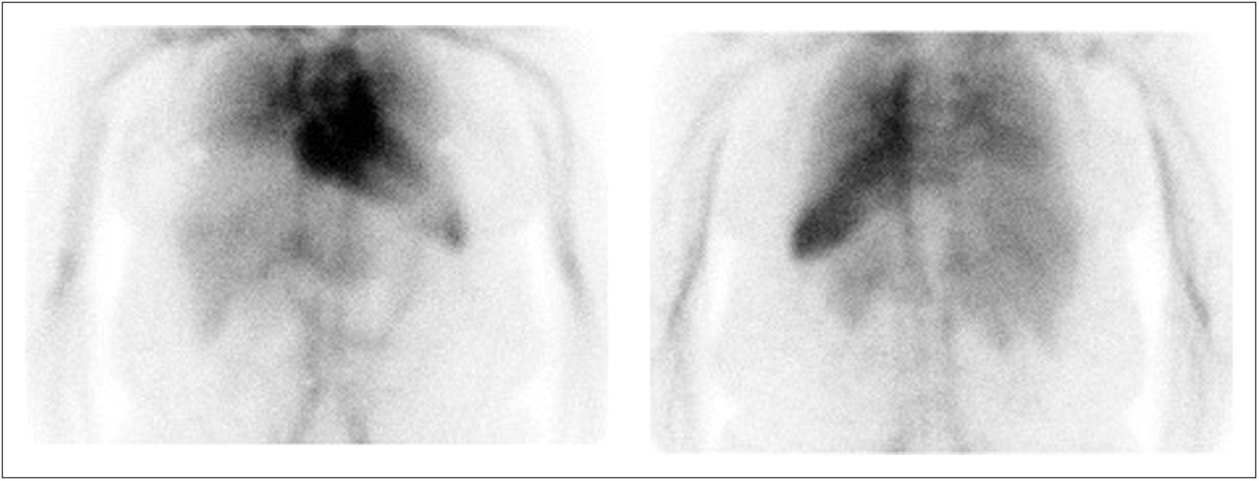
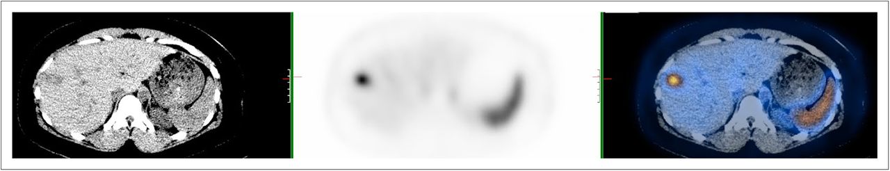
The imaging findings were typical for a cavernous hemangioma, as the radiotracer activity during the study was consistent with the physiologic activity of RBCs within a hemangioma (1,2). After the radiolabeled RBCs have had time to equilibrate, they start to enter the hemangioma and begin accumulating. The uptake may be equal to or slightly higher than in the surrounding normal liver but is still less than in the spleen, as seen on the early-phase planar images (Fig. 2) and the SPECT image (Fig. 3). On the delayed-phase images, the activity within the hemangioma exceeded that within the surrounding liver and was equal to the activity within the spleen (Fig. 4). SPECT/CT proved that the focal area of uptake corresponded to the low-density lesion found on the diagnostic CT scan (Fig. 5).

FIGURE 2.
  • Download figure
  • Open in new tab
  • Download powerpoint
FIGURE 2.

Anterior (left) and posterior (right) early planar images.

FIGURE 3.
  • Download figure
  • Open in new tab
  • Download powerpoint
FIGURE 3.

Early SPECT image.

FIGURE 4.
  • Download figure
  • Open in new tab
  • Download powerpoint
FIGURE 4.

Anterior (left) and posterior (right) 2-h delayed planar images.

FIGURE 5.
  • Download figure
  • Open in new tab
  • Download powerpoint
FIGURE 5.

Two-hour delayed CT, SPECT, and SPECT/CT images (from left to right).

SPECT alone was sufficient on the early images to localize the lesion; it was not necessary to deliver the additional radiation dose from CT (in accordance with the goal of keeping the dose as low as reasonably achievable). SPECT/CT should routinely be performed on the delayed images, as it greatly increases imaging sensitivity (3). Although the SNMMI guidelines recommend performing initial flow images, this is not part of the protocol at our institution, as they are frequently of low yield (4). Typically, there would be a photon-deficient area in the liver parenchyma on the initial flow images while the tracer is within the cardiac blood pool, until the hemangioma begins accumulating the labeled RBCs (1). However, if the hemangioma is in a difficult place to see on 2-dimensional planar images, the absence of this finding can lead the reader astray (3).

99mTc-RBC scintigraphy is highly specific for cavernous hemangioma (5); therefore, we consider here some differential diagnoses based on only the CT scan results. One possibility is focal nodular hyperplasia, another benign tumor that would be equivocal on 99mTc-RBC scintigraphy and better imaged by 99mTc-sulfur colloid scanning, on which it typically shows increased uptake. Focal nodular hyperplasia can incidentally be seen on hepatobiliary imaging as well (1,2).

Another possibility is hepatic adenoma. Although considered a benign tumor, hepatic adenoma is associated with life-threatening hemorrhages. Both hemangiomas and hepatic adenomas appear as photopenic defects on 99mTc-sulfur colloid scans, but adenomas will be photopenic on 99mTc-RBC scans as well (1,2,5). Suspected hepatic adenomas should be imaged by MRI, where in general, they will appear hyperintense relative to background liver on T1- and T2-weighted images (3).

A third possibility is metastatic or primary malignancy. Depending on the pathology, hepatic malignancies may or may not be imaged successfully by SPECT or PET techniques. On 99mTc-RBC scintigraphy, malignant lesions would have normal or decreased uptake, with the exception being the extremely rare angiosarcomas, which are the only notable cause for false-positive cases (1,5). MRI remains the modality of choice for characterizing suggestive hypodense lesions seen on staging CT scans (3).

QUESTION 1

For a potential hemangioma larger than 1.5 cm, what is the modality of choice?

  1. Ultrasound.

  2. MRI.

  3. 99mTc-RBC SPECT.

  4. Dynamic contrast-enhanced CT.

QUESTION 2

What is the typical appearance of a hemangioma on a liver–spleen sulfur colloid scan?

  1. Intense circumscribed lesion.

  2. Photopenic circumscribed defect.

  3. Normal hepatic uptake.

  4. Photopenic on early images, becoming intense on delayed.

CASE DISCUSSION

Cavernous hemangioma is the most common primary hepatic tumor. These tumors are frequently asymptomatic and incidentally discovered on imaging, surgery, or autopsy. They arise from ectasias in the endothelial lining of hepatic blood vessels and consist of multiple large, vascular channels lined by a single layer of endothelial cells and supported by collagenous walls. They are often associated with high estrogen states including puberty, pregnancy, oral contraceptive use, and steroid use. Though they are benign, hemangiomas need to be distinguished from other tumors. Rare complications include abdominal discomfort, fullness, and, rarer still, spontaneous rupture (1,6).

99mTc-RBC scintigraphy combined with multihead SPECT cameras has a nearly 100% positive predictive value. Although the sensitivity was previously dependent on size, the advent of SPECT/CT has allowed detection of hemangiomas as small as 0.5 cm with sensitivity of 88%. The high diagnostic accuracy, low cost, and relatively low radiation dose make 99mTc-RBC scintigraphy a useful, although underutilized, diagnostic study for the work-up of suspected benign hepatic lesions (3,5,6).

Our patient had a diagnosis of breast cancer without evidence of metastasis to the axillary lymph nodes. Therefore, the possibility of liver metastasis was considered less likely. 99mTc-scintigraphy was a low-cost, high-yield study to confirm the diagnosis of the hepatic lesion and spare the patient an unnecessary attempt at biopsy or surgical intervention.

Footnotes

  • Published online Oct. 9, 2012.

  • ↵* For the answers, see page 12A.

REFERENCES

  1. 1.↵
    1. Ziessman HA,
    2. O’Malley JP,
    3. Thrall JH
    . Nuclear Medicine (The Requisites). 3rd ed. Philadelphia, PA: Mosby; 2006.
  2. 2.↵
    1. Mettler FA,
    2. Guiberteau MJ
    . Essentials of Nuclear Medicine Imaging. 5th ed. Philadelphia, PA: Elsevier Inc.; 2006.
  3. 3.↵
    1. Vilgrain V,
    2. Boulos L,
    3. Vullierme MP,
    4. et al
    . Imaging of atypical hemangiomas of the liver with pathologic correlation. Radiographics. 2000;20:379–397.
    OpenUrlCrossRefPubMed
  4. 4.↵
    1. Gordhan A,
    2. Soliman J,
    3. Malpani A,
    4. Peg E III.
    . Symptomatic calvarial cavernous hemangioma. J Radiol Case Rep. 2009;3:25–29.
    OpenUrlPubMed
  5. 5.↵
    1. Mustafa Y,
    2. Erkan D,
    3. Oguz D
    . Evaluation of hepatic hemangioma with Tc-99m RBC scintigraphy: a review of 74 cases with liver masses. Turk J Gastroenterol. 2000;11:61–68.
    OpenUrl
  6. 6.↵
    1. Zheng JG,
    2. Yao ZM,
    3. Shu CY,
    4. et al
    . Role of SPECT/CT in diagnosis of hepatic cavernous hemangioma. World J Gastroenterol. 2005;11:5336–5341.
    OpenUrlPubMed
PreviousNext
Back to top

In this issue

Journal of Nuclear Medicine Technology: 40 (4)
Journal of Nuclear Medicine Technology
Vol. 40, Issue 4
December 1, 2012
  • Table of Contents
  • About the Cover
  • Index by author
Print
Download PDF
Article Alerts
Sign In to Email Alerts with your Email Address
Email Article

Thank you for your interest in spreading the word on Journal of Nuclear Medicine Technology.

NOTE: We only request your email address so that the person you are recommending the page to knows that you wanted them to see it, and that it is not junk mail. We do not capture any email address.

Enter multiple addresses on separate lines or separate them with commas.
Scintigraphic Evaluation of a Suspected Cavernous Hemangioma
(Your Name) has sent you a message from Journal of Nuclear Medicine Technology
(Your Name) thought you would like to see the Journal of Nuclear Medicine Technology web site.
Citation Tools
Scintigraphic Evaluation of a Suspected Cavernous Hemangioma
Prashant Jolepalem, Catalin Grigore, Jane C. Palka
Journal of Nuclear Medicine Technology Dec 2012, 40 (4) 290-292; DOI: 10.2967/jnmt.112.114280

Citation Manager Formats

  • BibTeX
  • Bookends
  • EasyBib
  • EndNote (tagged)
  • EndNote 8 (xml)
  • Medlars
  • Mendeley
  • Papers
  • RefWorks Tagged
  • Ref Manager
  • RIS
  • Zotero
Share
Scintigraphic Evaluation of a Suspected Cavernous Hemangioma
Prashant Jolepalem, Catalin Grigore, Jane C. Palka
Journal of Nuclear Medicine Technology Dec 2012, 40 (4) 290-292; DOI: 10.2967/jnmt.112.114280
Twitter logo Facebook logo LinkedIn logo Mendeley logo
  • Tweet Widget
  • Facebook Like
  • Google Plus One
Bookmark this article

Jump to section

  • Article
    • QUESTION 1
    • QUESTION 2
    • CASE DISCUSSION
    • Footnotes
    • REFERENCES
  • Figures & Data
  • Info & Metrics
  • PDF

Related Articles

  • NMTCB Report
  • PubMed
  • Google Scholar

Cited By...

  • No citing articles found.
  • Google Scholar

More in this TOC Section

  • Brain SPECT Imaging with Acetazolamide Challenge
  • Evaluation of Pleural-Based Nodules in a 52-Year-Old Woman with a History of Metastatic Breast Cancer
Show more Teaching File

Similar Articles

SNMMI

© 2025 SNMMI

Powered by HighWire