
Main menu

  • Home
  • Content
    • Current
      • JNMT Supplement
    • Ahead of print
    • Past Issues
    • Continuing Education
    • JNMT Podcast
    • SNMMI Annual Meeting Abstracts
  • Subscriptions
    • Subscribers
    • Rates
    • Journal Claims
    • Institutional and Non-member
  • Authors
    • Submit to JNMT
    • Information for Authors
    • Assignment of Copyright
    • AQARA Requirements
  • Info
    • Reviewers
    • Permissions
    • Advertisers
    • Corporate & Special Sales
  • About
    • About Us
    • Editorial Board
    • Contact Information
  • More
    • Alerts
    • Feedback
    • Help
    • SNMMI Journals
  • SNMMI
    • JNMT
    • JNM
    • SNMMI Journals
    • SNMMI

User menu

  • Subscribe
  • My alerts
  • Log in
  • Log out
  • My Cart

Search

  • Advanced search
Journal of Nuclear Medicine Technology
  • SNMMI
    • JNMT
    • JNM
    • SNMMI Journals
    • SNMMI
  • Subscribe
  • My alerts
  • Log in
  • Log out
  • My Cart
Journal of Nuclear Medicine Technology

Advanced Search

  • Home
  • Content
    • Current
    • Ahead of print
    • Past Issues
    • Continuing Education
    • JNMT Podcast
    • SNMMI Annual Meeting Abstracts
  • Subscriptions
    • Subscribers
    • Rates
    • Journal Claims
    • Institutional and Non-member
  • Authors
    • Submit to JNMT
    • Information for Authors
    • Assignment of Copyright
    • AQARA Requirements
  • Info
    • Reviewers
    • Permissions
    • Advertisers
    • Corporate & Special Sales
  • About
    • About Us
    • Editorial Board
    • Contact Information
  • More
    • Alerts
    • Feedback
    • Help
    • SNMMI Journals
  • Watch or Listen to JNMT Podcast
  • Visit SNMMI on Facebook
  • Join SNMMI on LinkedIn
  • Follow SNMMI on Twitter
  • Subscribe to JNMT RSS feeds
Review ArticleTEACHING FILE

Evaluation of Pleural-Based Nodules in a 52-Year-Old Woman with a History of Metastatic Breast Cancer

Blaine T. Mischen and Marques L. Bradshaw
Journal of Nuclear Medicine Technology September 2012, 40 (3) 204-206; DOI: https://doi.org/10.2967/jnmt.112.110452
Blaine T. Mischen
Nuclear Medicine, Radiology Department, Medical University of South Carolina, Charleston, South Carolina
  • Find this author on Google Scholar
  • Find this author on PubMed
  • Search for this author on this site
Marques L. Bradshaw
Nuclear Medicine, Radiology Department, Medical University of South Carolina, Charleston, South Carolina
  • Find this author on Google Scholar
  • Find this author on PubMed
  • Search for this author on this site
  • Article
  • Figures & Data
  • Info & Metrics
  • PDF
Loading

A 52-y-old woman with a history of invasive ductal carcinoma had undergone right mastectomy, right axillary lymph node dissection, and breast reconstruction. Two of six axillary lymph nodes were positive for ductal carcinoma at surgery, and the patient was staged pT2 pN1a Mx. A one-month follow-up chest CT scan showed multiple left-sided pleural-based nodules suggestive of metastatic disease. PET/CT, which was performed to further characterize these nodules, did not demonstrate significant 18F-FDG uptake within them or evidence of metastatic disease (Fig. 1). In the process of formulating a differential diagnosis for these nodules, it was discovered that, as a child, the patient had sustained an accidental gunshot wound. The possibility of thoracic splenosis was suggested, and a 99mTc-labeled heat-denatured red blood cell (DRBC) scan was ordered to confirm the origin of these conspicuous pleural-based nodules.

FIGURE 1.
  • Download figure
  • Open in new tab
  • Download powerpoint
FIGURE 1.

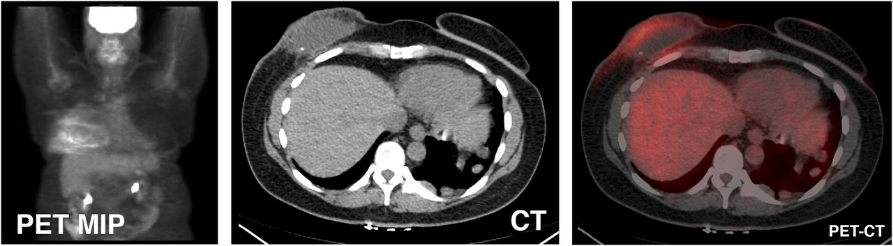
Initial maximum-intensity-projection (MIP) PET, CT, and PET/CT images.

A blood sample was drawn into a vial containing anticoagulant (acid citrate dextrose) labeled with 370 MBq (10 mCi) of 99mTc-pertechnetate and heat-denatured by submersion in a 49.5°C (±0.5°C) water bath for 20 min. Standard quality control analyses were completed. The radiolabeled autologous DRBCs were reinjected intravenously. Planar anterior and posterior images of the chest and abdomen were obtained 30 min after radiolabeled DRBC reinjection. Congruent SPECT and CT images of the chest and abdomen were also acquired and were displayed with and without fusion.

QUESTION 1

Localization of DRBCs takes place by what mechanism?

  • A. Active transport by splenocytes via the adenosine triphosphatase–driven Na-K pump.

  • B. Binding to transferrins, which localize to splenic tissue.

  • C. Ion exchange on the cell membrane of splenocytes.

  • D. Phagocytosis of the DRBCs by the reticuloendothelial system and size- and shape-dependent sequestration by the spleen.

QUESTION 2

Regarding the sensitivity and specificity of different imaging modalities to evaluate for splenosis, which statement is true?

  • A. 99mTc-DRBC scans have a higher sensitivity and specificity than sulfur colloid, CT, and MRI.

  • B. 99mTc-DRBC scans and sulfur colloid scans are equal in sensitivity and specificity, and both are superior to CT and MRI.

  • C. MRI is the gold standard in evaluating splenosis, having the highest sensitivity and specificity when compared with all other imaging modalities.

  • D. All imaging modalities are the same with regard to the sensitivity and specificity of splenosis detection.

CASE DISCUSSION

Given the patient’s history of locally metastatic breast cancer, the pleural-based nodules seen on CT were suspected of being new metastatic disease; however, the non–18F-FDG avidity of these nodules was a confounding result (Fig. 1). To formulate a more extensive differential diagnosis, additional past medical history was sought, and it was discovered the patient had sustained an accidental gunshot wound as a child. The trajectory of the bullet was from the left posterior chest wall to the diaphragm, spleen, and bowel and exiting through the anterior medial abdominal wall. This trauma required surgical repair of the bowel injuries, as well as a splenectomy.

The autotransplantation of splenic tissue to the thoracic cavity requires particular circumstances. Specifically, splenic trauma or surgery, violation of the diaphragm, and an appropriate seeding environment are necessary. Therefore, thoracic splenosis is a rare and usually incidental finding on various imaging studies (3). Differentiating splenosis from other, more common, pleural-based nodules, particularly when remote from the traumatic seeding event or without a pertinent past medical history, can be quite difficult. The diagnosis is further complicated when the patient has a known neoplasm, as in this case. However, if splenosis is suspected, the use of a noninvasive test with a high sensitivity and specificity can decrease radiation exposure from further imaging, avoid unnecessary biopsies and their associated sequelae, and give the patient and the clinician assurance of a definitive diagnosis. 99mTc-DRBC studies are highly sensitive at detecting even small volumes of splenic tissue and are very specific (2,4). Additionally, there is a higher target (splenic tissue) to background ratio in 99mTc-DRBC scans than in radiolabeled sulfur colloid scans due to the marked splenic reticuloendothelial system phagocytosis and pulp sequestration of damaged red blood cells (≤50%) compared with colloid distribution (10%–15% spleen, 80%–85% liver, and 5% bone marrow) (5,6).

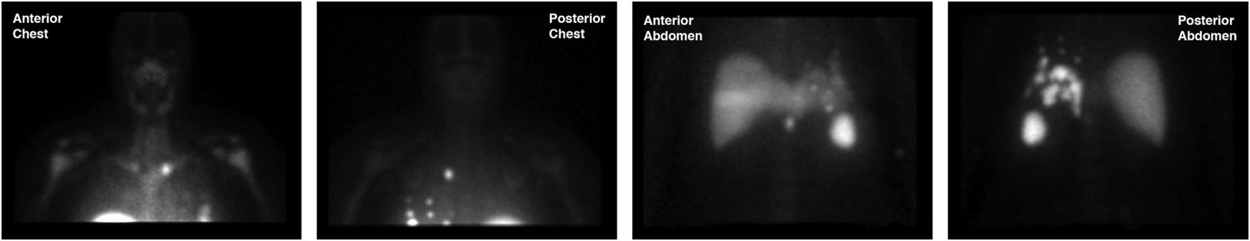
In this case, numerous foci of 99mTc-DRBCs were detected on the planar images (Fig. 2). Hybrid SPECT/CT images confirmed significant 99mTc-DRBC activity in all previously seen pleural-based nodules, thus supporting the diagnosis of thoracic splenosis (Fig. 3). Additionally, two islands of peritoneal splenosis were localized (Fig. 4).

FIGURE 2.
  • Download figure
  • Open in new tab
  • Download powerpoint
FIGURE 2.

Planar 99mTc-labeled DRBC images showing numerous foci of uptake.

FIGURE 3.
  • Download figure
  • Open in new tab
  • Download powerpoint
FIGURE 3.

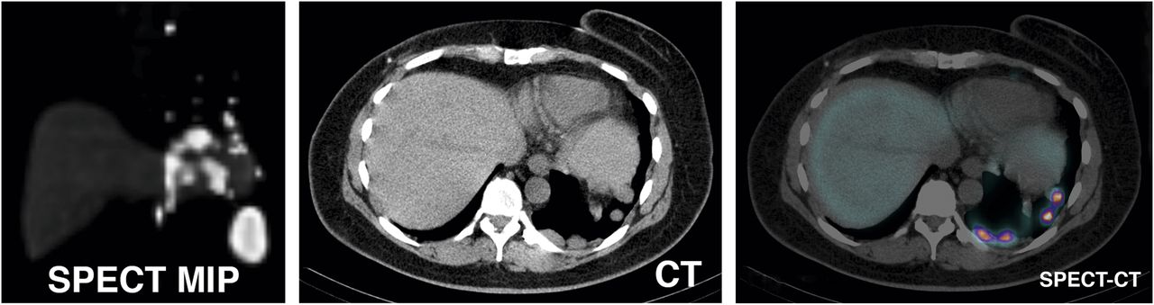
Maximum-intensity-projection (MIP) SPECT, CT, and SPECT/CT images confirming activity in all previously seen pleural-based nodules.

FIGURE 4.
  • Download figure
  • Open in new tab
  • Download powerpoint
FIGURE 4.

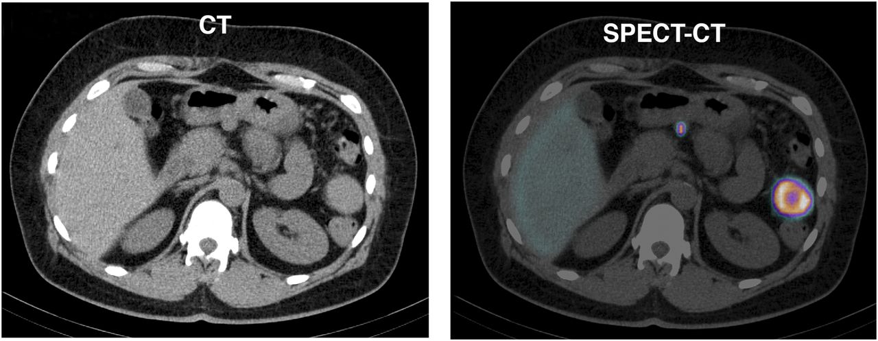
CT and SPECT/CT images showing two islands of peritoneal splenosis.

Footnotes

  • Published online Jul. 7, 2012.

  • * For the answers, see page 17A.

REFERENCES

  1. 1.
    1. Stewart CA,
    2. Sakimura ME
    . Scintigraphic demonstration of splenosis. Clin Nucl Med. 1986;11:161–164.
    OpenUrlCrossRefPubMed
  2. 2.↵
    1. Armas RR
    . Clinical studies with spleen-specific radiolabeled agents. Semin Nucl Med. 1985;15:260–275.
    OpenUrlCrossRefPubMed
  3. 3.↵
    1. Hagman TF,
    2. Winer-Muram HT,
    3. Meyer CA,
    4. Jennings SG
    . Intrathoracic splenosis: superiority of technetium Tc 99m heat-damaged RBC imaging. Chest. 2001;120:2097–2098.
    OpenUrlCrossRefPubMed
  4. 4.↵
    1. Yammine JN,
    2. Yatim A,
    3. Barbari A
    . Radionuclide imaging in thoracic splenosis and a review of the literature. Clin Nucl Med. 2003;28:121–123.
    OpenUrlCrossRefPubMed
  5. 5.↵
    1. Atkins HL,
    2. Goldman AG,
    3. Fairchild RG,
    4. et al
    . Splenic sequestration of 99mTc labeled, heat treated red blood cells. Radiology. 1980;136:501–503.
    OpenUrlPubMed
  6. 6.↵
    1. Massey MD,
    2. Stevens JS
    . Residual spleen found on denatured red blood cell scan following negative colloid scans. J Nucl Med. 1991;32:2286–2287.
    OpenUrlAbstract/FREE Full Text
PreviousNext
Back to top

In this issue

Journal of Nuclear Medicine Technology: 40 (3)
Journal of Nuclear Medicine Technology
Vol. 40, Issue 3
September 1, 2012
  • Table of Contents
  • About the Cover
  • Index by author
Print
Download PDF
Article Alerts
Sign In to Email Alerts with your Email Address
Email Article

Thank you for your interest in spreading the word on Journal of Nuclear Medicine Technology.

NOTE: We only request your email address so that the person you are recommending the page to knows that you wanted them to see it, and that it is not junk mail. We do not capture any email address.

Enter multiple addresses on separate lines or separate them with commas.
Evaluation of Pleural-Based Nodules in a 52-Year-Old Woman with a History of Metastatic Breast Cancer
(Your Name) has sent you a message from Journal of Nuclear Medicine Technology
(Your Name) thought you would like to see the Journal of Nuclear Medicine Technology web site.
Citation Tools
Evaluation of Pleural-Based Nodules in a 52-Year-Old Woman with a History of Metastatic Breast Cancer
Blaine T. Mischen, Marques L. Bradshaw
Journal of Nuclear Medicine Technology Sep 2012, 40 (3) 204-206; DOI: 10.2967/jnmt.112.110452

Citation Manager Formats

  • BibTeX
  • Bookends
  • EasyBib
  • EndNote (tagged)
  • EndNote 8 (xml)
  • Medlars
  • Mendeley
  • Papers
  • RefWorks Tagged
  • Ref Manager
  • RIS
  • Zotero
Share
Evaluation of Pleural-Based Nodules in a 52-Year-Old Woman with a History of Metastatic Breast Cancer
Blaine T. Mischen, Marques L. Bradshaw
Journal of Nuclear Medicine Technology Sep 2012, 40 (3) 204-206; DOI: 10.2967/jnmt.112.110452
Twitter logo Facebook logo LinkedIn logo Mendeley logo
  • Tweet Widget
  • Facebook Like
  • Google Plus One
Bookmark this article

Jump to section

  • Article
    • QUESTION 1
    • QUESTION 2
    • CASE DISCUSSION
    • Footnotes
    • REFERENCES
  • Figures & Data
  • Info & Metrics
  • PDF

Related Articles

  • No related articles found.
  • PubMed
  • Google Scholar

Cited By...

  • No citing articles found.
  • Google Scholar

More in this TOC Section

  • Brain SPECT Imaging with Acetazolamide Challenge
  • Scintigraphic Evaluation of a Suspected Cavernous Hemangioma
Show more Teaching File

Similar Articles

SNMMI

© 2025 SNMMI

Powered by HighWire