  • Main menu
  • User menu
  • Search
  • English ▼
    • English
    • Afrikaans
    • Albanian
    • Amharic
    • Arabic
    • Armenian
    • Azerbaijani
    • Basque
    • Belarusian
    • Bengali
    • Bosnian
    • Bulgarian
    • Catalan
    • Cebuano
    • Chichewa
    • Chinese (Simplified)
    • Chinese (Traditional)
    • Corsican
    • Croatian
    • Czech
    • Danish
    • Dutch
    • Esperanto
    • Estonian
    • Filipino
    • Finnish
    • French
    • Frisian
    • Galician
    • Georgian
    • German
    • Greek
    • Gujarati
    • Haitian Creole
    • Hausa
    • Hawaiian
    • Hebrew
    • Hindi
    • Hmong
    • Hungarian
    • Icelandic
    • Igbo
    • Indonesian
    • Irish
    • Italian
    • Japanese
    • Javanese
    • Kannada
    • Kazakh
    • Khmer
    • Korean
    • Kurdish (Kurmanji)
    • Kyrgyz
    • Lao
    • Latin
    • Latvian
    • Lithuanian
    • Luxembourgish
    • Macedonian
    • Malagasy
    • Malay
    • Malayalam
    • Maltese
    • Maori
    • Marathi
    • Mongolian
    • Myanmar (Burmese)
    • Nepali
    • Norwegian
    • Pashto
    • Persian
    • Polish
    • Portuguese
    • Punjabi
    • Romanian
    • Russian
    • Samoan
    • Scottish Gaelic
    • Serbian
    • Sesotho
    • Shona
    • Sindhi
    • Sinhala
    • Slovak
    • Slovenian
    • Somali
    • Spanish
    • Sudanese
    • Swahili
    • Swedish
    • Tajik
    • Tamil
    • Telugu
    • Thai
    • Turkish
    • Ukrainian
    • Urdu
    • Uzbek
    • Vietnamese
    • Welsh
    • Xhosa
    • Yiddish
    • Yoruba
    • Zulu

Main menu

  • Home
  • Content
    • Current
      • JNMT Supplement
    • Ahead of print
    • Past Issues
    • Continuing Education
    • JNMT Podcast
    • SNMMI Annual Meeting Abstracts
  • Subscriptions
    • Subscribers
    • Rates
    • Journal Claims
    • Institutional and Non-member
  • Authors
    • Submit to JNMT
    • Information for Authors
    • Assignment of Copyright
    • AQARA Requirements
  • Info
    • Reviewers
    • Permissions
    • Advertisers
    • Corporate & Special Sales
  • About
    • About Us
    • Editorial Board
    • Contact Information
  • More
    • Alerts
    • Feedback
    • Help
    • SNMMI Journals
  • SNMMI
    • JNMT
    • JNM
    • SNMMI Journals
    • SNMMI

User menu

  • Subscribe
  • My alerts
  • Log in
  • Log out
  • My Cart

Search

  • Advanced search
Journal of Nuclear Medicine Technology
  • SNMMI
    • JNMT
    • JNM
    • SNMMI Journals
    • SNMMI
  • Subscribe
  • My alerts
  • Log in
  • Log out
  • My Cart
Journal of Nuclear Medicine Technology

Advanced Search

English ▼
  • English
  • Afrikaans
  • Albanian
  • Amharic
  • Arabic
  • Armenian
  • Azerbaijani
  • Basque
  • Belarusian
  • Bengali
  • Bosnian
  • Bulgarian
  • Catalan
  • Cebuano
  • Chichewa
  • Chinese (Simplified)
  • Chinese (Traditional)
  • Corsican
  • Croatian
  • Czech
  • Danish
  • Dutch
  • Esperanto
  • Estonian
  • Filipino
  • Finnish
  • French
  • Frisian
  • Galician
  • Georgian
  • German
  • Greek
  • Gujarati
  • Haitian Creole
  • Hausa
  • Hawaiian
  • Hebrew
  • Hindi
  • Hmong
  • Hungarian
  • Icelandic
  • Igbo
  • Indonesian
  • Irish
  • Italian
  • Japanese
  • Javanese
  • Kannada
  • Kazakh
  • Khmer
  • Korean
  • Kurdish (Kurmanji)
  • Kyrgyz
  • Lao
  • Latin
  • Latvian
  • Lithuanian
  • Luxembourgish
  • Macedonian
  • Malagasy
  • Malay
  • Malayalam
  • Maltese
  • Maori
  • Marathi
  • Mongolian
  • Myanmar (Burmese)
  • Nepali
  • Norwegian
  • Pashto
  • Persian
  • Polish
  • Portuguese
  • Punjabi
  • Romanian
  • Russian
  • Samoan
  • Scottish Gaelic
  • Serbian
  • Sesotho
  • Shona
  • Sindhi
  • Sinhala
  • Slovak
  • Slovenian
  • Somali
  • Spanish
  • Sudanese
  • Swahili
  • Swedish
  • Tajik
  • Tamil
  • Telugu
  • Thai
  • Turkish
  • Ukrainian
  • Urdu
  • Uzbek
  • Vietnamese
  • Welsh
  • Xhosa
  • Yiddish
  • Yoruba
  • Zulu
  • Home
  • Content
    • Current
    • Ahead of print
    • Past Issues
    • Continuing Education
    • JNMT Podcast
    • SNMMI Annual Meeting Abstracts
  • Subscriptions
    • Subscribers
    • Rates
    • Journal Claims
    • Institutional and Non-member
  • Authors
    • Submit to JNMT
    • Information for Authors
    • Assignment of Copyright
    • AQARA Requirements
  • Info
    • Reviewers
    • Permissions
    • Advertisers
    • Corporate & Special Sales
  • About
    • About Us
    • Editorial Board
    • Contact Information
  • More
    • Alerts
    • Feedback
    • Help
    • SNMMI Journals
  • Watch or Listen to JNMT Podcast
  • Visit SNMMI on Facebook
  • Join SNMMI on LinkedIn
  • Follow SNMMI on Twitter
  • Subscribe to JNMT RSS feeds
Review ArticleTeaching Case Studies

A Rare Case of Giant Intraperitoneal Lipoma Found Incidentally on 18F-FDG PET/CT in a Patient with Nodular Fasciitis

María J. Ibáñez, Rafael H. Reyes, Laura Frutos and María A. Claver
Journal of Nuclear Medicine Technology June 2019, 47 (2) 173-174; DOI: https://doi.org/10.2967/jnmt.118.220152
María J. Ibáñez
Department of Nuclear Medicine, Clinic University Virgen de la Arrixaca Hospital, Murcia, Spain
  • Find this author on Google Scholar
  • Find this author on PubMed
  • Search for this author on this site
Rafael H. Reyes
Department of Nuclear Medicine, Clinic University Virgen de la Arrixaca Hospital, Murcia, Spain
  • Find this author on Google Scholar
  • Find this author on PubMed
  • Search for this author on this site
Laura Frutos
Department of Nuclear Medicine, Clinic University Virgen de la Arrixaca Hospital, Murcia, Spain
  • Find this author on Google Scholar
  • Find this author on PubMed
  • Search for this author on this site
María A. Claver
Department of Nuclear Medicine, Clinic University Virgen de la Arrixaca Hospital, Murcia, Spain
  • Find this author on Google Scholar
  • Find this author on PubMed
  • Search for this author on this site
  • Article
  • Figures & Data
  • Info & Metrics
  • PDF
Loading

Abstract

We report a case of concurrent nodular fasciitis and incidental giant intraperitoneal lipoma diagnosed on whole-body 18F-FDG PET/CT, an imaging modality that has the advantage of being able to detect unsuspected lesions.

  • 18F-FDG PET/CT
  • giant intraperitoneal lipoma
  • nodular fasciitis
  • pseudosarcomatous

Nodular fasciitis is a benign reactive proliferation of fibroblasts sometimes misdiagnosed as sarcoma on the basis of cellularity and rapid growth. Giant intraperitoneal lipomas are quite rare, and most are reported as liposarcoma because of their large size. Although liposarcomas make up only 0.1% of all human neoplasms, they are the most common histologic subtype of retroperitoneal soft-tissue sarcoma. This paper reports a case in which nodular fasciitis and incidental giant intraperitoneal lipoma concurred in a patient who underwent whole-body 18F-FDG PET/CT. 18F-FDG PET/CT plays a fundamental role in the differentiation of benign and malignant lesions and in the detection of unsuspected lesions.

CASE REPORT

We present a 33-y-old woman with no relevant medical or surgical history in whom a tumor in the right axillary area had been evolving for several months. Right axillary sonography, which was performed first, showed a heterogeneous solid mass measuring approximately 2 × 2.5 cm in the external region of the right axilla. The mass had no Doppler flow and was located between muscle masses. Because these findings might be compatible with intramuscular hematoma but cannot exclude underlying tumor, MRI was performed. It showed a nodular, lobulated, well-defined lesion in the right quadrilateral space, compatible with a neural tumor or paralabral cyst.

It was decided to perform sonography and fine-needle aspiration of the lesion, which showed a mesenchymal proliferation with scarce-to-moderate cellularity and a myxoid stroma, potentially compatible with myxoid sarcoma. Surgical exeresis was planned, and 18F-FDG PET/CT was requested to determine the extent of the lesion before surgery. PET/CT images taken 60 min after the intravenous administration of 370 MBq of 18F-FDG showed a slight metabolic increase (SUVmax of 1.9) in the right brachial lesion, suggesting a benign lesion but not being able to totally dismiss the possibility of a malignant lesion of low histologic grade and low affinity for 18F-FDG (Fig. 1). Likewise, as an incidental finding on the PET/CT images, a giant abdominopelvic mass of fat density and more than 30 cm in craniocaudal length was observed displacing the abdominal contents toward the left hypochondrium and flank; this mass did not present a metabolic increase (Figs. 2 and 3).

FIGURE 1.
  • Download figure
  • Open in new tab
  • Download powerpoint
FIGURE 1.

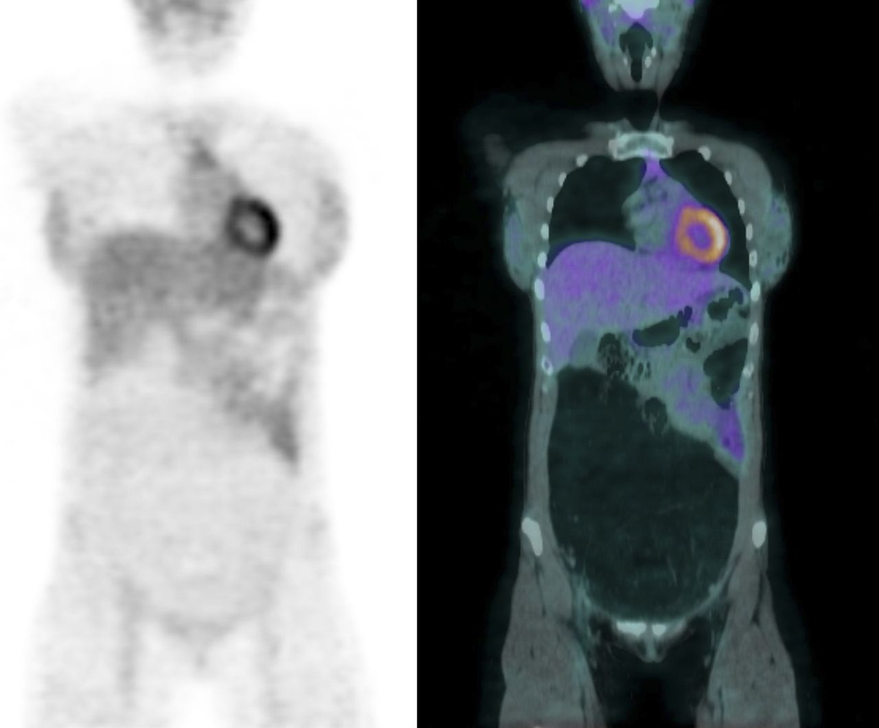
Right brachial nodular fasciitis on axial PET (top), CT (middle), and PET/CT (bottom). Slight focal metabolic increase, with SUVmax of 1.9, is seen in posterior region of right shoulder.

FIGURE 2.
  • Download figure
  • Open in new tab
  • Download powerpoint
FIGURE 2.

Giant intraperitoneal lipoma on coronal PET (left) and PET/CT (right). Giant intraabdominal mass of fat density is seen displacing abdominal content toward left hypochondrium and flank, without uptake on PET.

FIGURE 3.
  • Download figure
  • Open in new tab
  • Download powerpoint
FIGURE 3.

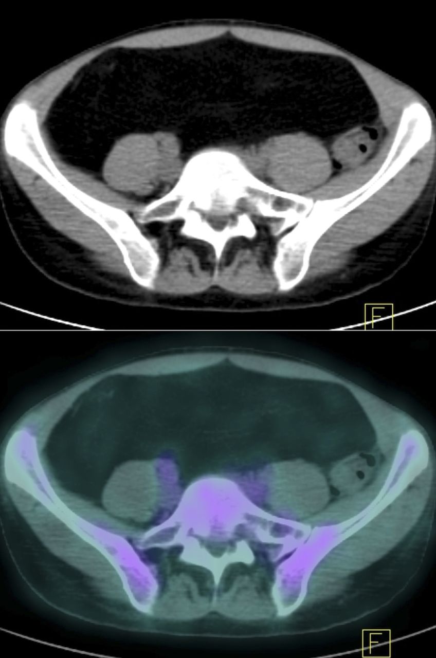
Giant intraperitoneal lipoma as seen on axial CT (top) and PET/CT (bottom).

In view of these findings, we took a deeper medical history, and this time the patient recalled experiencing a progressive abdominal distension occurring over a span of years, which she had interpreted as digestive intolerance. Abdominal MRI was performed and showed a large fatty mass with no large vessels or infiltration into adjacent organs.

In view of the diagnostic doubt about the atypical lipomatous tumor, which because of its enormous size might have been a liposarcoma, as well as the diagnosis of a possible brachial myxoid sarcoma, the patient underwent surgery. The brachial lesion was accessed with radical excision, and in the abdomen, a supraumbilical–infraumbilical midline incision revealed a large lipomatous pedunculated mass depending from subperitoneal fatty tissue at the level of the right iliac wall.

The definitive anatomic–pathologic diagnosis was nodular fasciitis for the brachial lesion and giant intraperitoneal lipoma for the abdominal lesion, with no malignant cells found in either of the 2 tumors.

DISCUSSION

Nodular fasciitis is a benign, rapidly growing, self-limiting proliferation of fibroblasts that originates in subcutaneous tissue or skeletal muscle and frequently undergoes spontaneous regression (1). Histologically, it can simulate a sarcoma because of the irregularity of the fibroblasts, which are randomly arranged in a myxoid stroma. Regarding imaging diagnosis, the appearance on MRI and sonography is quite variable and difficult to characterize. PET shows a mild hypermetabolic focus, ovoid and well circumscribed, at the muscular level, between muscles, or adjacent to tendons. Rapid growth, high cellularity, and high mitotic activity may be responsible for the metabolic increase on PET; however, both malignant and benign masses can show hypermetabolic activity; likewise, some malignant lesions may show only slight uptake because of acellularity or avascularity (2).

In our patient, giant intraperitoneal lipoma was an incidental finding on PET/CT. Lipomas are benign mesenchymal tumors that appear similar to normal fat. They can be found at any location, although intraperitoneal, omental, and mesenteric locations are exceptional (3). In this patient, the main concern in the diagnosis was potential malignancy because the large size of the lesion did not eliminate the possibility of well-differentiated liposarcoma, which can look similar to lipomas on conventional imaging (4,5).

CONCLUSION

18F-FDG PET/CT is a useful tool for discriminating the benign nature of 2 independent lesions that are creating diagnostic doubt. When used for full-body imaging, 18F-FDG PET/CT has the advantage of being able to detect unsuspected lesions—a capability that, in this case, led to the diagnosis of a huge, asymptomatic abdominal lesion.

DISCLOSURE

No potential conflict of interest relevant to this article was reported.

Footnotes

  • Published online Apr. 24, 2019.

REFERENCES

  1. 1.↵
    1. Tajima S,
    2. Zuki T,
    3. Koda K
    . Articular nodular fasciitis of the right shoulder joint: report of an unusual case with focus on immunohistochemical differential diagnosis. Int J Clin Exp Pathol. 2015;8:4242–4247.
    OpenUrlGoogle Scholar
  2. 2.↵
    1. Kim JY,
    2. Park J,
    3. Choi YY,
    4. Lee S,
    5. Paik SS
    . Nodular fasciitis mimicking soft tissue metastasis on 18F-FDG PET/CT during surveillance. Clin Nucl Med. 2015;40:172–174.
    OpenUrlGoogle Scholar
  3. 3.↵
    1. Kransdorf MJ,
    2. Bancroft LW,
    3. Peterson JJ,
    4. Murphey MD,
    5. Foster WC,
    6. Temple HT
    . Imaging of fatty tumors: distinction of lipoma and well-differentiated liposarcoma. Radiology. 2002;224:99–104.
    OpenUrlCrossRefPubMedGoogle Scholar
  4. 4.↵
    1. Weniger M,
    2. D’Haese JG,
    3. Kunz W,
    4. et al
    . En-bloc resection of a giant retroperitoneal lipoma: a case report and review of the literature. BMC Res Notes. 2015;8:75.
    OpenUrlGoogle Scholar
  5. 5.↵
    1. Metser U,
    2. Miller E,
    3. Lerman H,
    4. Even-Sapir E
    . Benign nonphysiologic lesions with increased 18F-FDG uptake on PET/CT: characterization and incidence. AJR. 2007;189:1203–1210.
    OpenUrlCrossRefPubMedGoogle Scholar
  • Received for publication September 13, 2018.
  • Accepted for publication January 7, 2019.
PreviousNext
Back to top

In this issue

Journal of Nuclear Medicine Technology: 47 (2)
Journal of Nuclear Medicine Technology
Vol. 47, Issue 2
June 1, 2019
  • Table of Contents
  • About the Cover
  • Index by author
Print
Download PDF
Article Alerts
Email Article
Citation Tools
Share
A Rare Case of Giant Intraperitoneal Lipoma Found Incidentally on 18F-FDG PET/CT in a Patient with Nodular Fasciitis
María J. Ibáñez, Rafael H. Reyes, Laura Frutos, María A. Claver
Journal of Nuclear Medicine Technology Jun 2019, 47 (2) 173-174; DOI: 10.2967/jnmt.118.220152
Twitter logo Facebook logo LinkedIn logo Mendeley logo
  • Tweet Widget
Bookmark this article

Jump to section

  • Article
    • Abstract
    • CASE REPORT
    • DISCUSSION
    • CONCLUSION
    • DISCLOSURE
    • Footnotes
    • REFERENCES
  • Figures & Data
  • Info & Metrics
  • PDF

Related Articles

  • No related articles found.
  • PubMed
  • Google Scholar

Cited By...

  • No citing articles found.
  • Google Scholar

More in this TOC Section

  • High-Sensitivity Troponin Elevation in a Young Woman with Typical Chest Pain: The Heart of the Matter
  • Pulmonary Adenocarcinoma Revealed by Parathyroid Scintigraphy: An Incidental Case to Remember
  • Prominent Right Ventricular Tracer Uptake: A Harbinger of Multivessel Coronary Artery Disease
Show more Teaching Case Studies

Similar Articles

  • A Season of Celebration
  • Professional Celebration
  • PET Imaging in Alzheimer Disease: Pathology and Research Insights for Technologists
  • Navigating a Transatlantic Career Shift: Guidance for U.S. Nuclear Medicine Technologists Looking to Relocate to the U.K.
  • SNMMI Clinical Trials Network Research Series for Technologists: An Introduction to Conducting Theranostic Clinical Trials
See more

Keywords

  • 18F-FDG PET/CT
  • GIANT INTRAPERITONEAL LIPOMA
  • NODULAR FASCIITIS
  • PSEUDOSARCOMATOUS
SNMMI

© 2025 SNMMI

Powered by HighWire
Alerts for this Article
Sign In to Email Alerts with your Email Address
Email this Article

Thank you for your interest in spreading the word on Journal of Nuclear Medicine Technology.

NOTE: We only request your email address so that the person you are recommending the page to knows that you wanted them to see it, and that it is not junk mail. We do not capture any email address.

Enter multiple addresses on separate lines or separate them with commas.
A Rare Case of Giant Intraperitoneal Lipoma Found Incidentally on 18F-FDG PET/CT in a Patient with Nodular Fasciitis
(Your Name) has sent you a message from Journal of Nuclear Medicine Technology
(Your Name) thought you would like to see the Journal of Nuclear Medicine Technology web site.
Citation Tools
A Rare Case of Giant Intraperitoneal Lipoma Found Incidentally on 18F-FDG PET/CT in a Patient with Nodular Fasciitis
María J. Ibáñez, Rafael H. Reyes, Laura Frutos, María A. Claver
Journal of Nuclear Medicine Technology Jun 2019, 47 (2) 173-174; DOI: 10.2967/jnmt.118.220152

Citation Manager Formats

  • BibTeX
  • Bookends
  • EasyBib
  • EndNote (tagged)
  • EndNote 8 (xml)
  • Medlars
  • Mendeley
  • Papers
  • RefWorks Tagged
  • Ref Manager
  • RIS
  • Zotero

We use cookies on this site to enhance your user experience

By clicking any link on this page you are giving your consent for us to set cookies.