
Main menu

  • Home
  • Content
    • Current
      • JNMT Supplement
    • Ahead of print
    • Past Issues
    • Continuing Education
    • JNMT Podcast
    • SNMMI Annual Meeting Abstracts
  • Subscriptions
    • Subscribers
    • Rates
    • Journal Claims
    • Institutional and Non-member
  • Authors
    • Submit to JNMT
    • Information for Authors
    • Assignment of Copyright
    • AQARA Requirements
  • Info
    • Reviewers
    • Permissions
    • Advertisers
    • Corporate & Special Sales
  • About
    • About Us
    • Editorial Board
    • Contact Information
  • More
    • Alerts
    • Feedback
    • Help
    • SNMMI Journals
  • SNMMI
    • JNMT
    • JNM
    • SNMMI Journals
    • SNMMI

User menu

  • Subscribe
  • My alerts
  • Log in
  • My Cart

Search

  • Advanced search
Journal of Nuclear Medicine Technology
  • SNMMI
    • JNMT
    • JNM
    • SNMMI Journals
    • SNMMI
  • Subscribe
  • My alerts
  • Log in
  • My Cart
Journal of Nuclear Medicine Technology

Advanced Search

  • Home
  • Content
    • Current
    • Ahead of print
    • Past Issues
    • Continuing Education
    • JNMT Podcast
    • SNMMI Annual Meeting Abstracts
  • Subscriptions
    • Subscribers
    • Rates
    • Journal Claims
    • Institutional and Non-member
  • Authors
    • Submit to JNMT
    • Information for Authors
    • Assignment of Copyright
    • AQARA Requirements
  • Info
    • Reviewers
    • Permissions
    • Advertisers
    • Corporate & Special Sales
  • About
    • About Us
    • Editorial Board
    • Contact Information
  • More
    • Alerts
    • Feedback
    • Help
    • SNMMI Journals
  • Watch or Listen to JNMT Podcast
  • Visit SNMMI on Facebook
  • Join SNMMI on LinkedIn
  • Follow SNMMI on Twitter
  • Subscribe to JNMT RSS feeds
Review ArticleTeaching Case Studies

The Appearance of Maffucci Syndrome on 18F-FDG PET/CT

Sayf Al-katib, Zaid Al-faham, Paul Grant and Jane C. Palka
Journal of Nuclear Medicine Technology June 2015, 43 (2) 131-132; DOI: https://doi.org/10.2967/jnmt.114.146480
Sayf Al-katib
Department of Diagnostic Radiology and Molecular Imaging, William Beaumont School of Medicine, Oakland University, Royal Oak, Michigan
  • Find this author on Google Scholar
  • Find this author on PubMed
  • Search for this author on this site
Zaid Al-faham
Department of Diagnostic Radiology and Molecular Imaging, William Beaumont School of Medicine, Oakland University, Royal Oak, Michigan
  • Find this author on Google Scholar
  • Find this author on PubMed
  • Search for this author on this site
Paul Grant
Department of Diagnostic Radiology and Molecular Imaging, William Beaumont School of Medicine, Oakland University, Royal Oak, Michigan
  • Find this author on Google Scholar
  • Find this author on PubMed
  • Search for this author on this site
Jane C. Palka
Department of Diagnostic Radiology and Molecular Imaging, William Beaumont School of Medicine, Oakland University, Royal Oak, Michigan
  • Find this author on Google Scholar
  • Find this author on PubMed
  • Search for this author on this site
  • Article
  • Figures & Data
  • Info & Metrics
  • PDF
Loading

Abstract

Maffucci syndrome is a rare condition with multiple enchondromas and hemangiomas. Fewer than 200 cases have been reported in the United States. There is a high predilection for neoplastic changes, and PET/CT has an important role in detecting these changes.

  • Maffucci syndrome
  • 18F FDG PET/CT
  • enchondromas

Maffucci syndrome is a rare condition with multiple enchondromas and hemangiomas. Fewer than 200 cases have been reported in the United States. To our knowledge, this is only the second case report to show the appearance of Maffucci syndrome on PET/CT scans.

CASE REPORT

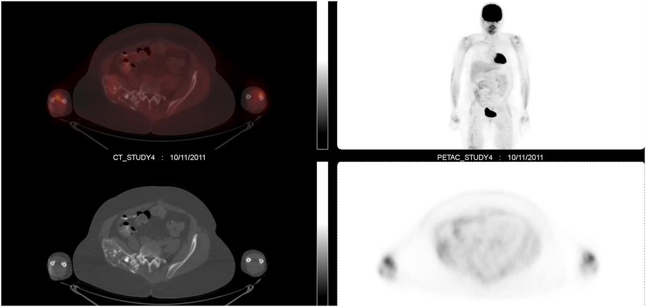
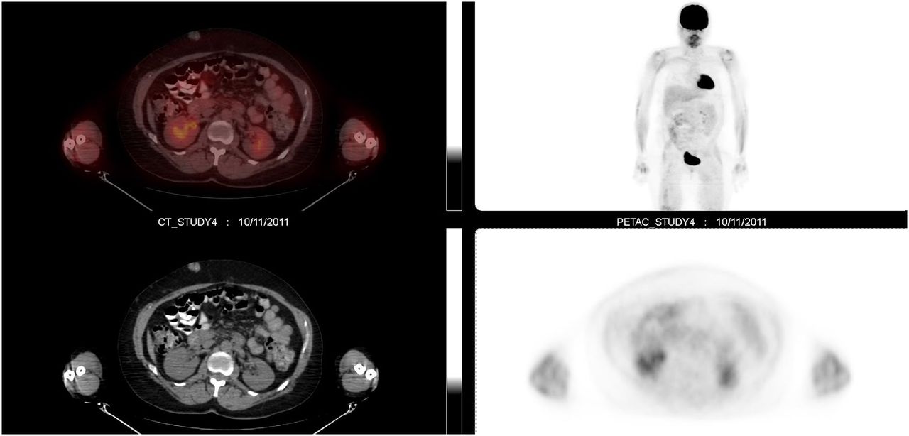
A 45-y-old man with a history of Maffucci syndrome underwent right-knee radiography because of knee pain (Fig. 1). Restaging 18F-FDG PET/CT for chondrosarcoma was ordered. Sixty minutes after intravenous administration of 612.72 MBq (16.56 mCi) of 18F-FDG, sequential unenhanced CT and then PET images were acquired. The PET/CT scan showed multiple osseous lesions similar in appearance to one another (Fig. 2). In such a case, a benign etiology, such as an enchondroma, would be highly favored over an aggressive tumor. Also, throughout the subcutaneous tissues there were multiple soft-tissue nodular densities lacking abnormal 18F-FDG uptake, favoring the benign etiology hemangioma (Fig. 3). Several of the subcutaneous nodules on the right side of the abdominal wall had punctate calcifications. These represent phleboliths, a typical finding in hemangioma. Abnormal 18F-FDG uptake within a subcutaneous nodule would raise concern about malignant transformation to hemangiosarcoma.

FIGURE 1.
  • Download figure
  • Open in new tab
  • Download powerpoint
FIGURE 1.

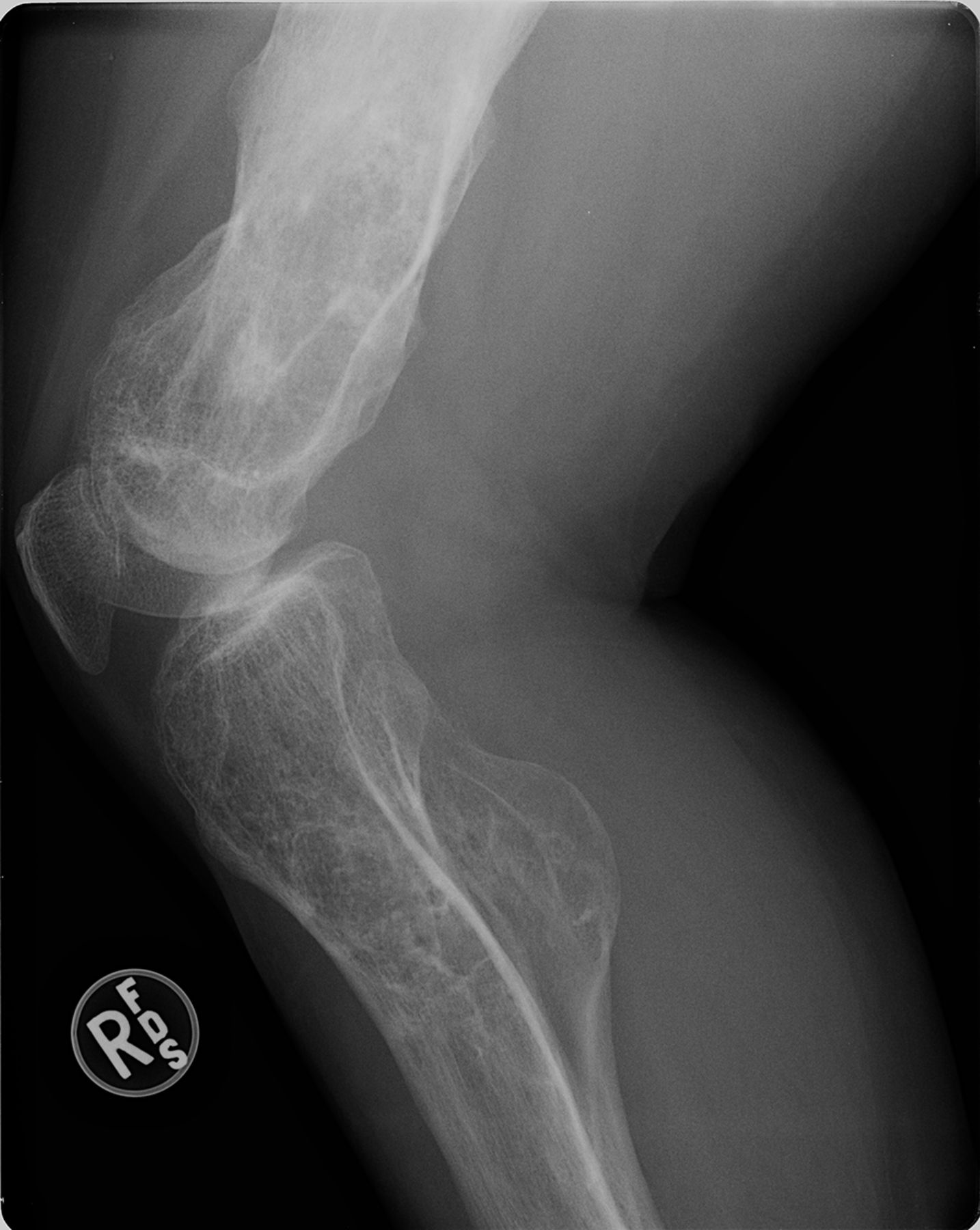
Right-knee radiograph demonstrating marked deformity of distal femur and proximal tibia and fibula. There are expansile lucent lesions with cortical thinning and calcified matrix that are consistent with chondroid-type lesions. Presence of multiple chondroid lesions is consistent with Maffucci syndrome in patients who have characteristic bluish subcutaneous hemangiomas on physical examination.

FIGURE 2.
  • Download figure
  • Open in new tab
  • Download powerpoint
FIGURE 2.

18F-FDG PET/CT scan demonstrating only mild 18F-FDG uptake in right hemipelvis. Maximum standardized uptake value is 1.8, similar to surrounding osseous structure.

FIGURE 3.
  • Download figure
  • Open in new tab
  • Download powerpoint
FIGURE 3.

18F-FDG PET/CT scan demonstrating multiple soft-tissue nodular densities throughout subcutaneous tissues (phleboliths).

DISCUSSION

Maffucci syndrome is a rare condition with multiple enchondromas and hemangiomas. Fewer than 200 cases have been reported in the United States (1). If a patient has multiple enchondromas without hemangiomas, the condition is known as Ollier disease. Maffucci syndrome is a congenital nonhereditary condition. It usually presents before the onset of puberty and is often located within the long bones. An estimated 25%–30% of enchondromas develop into chondrosarcoma (2,3).

The average age for neoplastic change from enchondroma to chondrosarcoma in Maffucci syndrome patients is 40 y. There is also an increased risk of malignant transformation of hemangiomas into hemangiosarcomas and hemangioendotheliomas (4). A maximum standardized uptake value of more than 2.0 can be used to distinguish benign from malignant cartilaginous tumors with 90.9% sensitivity, 100% specificity, and 96.6% accuracy (5). Maffucci syndrome is often distinguished from Ollier disease by physical examination. The presence of red or purplish growths in the skin would be consistent with a hemangioma. Affected individuals can also have lymphangiomas.

CONCLUSION

Maffucci syndrome is a rare congenital nonhereditary condition. There is a high predilection for neoplastic changes, and PET/CT has an important role in detecting these changes.

DISCLOSURE

No potential conflict of interest relevant to this article was reported.

Footnotes

  • Published online Dec. 23, 2014.

REFERENCES

  1. 1. ↵
    1. Faik A ,
    2. Allali F ,
    3. El Hassani S ,
    4. Hajjaj-Hassouni N
    . Maffucci’s syndrome: a case report. Clin Rheumatol. 2006;25:88–91.
    OpenUrlCrossRefPubMed
  2. 2. ↵
    1. Lewis RJ ,
    2. Ketcham AS
    . Maffucci’s syndrome: functional and neoplastic significance—case report and review of the literature. J Bone Joint Surg Am. 1973;55:1465–1479.
    OpenUrlAbstract/FREE Full Text
  3. 3. ↵
    1. Makis W ,
    2. Hickeson M ,
    3. Lisbona R
    . Maffucci syndrome with extraosseous chondrosarcoma imaged with F-18 FDG PET-CT. Clin Nucl Med. 2010;35:29–31.
    OpenUrlCrossRefPubMed
  4. 4. ↵
    1. Zwenneke Flach H ,
    2. Ginai AZ ,
    3. Wolter Oosterhuis J
    . Best cases from the AFIP: Maffucci syndrome—radiologic and pathologic findings. Radiographics. 2001;21:1311–1316.
    OpenUrlCrossRefPubMed
  5. 5. ↵
    1. Morimoto S ,
    2. Futani H ,
    3. Tsuchiyama K ,
    4. Fukunaga S ,
    5. Tsukamoto Y ,
    6. Yoshiya S
    . Usefulness of PET/CT for diagnosis of periosteal chondrosarcoma of the femur: a case report. Oncol Lett. 2014;7:1826–1828.
    OpenUrlPubMed
  • Received for publication July 30, 2014.
  • Accepted for publication October 9, 2014.
PreviousNext
Back to top

In this issue

Journal of Nuclear Medicine Technology: 43 (2)
Journal of Nuclear Medicine Technology
Vol. 43, Issue 2
June 1, 2015
  • Table of Contents
  • About the Cover
  • Index by author
Print
Download PDF
Article Alerts
Sign In to Email Alerts with your Email Address
Email Article

Thank you for your interest in spreading the word on Journal of Nuclear Medicine Technology.

NOTE: We only request your email address so that the person you are recommending the page to knows that you wanted them to see it, and that it is not junk mail. We do not capture any email address.

Enter multiple addresses on separate lines or separate them with commas.
The Appearance of Maffucci Syndrome on 18F-FDG PET/CT
(Your Name) has sent you a message from Journal of Nuclear Medicine Technology
(Your Name) thought you would like to see the Journal of Nuclear Medicine Technology web site.
Citation Tools
The Appearance of Maffucci Syndrome on 18F-FDG PET/CT
Sayf Al-katib, Zaid Al-faham, Paul Grant, Jane C. Palka
Journal of Nuclear Medicine Technology Jun 2015, 43 (2) 131-132; DOI: 10.2967/jnmt.114.146480

Citation Manager Formats

  • BibTeX
  • Bookends
  • EasyBib
  • EndNote (tagged)
  • EndNote 8 (xml)
  • Medlars
  • Mendeley
  • Papers
  • RefWorks Tagged
  • Ref Manager
  • RIS
  • Zotero
Share
The Appearance of Maffucci Syndrome on 18F-FDG PET/CT
Sayf Al-katib, Zaid Al-faham, Paul Grant, Jane C. Palka
Journal of Nuclear Medicine Technology Jun 2015, 43 (2) 131-132; DOI: 10.2967/jnmt.114.146480
Twitter logo Facebook logo LinkedIn logo Mendeley logo
  • Tweet Widget
  • Facebook Like
  • Google Plus One
Bookmark this article

Jump to section

  • Article
    • Abstract
    • CASE REPORT
    • DISCUSSION
    • CONCLUSION
    • DISCLOSURE
    • Footnotes
    • REFERENCES
  • Figures & Data
  • Info & Metrics
  • PDF

Related Articles

  • No related articles found.
  • PubMed
  • Google Scholar

Cited By...

  • No citing articles found.
  • Google Scholar

More in this TOC Section

  • High-Sensitivity Troponin Elevation in a Young Woman with Typical Chest Pain: The Heart of the Matter
  • Pulmonary Adenocarcinoma Revealed by Parathyroid Scintigraphy: An Incidental Case to Remember
  • Prominent Right Ventricular Tracer Uptake: A Harbinger of Multivessel Coronary Artery Disease
Show more Teaching Case Studies

Similar Articles

Keywords

  • Maffucci syndrome
  • 18F FDG PET/CT
  • enchondromas
SNMMI

© 2025 SNMMI

Powered by HighWire