  • Main menu
  • User menu
  • Search
  • English ▼
    • English
    • Afrikaans
    • Albanian
    • Amharic
    • Arabic
    • Armenian
    • Azerbaijani
    • Basque
    • Belarusian
    • Bengali
    • Bosnian
    • Bulgarian
    • Catalan
    • Cebuano
    • Chichewa
    • Chinese (Simplified)
    • Chinese (Traditional)
    • Corsican
    • Croatian
    • Czech
    • Danish
    • Dutch
    • Esperanto
    • Estonian
    • Filipino
    • Finnish
    • French
    • Frisian
    • Galician
    • Georgian
    • German
    • Greek
    • Gujarati
    • Haitian Creole
    • Hausa
    • Hawaiian
    • Hebrew
    • Hindi
    • Hmong
    • Hungarian
    • Icelandic
    • Igbo
    • Indonesian
    • Irish
    • Italian
    • Japanese
    • Javanese
    • Kannada
    • Kazakh
    • Khmer
    • Korean
    • Kurdish (Kurmanji)
    • Kyrgyz
    • Lao
    • Latin
    • Latvian
    • Lithuanian
    • Luxembourgish
    • Macedonian
    • Malagasy
    • Malay
    • Malayalam
    • Maltese
    • Maori
    • Marathi
    • Mongolian
    • Myanmar (Burmese)
    • Nepali
    • Norwegian
    • Pashto
    • Persian
    • Polish
    • Portuguese
    • Punjabi
    • Romanian
    • Russian
    • Samoan
    • Scottish Gaelic
    • Serbian
    • Sesotho
    • Shona
    • Sindhi
    • Sinhala
    • Slovak
    • Slovenian
    • Somali
    • Spanish
    • Sudanese
    • Swahili
    • Swedish
    • Tajik
    • Tamil
    • Telugu
    • Thai
    • Turkish
    • Ukrainian
    • Urdu
    • Uzbek
    • Vietnamese
    • Welsh
    • Xhosa
    • Yiddish
    • Yoruba
    • Zulu

Main menu

  • Home
  • Content
    • Current
      • JNMT Supplement
    • Ahead of print
    • Past Issues
    • Continuing Education
    • JNMT Podcast
    • SNMMI Annual Meeting Abstracts
  • Subscriptions
    • Subscribers
    • Rates
    • Journal Claims
    • Institutional and Non-member
  • Authors
    • Submit to JNMT
    • Information for Authors
    • Assignment of Copyright
    • AQARA Requirements
  • Info
    • Reviewers
    • Permissions
    • Advertisers
    • Corporate & Special Sales
  • About
    • About Us
    • Editorial Board
    • Contact Information
  • More
    • Alerts
    • Feedback
    • Help
    • SNMMI Journals
  • SNMMI
    • JNMT
    • JNM
    • SNMMI Journals
    • SNMMI

User menu

  • Subscribe
  • My alerts
  • Log in
  • My Cart

Search

  • Advanced search
Journal of Nuclear Medicine Technology
  • SNMMI
    • JNMT
    • JNM
    • SNMMI Journals
    • SNMMI
  • Subscribe
  • My alerts
  • Log in
  • My Cart
Journal of Nuclear Medicine Technology

Advanced Search

English ▼
  • English
  • Afrikaans
  • Albanian
  • Amharic
  • Arabic
  • Armenian
  • Azerbaijani
  • Basque
  • Belarusian
  • Bengali
  • Bosnian
  • Bulgarian
  • Catalan
  • Cebuano
  • Chichewa
  • Chinese (Simplified)
  • Chinese (Traditional)
  • Corsican
  • Croatian
  • Czech
  • Danish
  • Dutch
  • Esperanto
  • Estonian
  • Filipino
  • Finnish
  • French
  • Frisian
  • Galician
  • Georgian
  • German
  • Greek
  • Gujarati
  • Haitian Creole
  • Hausa
  • Hawaiian
  • Hebrew
  • Hindi
  • Hmong
  • Hungarian
  • Icelandic
  • Igbo
  • Indonesian
  • Irish
  • Italian
  • Japanese
  • Javanese
  • Kannada
  • Kazakh
  • Khmer
  • Korean
  • Kurdish (Kurmanji)
  • Kyrgyz
  • Lao
  • Latin
  • Latvian
  • Lithuanian
  • Luxembourgish
  • Macedonian
  • Malagasy
  • Malay
  • Malayalam
  • Maltese
  • Maori
  • Marathi
  • Mongolian
  • Myanmar (Burmese)
  • Nepali
  • Norwegian
  • Pashto
  • Persian
  • Polish
  • Portuguese
  • Punjabi
  • Romanian
  • Russian
  • Samoan
  • Scottish Gaelic
  • Serbian
  • Sesotho
  • Shona
  • Sindhi
  • Sinhala
  • Slovak
  • Slovenian
  • Somali
  • Spanish
  • Sudanese
  • Swahili
  • Swedish
  • Tajik
  • Tamil
  • Telugu
  • Thai
  • Turkish
  • Ukrainian
  • Urdu
  • Uzbek
  • Vietnamese
  • Welsh
  • Xhosa
  • Yiddish
  • Yoruba
  • Zulu
  • Home
  • Content
    • Current
    • Ahead of print
    • Past Issues
    • Continuing Education
    • JNMT Podcast
    • SNMMI Annual Meeting Abstracts
  • Subscriptions
    • Subscribers
    • Rates
    • Journal Claims
    • Institutional and Non-member
  • Authors
    • Submit to JNMT
    • Information for Authors
    • Assignment of Copyright
    • AQARA Requirements
  • Info
    • Reviewers
    • Permissions
    • Advertisers
    • Corporate & Special Sales
  • About
    • About Us
    • Editorial Board
    • Contact Information
  • More
    • Alerts
    • Feedback
    • Help
    • SNMMI Journals
  • Watch or Listen to JNMT Podcast
  • Visit SNMMI on Facebook
  • Join SNMMI on LinkedIn
  • Follow SNMMI on Twitter
  • Subscribe to JNMT RSS feeds
Review ArticleTEACHING FILES

Patient with Acute Shortness of Breath, Elevated D-Dimer, and a Previous History of Cancer

Katie Traylor and Paresh Mahajan
Journal of Nuclear Medicine Technology September 2013, 41 (3) 238-240; DOI: https://doi.org/10.2967/jnmt.113.126755
Katie Traylor
Department of Diagnostic Radiology and Molecular Imaging, Oakland University William Beaumont School of Medicine, Royal Oak, Michigan
  • Find this author on Google Scholar
  • Find this author on PubMed
  • Search for this author on this site
Paresh Mahajan
Department of Diagnostic Radiology and Molecular Imaging, Oakland University William Beaumont School of Medicine, Royal Oak, Michigan
  • Find this author on Google Scholar
  • Find this author on PubMed
  • Search for this author on this site
  • Article
  • Figures & Data
  • Info & Metrics
  • PDF
Loading

A 69-y-old man with a history of small cell lung cancer, esophageal cancer, and pulmonary hypertension stated that he had shortness of breath. The D-dimer level was elevated at 1,385.

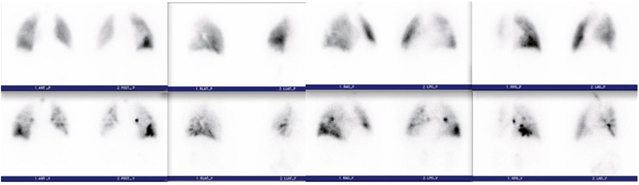
The patient inhaled a calculated 51.8 MBq (1.4 mCi) of 99mTc-diethylenetriminepentaacetic acid aerosol, after which planar images of the lungs in the standard 8 projections were obtained. After intravenous administration of 151.7 MBq (4.1 mCi) of 99mTc-labeled macroaggregated albumin (MAA), a set of projections corresponding to those obtained on the ventilation study was obtained (Fig. 1).

FIGURE 1.
  • Download figure
  • Open in new tab
  • Download powerpoint
FIGURE 1.

(Top) Perfusion images of lungs. Views from left to right are anterior/posterior, lateral, right anterior oblique/left posterior oblique, and left anterior oblique/right posterior oblique. (Bottom) Ventilation images in projections corresponding to perfusion images.

QUESTION 1

This patient has a history of pulmonary hypertension. What difference in the technique had to be addressed before the study was performed? Why?

  1. Reduction of the number of 99mTc-MAA particles to less than 100,000 to lessen the likelihood of blockade of the capillaries.

  2. Reduction of the 99mTc dose to decrease the radiation dose to the patient.

  3. Completion of perfusion images first. If these are normal, the ventilation images do not need to be completed, thus decreasing the radiation dose to the patient.

  4. Reduction of the number of 99mTc-MAA particles to 100,000–200,000 to lessen the likelihood of blockade of the capillaries.

QUESTION 2

After reviewing the lung scan images and comparing them to the chest radiograph (Fig. 2), what is your assessment of the findings based on the modified PIOPED II interpretation criteria (Table 1)?

  1. High probability of pulmonary embolism, as there are 2 large segmental mismatched defects in the right lower lobe.

  2. Nondiagnostic scan for pulmonary embolism, as there is 1 moderate segmental triple match in the superior segment of the right lower lobe.

  3. Low probability of pulmonary embolism.

  4. Nondiagnostic scan for pulmonary embolism, as there is 1 large segmental triple match in the superior segment of the right lower lobe, although more information would be valuable for an accurate interpretation.

FIGURE 2.
  • Download figure
  • Open in new tab
  • Download powerpoint
FIGURE 2.

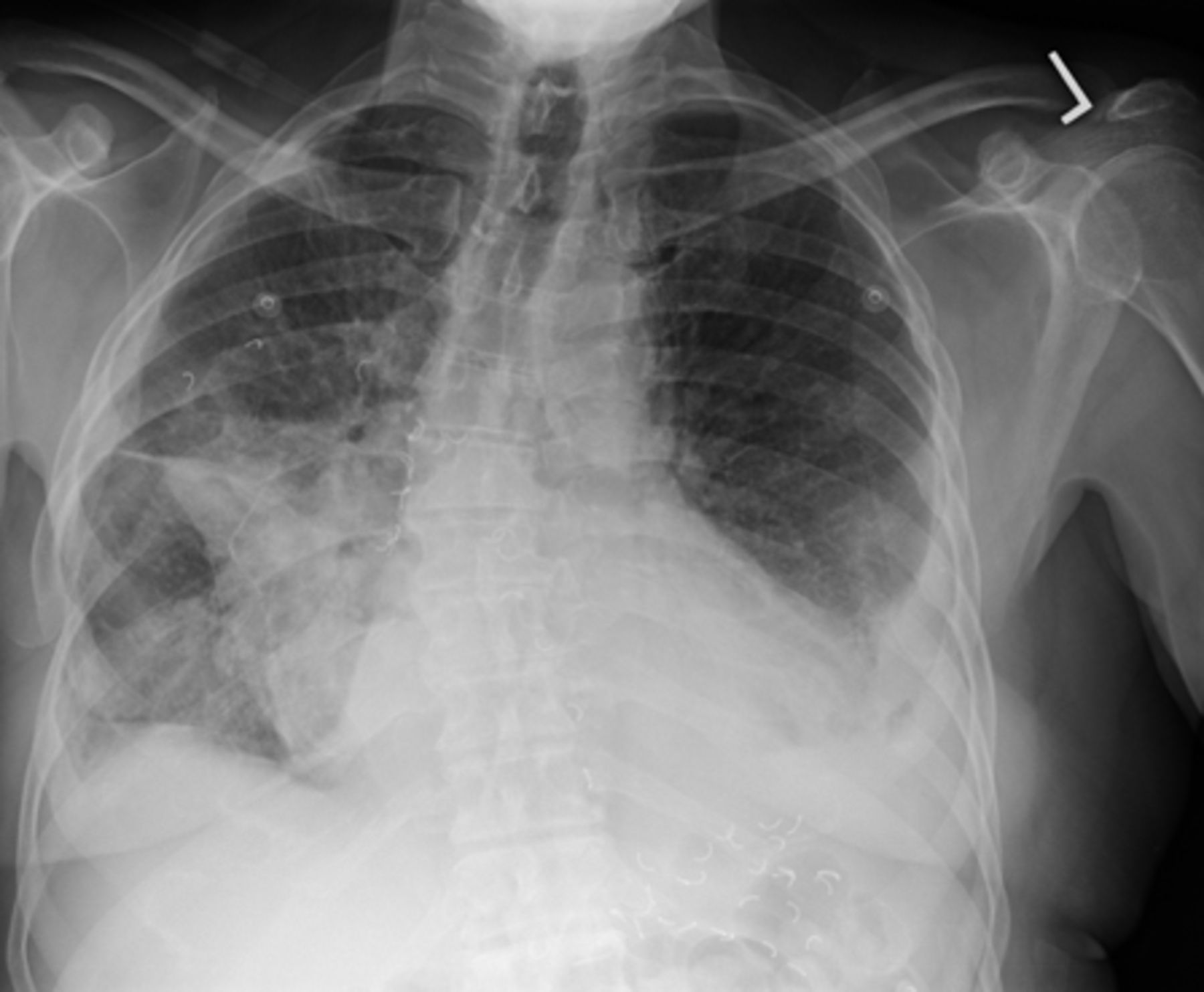
Chest posteroanterior radiograph that was completed approximately 12 h before lung scan.

View this table:
  • View inline
  • View popup
TABLE 1

Ventilation, Perfusion, and Radiographic Interpretive Criteria for Pulmonary Embolism

QUESTION 3

The patient also underwent CT of the chest with intravenous contrast material (Fig. 3) within the last week. Do the CT findings change your previous diagnosis?

  1. No, the patient still has a high probability of pulmonary embolism.

  2. No, the patient still has a nondiagnostic scan for pulmonary embolism.

  3. The scan is technically nondiagnostic; however, it appears that the patient has postsurgical changes within the opaque region identified on the chest radiograph.

  4. No, the patient still has a high probability for pulmonary embolism even though there are postsurgical changes within the opaque region identified on the chest radiograph.

FIGURE 3.
  • Download figure
  • Open in new tab
  • Download powerpoint
FIGURE 3.

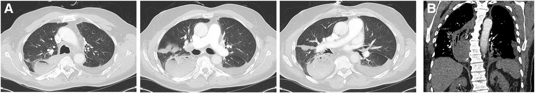
Contrast-enhanced chest CT scan. (A) From left to right: 3 axial slices (lung window) from cranial to caudal. (B) Coronal slice (soft-tissue window) illustrating extent of gastric pull-through, which caused large defect on ventilation–perfusion images.

QUESTION 4

Besides lymph node involvement and distant metastatic disease, what additional information is needed for staging purposes?

  1. The lymph node location is needed because lymph nodes above and below the diaphragm may change the stage of the disease.

  2. The endoscopic ultrasonography and pathology report is needed because the extent of invasion of the cancer can change the stage of the disease.

  3. Whether a lymph node is present in the supraclavicular regions needs to be known because the patient would be deemed a nonsurgical candidate.

  4. The location of the distant metastatic disease is needed because this information may change the stage of the patient.

DISCUSSION

Knowing the entire history of a patient is important when reviewing studies, especially when the patient may have postsurgical changes that may interfere with one’s interpretation. Patients with benign and malignant disease of their esophagus may undergo radical resection and reconstruction with an intrathoracic stomach, colon, or jejunum pulled up through the mediastinum, retrosternally or subcutaneously. These postsurgical changes will appear as a widened mediastinum, usually on the right side. The history is important, as air-space disease, mass lesions, pleural effusion, or lobar collapse may look the same as one another on a chest radiograph. Knowing the history of our patient, we were able to relay this information to the referring physician and communicate that the mismatch was most likely due to the patient’s gastric pull-through and radiotracer activity located within the stomach. Strictly speaking, with the PIOPED criteria the patient would still remain at intermediate probability of pulmonary embolism because the corresponding ventilation–perfusion defects correlated with the parenchymal opacity seen in the lower lung zone on the chest radiograph. The PIOPED criteria look only at comparing the lung scan to the chest radiograph and not any other helpful imaging, such as the CT scan of the chest.

Footnotes

  • Published online Jul. 18, 2013.

  • ↵* For the answers, see page 242.

BIBLIOGRAPHY

    1. Brunicardi FC,
    2. Anderson DK,
    3. Billiar T,
    4. et al
    . Schwartz’s Principles of Surgery. 8th ed. New York, NY: McGraw-Hill Professional; 2005:888–893.
    Google Scholar
    1. Cameron JL
    . Current Surgical Therapy. 8th ed. Maryland Heights, MO: Mosby; 2004:44–49.
    Google Scholar
    1. Cole TJ,
    2. Turner MA
    . Manifestations of gastrointestinal disease on chest radiographs. Radiographics. 1993;13:1013–1034.
    OpenUrlPubMedGoogle Scholar
    1. Krasna MJ
    . Multimodality therapy for esophageal cancer. Oncology. 2010;24:1134–1138.
    OpenUrlPubMedGoogle Scholar
    1. Mettler FA,
    2. Guiberteau MJ
    . Essentials of Nuclear Medicine Imaging. 6th ed. Philadelphia, PA: Elsevier Saunders; 2012:197, 213–216, 223.
    Google Scholar
  1. Society of Nuclear Medicine Procedure Guideline for Lung Scintigraphy. SNMMI Web site. http://interactive.snm.org/docs/Lung_Scintigraphy_V4_Final.pdf. Published  March 2012. Accessed June 24, 2013.
    Google Scholar
PreviousNext
Back to top

In this issue

Journal of Nuclear Medicine Technology: 41 (3)
Journal of Nuclear Medicine Technology
Vol. 41, Issue 3
September 1, 2013
  • Table of Contents
  • About the Cover
  • Index by author
Print
Download PDF
Article Alerts
Email Article
Citation Tools
Share
Patient with Acute Shortness of Breath, Elevated D-Dimer, and a Previous History of Cancer
Katie Traylor, Paresh Mahajan
Journal of Nuclear Medicine Technology Sep 2013, 41 (3) 238-240; DOI: 10.2967/jnmt.113.126755
Twitter logo Facebook logo LinkedIn logo Mendeley logo
  • Tweet Widget
Bookmark this article

Jump to section

  • Article
    • QUESTION 1
    • QUESTION 2
    • QUESTION 3
    • QUESTION 4
    • DISCUSSION
    • Footnotes
    • BIBLIOGRAPHY
  • Figures & Data
  • Info & Metrics
  • PDF

Related Articles

  • No related articles found.
  • PubMed
  • Google Scholar

Cited By...

  • No citing articles found.
  • Google Scholar

More in this TOC Section

  • Bronchopleural Fistula Mimicking Chronic Obstructive Lung Disease on 133Xe Scan
  • Increased Sacral Uptake on a Bone Scan with SPECT/CT in a Patient with Achondroplasia: Normal or Abnormal?
Show more TEACHING FILES

Similar Articles

SNMMI

© 2025 SNMMI

Powered by HighWire
Alerts for this Article
Sign In to Email Alerts with your Email Address
Email this Article

Thank you for your interest in spreading the word on Journal of Nuclear Medicine Technology.

NOTE: We only request your email address so that the person you are recommending the page to knows that you wanted them to see it, and that it is not junk mail. We do not capture any email address.

Enter multiple addresses on separate lines or separate them with commas.
Patient with Acute Shortness of Breath, Elevated D-Dimer, and a Previous History of Cancer
(Your Name) has sent you a message from Journal of Nuclear Medicine Technology
(Your Name) thought you would like to see the Journal of Nuclear Medicine Technology web site.
Citation Tools
Patient with Acute Shortness of Breath, Elevated D-Dimer, and a Previous History of Cancer
Katie Traylor, Paresh Mahajan
Journal of Nuclear Medicine Technology Sep 2013, 41 (3) 238-240; DOI: 10.2967/jnmt.113.126755

Citation Manager Formats

  • BibTeX
  • Bookends
  • EasyBib
  • EndNote (tagged)
  • EndNote 8 (xml)
  • Medlars
  • Mendeley
  • Papers
  • RefWorks Tagged
  • Ref Manager
  • RIS
  • Zotero

We use cookies on this site to enhance your user experience

By clicking any link on this page you are giving your consent for us to set cookies.