
Main menu

  • Home
  • Content
    • Current
      • JNMT Supplement
    • Ahead of print
    • Past Issues
    • Continuing Education
    • JNMT Podcast
    • SNMMI Annual Meeting Abstracts
  • Subscriptions
    • Subscribers
    • Rates
    • Journal Claims
    • Institutional and Non-member
  • Authors
    • Submit to JNMT
    • Information for Authors
    • Assignment of Copyright
    • AQARA Requirements
  • Info
    • Reviewers
    • Permissions
    • Advertisers
    • Corporate & Special Sales
  • About
    • About Us
    • Editorial Board
    • Contact Information
  • More
    • Alerts
    • Feedback
    • Help
    • SNMMI Journals
  • SNMMI
    • JNMT
    • JNM
    • SNMMI Journals
    • SNMMI

User menu

  • Subscribe
  • My alerts
  • Log in
  • My Cart

Search

  • Advanced search
Journal of Nuclear Medicine Technology
  • SNMMI
    • JNMT
    • JNM
    • SNMMI Journals
    • SNMMI
  • Subscribe
  • My alerts
  • Log in
  • My Cart
Journal of Nuclear Medicine Technology

Advanced Search

  • Home
  • Content
    • Current
    • Ahead of print
    • Past Issues
    • Continuing Education
    • JNMT Podcast
    • SNMMI Annual Meeting Abstracts
  • Subscriptions
    • Subscribers
    • Rates
    • Journal Claims
    • Institutional and Non-member
  • Authors
    • Submit to JNMT
    • Information for Authors
    • Assignment of Copyright
    • AQARA Requirements
  • Info
    • Reviewers
    • Permissions
    • Advertisers
    • Corporate & Special Sales
  • About
    • About Us
    • Editorial Board
    • Contact Information
  • More
    • Alerts
    • Feedback
    • Help
    • SNMMI Journals
  • Watch or Listen to JNMT Podcast
  • Visit SNMMI on Facebook
  • Join SNMMI on LinkedIn
  • Follow SNMMI on Twitter
  • Subscribe to JNMT RSS feeds
Review ArticleTEACHING FILES

Bronchopleural Fistula Mimicking Chronic Obstructive Lung Disease on 133Xe Scan

Sumina Goel
Journal of Nuclear Medicine Technology September 2013, 41 (3) 236-237; DOI: https://doi.org/10.2967/jnmt.113.126425
Sumina Goel
Division of Nuclear Medicine, Department of Radiology, Hospital of University of Pennsylvania, Philadelphia, Pennsylvania
  • Find this author on Google Scholar
  • Find this author on PubMed
  • Search for this author on this site
  • Article
  • Figures & Data
  • Info & Metrics
  • PDF
Loading

A 56-y-old woman with a history of non–small cell lung cancer presented with a persistent left pneumothorax and a left bronchopleural fistula after bronchial balloon occlusion. A ventilation–perfusion scan using 133Xe and 99mTc-macroaggregated albumin (MAA) was requested to evaluate for possible lung reduction surgery.

With 428.46 MBq (11.58 mCi) of 133Xe gas, ventilation images were acquired in the posterior projection during a single breath-hold in the equilibrium and washout phases. Regions of interest were drawn around the lungs to generate ventilation time–activity curves. Then, 162.06 MBq (4.38 mCi) of 99mTc-MAA were injected intravenously and 8 standard projections of the lungs were acquired. Regions of interest were drawn around the lungs to calculate differential perfusion.

QUESTION 1

Based on the 133Xe ventilation and 99mTc-MAA perfusion images shown (Fig. 1), what is the best interpretation of this study?

  1. Chronic obstructive pulmonary disease.

  2. Bronchopleural fistula.

  3. Normal scan findings.

  4. A or B.

FIGURE 1.
  • Download figure
  • Open in new tab
  • Download powerpoint
FIGURE 1.

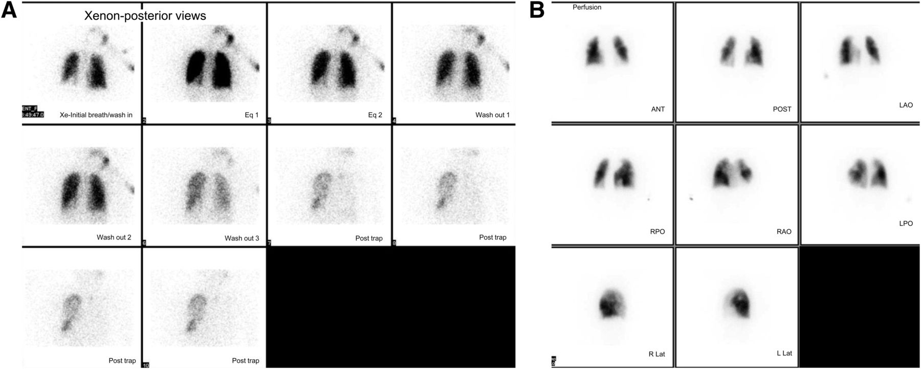
133Xe ventilation images (A) and 99mTc-MAA perfusion images (B).

QUESTION 2

What are the physical half-life and biologic half-life of 133Xe, respectively?

  1. 30 min and 20 s.

  2. 2 d and 50 s.

  3. 5.3 d and 30 s.

  4. 7 d and 60 s.

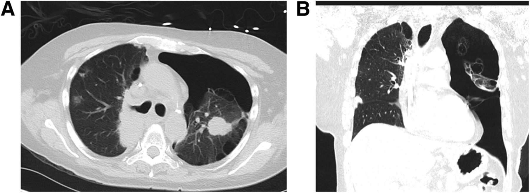
Correlative CT images of the chest are shown (Fig. 2).

FIGURE 2.
  • Download figure
  • Open in new tab
  • Download powerpoint
FIGURE 2.

Correlative CT images of chest in cross-sectional (A) and coronal (B) views showing mild bilateral emphysema and large left-sided pneumothorax.

QUESTION 3

What are the CT imaging features of a bronchopleural fistula?

  1. Pneumothorax or air trapping on the side of the fistula.

  2. No abnormality on CT images.

  3. Return to midline of a previously shifted mediastinum or a drop in the fluid level by more than 2 cm in patients after pneumonectomy.

  4. A and C.

DISCUSSION

Bronchopleural fistula is a process that can be caused by several lung diseases or by injury. The most common causes are pulmonary infections, trauma, surgical complications, malignancy, and therapy of malignancy. Among various imaging techniques such as plain radiography, CT, 133Xe scanning, and ultrasound, CT and 133Xe scans are the most promising. CT is the anatomic imaging technique of choice for visualization and characterization of a bronchopleural fistula. Most authors recommend CT as the first diagnostic examination. 133Xe ventilation scanning is a simple and accurate method for diagnosis and follow-up of bronchopleural fistula. 133Xe images will show trapping of the tracer on the affected side during the washout phase. A study by Raja et al. in 1999 showed the sensitivity of 133Xe to be 83% and that of 99mTc-DTPA to be 0% in the diagnosis of bronchopleural fistula. In this case, the 133Xe scan findings can be seen in bronchopleural fistula as well as in chronic obstructive pulmonary disease. The distinction could be made by correlating with the history and CT imaging. According to the present literature, CT should be the first modality for the diagnosis and characterization of bronchopleural fistula, and 133Xe scanning can be used as an inexpensive and simple follow-up imaging modality.

Footnotes

  • Published online Jul. 18, 2013.

  • ↵* For the answers, see page 241.

BIBLIOGRAPHY

    1. Raja S,
    2. Rice TW,
    3. Neumann DR,
    4. et al
    . Scintigraphic detection of post-pneumonectomy bronchopleural fistulae. Eur J Nucl Med. 1999;26:215–219.
    OpenUrlCrossRefPubMed
    1. Stern EJ,
    2. Sun H,
    3. Haramati LB
    . Peripheral bronchopleural fistulas: CT imaging features. AJR. 1996;167:117–120.
    OpenUrlPubMed
PreviousNext
Back to top

In this issue

Journal of Nuclear Medicine Technology: 41 (3)
Journal of Nuclear Medicine Technology
Vol. 41, Issue 3
September 1, 2013
  • Table of Contents
  • About the Cover
  • Index by author
Print
Download PDF
Article Alerts
Sign In to Email Alerts with your Email Address
Email Article

Thank you for your interest in spreading the word on Journal of Nuclear Medicine Technology.

NOTE: We only request your email address so that the person you are recommending the page to knows that you wanted them to see it, and that it is not junk mail. We do not capture any email address.

Enter multiple addresses on separate lines or separate them with commas.
Bronchopleural Fistula Mimicking Chronic Obstructive Lung Disease on 133Xe Scan
(Your Name) has sent you a message from Journal of Nuclear Medicine Technology
(Your Name) thought you would like to see the Journal of Nuclear Medicine Technology web site.
Citation Tools
Bronchopleural Fistula Mimicking Chronic Obstructive Lung Disease on 133Xe Scan
Sumina Goel
Journal of Nuclear Medicine Technology Sep 2013, 41 (3) 236-237; DOI: 10.2967/jnmt.113.126425

Citation Manager Formats

  • BibTeX
  • Bookends
  • EasyBib
  • EndNote (tagged)
  • EndNote 8 (xml)
  • Medlars
  • Mendeley
  • Papers
  • RefWorks Tagged
  • Ref Manager
  • RIS
  • Zotero
Share
Bronchopleural Fistula Mimicking Chronic Obstructive Lung Disease on 133Xe Scan
Sumina Goel
Journal of Nuclear Medicine Technology Sep 2013, 41 (3) 236-237; DOI: 10.2967/jnmt.113.126425
Twitter logo Facebook logo LinkedIn logo Mendeley logo
  • Tweet Widget
  • Facebook Like
  • Google Plus One
Bookmark this article

Jump to section

  • Article
    • QUESTION 1
    • QUESTION 2
    • QUESTION 3
    • DISCUSSION
    • Footnotes
    • BIBLIOGRAPHY
  • Figures & Data
  • Info & Metrics
  • PDF

Related Articles

  • No related articles found.
  • PubMed
  • Google Scholar

Cited By...

  • No citing articles found.
  • Google Scholar

More in this TOC Section

  • Increased Sacral Uptake on a Bone Scan with SPECT/CT in a Patient with Achondroplasia: Normal or Abnormal?
  • Patient with Acute Shortness of Breath, Elevated D-Dimer, and a Previous History of Cancer
Show more TEACHING FILES

Similar Articles

SNMMI

© 2025 SNMMI

Powered by HighWire