
Main menu

  • Home
  • Content
    • Current
      • JNMT Supplement
    • Ahead of print
    • Past Issues
    • Continuing Education
    • JNMT Podcast
    • SNMMI Annual Meeting Abstracts
  • Subscriptions
    • Subscribers
    • Rates
    • Journal Claims
    • Institutional and Non-member
  • Authors
    • Submit to JNMT
    • Information for Authors
    • Assignment of Copyright
    • AQARA Requirements
  • Info
    • Reviewers
    • Permissions
    • Advertisers
    • Corporate & Special Sales
  • About
    • About Us
    • Editorial Board
    • Contact Information
  • More
    • Alerts
    • Feedback
    • Help
    • SNMMI Journals
  • SNMMI
    • JNMT
    • JNM
    • SNMMI Journals
    • SNMMI

User menu

  • Subscribe
  • My alerts
  • Log in
  • My Cart

Search

  • Advanced search
Journal of Nuclear Medicine Technology
  • SNMMI
    • JNMT
    • JNM
    • SNMMI Journals
    • SNMMI
  • Subscribe
  • My alerts
  • Log in
  • My Cart
Journal of Nuclear Medicine Technology

Advanced Search

  • Home
  • Content
    • Current
    • Ahead of print
    • Past Issues
    • Continuing Education
    • JNMT Podcast
    • SNMMI Annual Meeting Abstracts
  • Subscriptions
    • Subscribers
    • Rates
    • Journal Claims
    • Institutional and Non-member
  • Authors
    • Submit to JNMT
    • Information for Authors
    • Assignment of Copyright
    • AQARA Requirements
  • Info
    • Reviewers
    • Permissions
    • Advertisers
    • Corporate & Special Sales
  • About
    • About Us
    • Editorial Board
    • Contact Information
  • More
    • Alerts
    • Feedback
    • Help
    • SNMMI Journals
  • Watch or Listen to JNMT Podcast
  • Visit SNMMI on Facebook
  • Join SNMMI on LinkedIn
  • Follow SNMMI on Twitter
  • Subscribe to JNMT RSS feeds
Research ArticleTeaching Case Study

Detection of Esophageal Tuberculosis, a Rare Cause of Abdominal Pain, on 18F-FDG PET/CT

Tarun Kumar Jain, Hemant Malhotra, Subhash Nepalia and Ganesh Narayan Saxena
Journal of Nuclear Medicine Technology February 2024, jnmt.123.266428; DOI: https://doi.org/10.2967/jnmt.123.266428
Tarun Kumar Jain
1Department of Nuclear Medicine, Mahatma Gandhi Medical College and Hospital, Jaipur, India;
  • Find this author on Google Scholar
  • Find this author on PubMed
  • Search for this author on this site
Hemant Malhotra
2Department of Medical Oncology, Mahatma Gandhi Medical College and Hospital, Jaipur, India;
  • Find this author on Google Scholar
  • Find this author on PubMed
  • Search for this author on this site
Subhash Nepalia
3Department of Gastroenterology, Mahatma Gandhi Medical College and Hospital, Jaipur, India; and
  • Find this author on Google Scholar
  • Find this author on PubMed
  • Search for this author on this site
Ganesh Narayan Saxena
4Department of Internal Medicine, Mahatma Gandhi Medical College and Hospital, Jaipur, India
  • Find this author on Google Scholar
  • Find this author on PubMed
  • Search for this author on this site
  • Article
  • Figures & Data
  • Info & Metrics
  • PDF
Loading

Abstract

The esophagus is rarely affected by Mycobacterium. A 75-y-old man presented with upper abdominal pain and significant weight loss for 2 mo. Contrast-enhanced CT, upper gastrointestinal endoscopy, and abdominal vessel angiography gave normal results. To clarify the facts, 18F-FDG PET/CT was performed, revealing an 18F-FDG–avid lesion in the posterior wall of the lower thoracic esophagus. On endoscopic ultrasound–guided fine-needle aspiration of this lesion, puslike material was released. On microscopic examination, acid-fast bacilli were noted. The patient then began receiving standard antitubercular therapy.

  • esophageal tuberculosis
  • endoscopic ultrasound
  • 18F-FDG PET/CT

The esophagus is rarely affected by Mycobacterium, and esophageal tuberculosis in the absence of mucosal changes is an uncommon cause for abdominal pain. We present the case of a patient with upper abdominal pain and weight loss in whom tuberculosis was revealed through 18F-FDG PET/CT followed by lesion aspiration.

CASE REPORT

A 75-y-old man complained of upper abdominal pain and significant weight loss for 2 mo. The pain was dull, constant, and of varying intensity. He was on regular analgesics, but no significant improvement had been noted. He had no difficulty swallowing. There was no significant contributory past history or family history. On general and physical examination, no abnormality was noted. The results of laboratory assessment and biochemical profile were also normal, as were the results of contrast-enhanced CT of the chest and abdomen, upper gastrointestinal endoscopy, and abdominal vessel angiography.

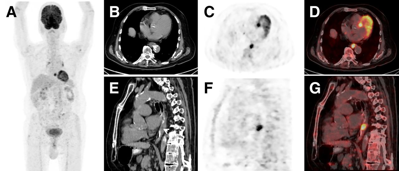
Because PET may identify early functional changes in an organ or tissue related to early onset of a disease before anatomic changes appear, we performed 18F-FDG PET/CT to search for an early pathologic cause of the patient’s discomfort. The images (Fig. 1) revealed an 18F-FDG–avid (SUVmax, 9.8) heterogeneously enhancing soft-tissue-density lesion (∼1.3 × 1.2 × 1.5 cm) in the posterior wall of the lower thoracic esophagus. The lesion was causing mild narrowing of the esophageal lumen. Additionally, a few mildly 18F-FDG–avid mediastinal lymph nodes with foci of calcification were noted.

FIGURE 1.
  • Download figure
  • Open in new tab
  • Download powerpoint
FIGURE 1.

(A) 18F-FDG PET maximum-intensity projection shows abnormal hot spot in midline lower thoracic region. (B–G) Corresponding transaxial (top) and sagittal (bottom) contrast-enhanced CT (B and E), PET (C and F), and PET/CT (D and G) images reveal 18F-FDG–avid (SUVmax, 9.8) heterogeneously enhancing soft-tissue-density lesion (∼1.3 × 1.2 × 1.5 cm) in posterior wall of lower thoracic esophagus (arrowheads). Lesion was causing mild narrowing of esophageal lumen.

On endoscopic ultrasound examination, the inner esophageal mucosal lining was normal. Endoscopic ultrasound–guided fine-needle aspiration of the esophageal lesion was then performed, extracting dense, puslike material that, on microscopic examination, revealed acid-fast bacilli. The patient was subsequently scheduled to receive standard antitubercular therapy, and after 2 mo of treatment, the abdominal pain had improved significantly.

DISCUSSION

Esophageal tuberculosis is extremely rare, accounting for only 0.15% of tuberculosis cases (1,2), and usually affects the mid esophagus because of direct extension from mediastinal structures. Common symptoms of esophageal tuberculosis are dysphasia and retrosternal pain; however, histopathology is needed to confirm the diagnosis (3–5). Esophageal tuberculosis is difficult to identify in the absence of a mucosal lesion or at an early stage (3). Clinical, radiologic, and endoscopic findings are not well defined because of the rarity of the disease (6,7). Because 18F-FDG PET/CT is a well-established modality for detection of metabolically active lesions, it is a good modality for clarifying uncertain cases (8–10).

CONCLUSION

Esophageal Mycobacterium tuberculosis may be missed in the absence of mucosal changes and is an uncommon cause for abdominal pain. In the present case, 18F-FDG PET/CT proved used in detecting an esophageal tuberculosis lesion even after upper gastrointestinal endoscopy gave normal results.

DISCLOSURE

No potential conflict of interest relevant to this article was reported.

ACKNOWLEDGMENTS

We thank all technologists of the PET/CT department for acquiring the data.

Footnotes

  • Published online Feb. 13, 2024.

REFERENCES

  1. 1.↵
    1. Dahale AS,
    2. Kumar A,
    3. Srivastava S,
    4. Varakanahalli S,
    5. Sachdeva S,
    6. Puri AS
    . Esophageal tuberculosis: uncommon of common. JGH Open. 2018;2:34–38.
    OpenUrl
  2. 2.↵
    1. Jain SK,
    2. Jain S,
    3. Jain M,
    4. Yaduvanshi A
    . Esophageal tuberculosis: is it so rare? Report of 12 cases and review of the literature. Am J Gastroenterol. 2002;97:287–291.
    OpenUrlCrossRefPubMed
  3. 3.↵
    1. Kim D,
    2. Kim J,
    3. Lee D,
    4. et al
    . Multidrug-resistant tuberculous mediastinal lymphadenitis, with an esophagomediastinal fistula, mimicking an esophageal submucosal tumor. Clin Endosc. 2016;49:564–569.
    OpenUrl
  4. 4.
    1. Huang YK,
    2. Wu YC,
    3. Liu YH,
    4. Liu HP
    . Esophageal tuberculosis mimicking submucosal tumor. Interact Cardiovasc Thorac Surg. 2004;3:274–276.
    OpenUrlCrossRefPubMed
  5. 5.↵
    1. Debi U,
    2. Ravisankar V,
    3. Prasad KK,
    4. Sinha SK,
    5. Sharma AK
    . Abdominal tuberculosis of the gastrointestinal tract: revisited. World J Gastroenterol. 2014;20:14831–14840.
    OpenUrlPubMed
  6. 6.↵
    1. Prasant P,
    2. Kajal NC,
    3. Dadra R,
    4. Nithin KT,
    5. Kaur J
    . Esophageal tuberculosis: a rare case report. Int J Mycobacteriol. 2019;8:409–411.
    OpenUrl
  7. 7.↵
    1. Khanna V,
    2. Kumar A,
    3. Alexander N,
    4. Surendran P
    . A case report on esophageal tuberculosis: a rare entity. Int J Surg Case Rep. 2017;35:41–43.
    OpenUrl
  8. 8.↵
    1. Ankrah AO,
    2. van der Werf TS,
    3. de Vries EF,
    4. Dierckx RA,
    5. Sathekge MM,
    6. Glaudemans AW
    . PET/CT imaging of Mycobacterium tuberculosis infection. Clin Transl Imaging. 2016;4:131–144.
    OpenUrl
  9. 9.
    1. Jain TK
    . Tuberculosis presenting as cutaneous erythema nodosum on 18F-FDG PET/CT. Clin Nucl Med. 2019;44:e406–e408.
    OpenUrl
  10. 10.↵
    1. Stelzmueller I,
    2. Huber H,
    3. Wunn R,
    4. et al
    . 18F-FDG PET/CT in the initial assessment and for follow-up in patients with tuberculosis. Clin Nucl Med. 2016;41:e187–e194.
    OpenUrlPubMed
  • Received for publication July 25, 2023.
  • Revision received November 18, 2023.
PreviousNext
Back to top

In this issue

Journal of Nuclear Medicine Technology: 53 (1)
Journal of Nuclear Medicine Technology
Vol. 53, Issue 1
March 1, 2025
  • Table of Contents
  • About the Cover
  • Index by author
  • Complete Issue (PDF)
Print
Download PDF
Article Alerts
Sign In to Email Alerts with your Email Address
Email Article

Thank you for your interest in spreading the word on Journal of Nuclear Medicine Technology.

NOTE: We only request your email address so that the person you are recommending the page to knows that you wanted them to see it, and that it is not junk mail. We do not capture any email address.

Enter multiple addresses on separate lines or separate them with commas.
Detection of Esophageal Tuberculosis, a Rare Cause of Abdominal Pain, on 18F-FDG PET/CT
(Your Name) has sent you a message from Journal of Nuclear Medicine Technology
(Your Name) thought you would like to see the Journal of Nuclear Medicine Technology web site.
Citation Tools
Detection of Esophageal Tuberculosis, a Rare Cause of Abdominal Pain, on 18F-FDG PET/CT
Tarun Kumar Jain, Hemant Malhotra, Subhash Nepalia, Ganesh Narayan Saxena
Journal of Nuclear Medicine Technology Feb 2024, jnmt.123.266428; DOI: 10.2967/jnmt.123.266428

Citation Manager Formats

  • BibTeX
  • Bookends
  • EasyBib
  • EndNote (tagged)
  • EndNote 8 (xml)
  • Medlars
  • Mendeley
  • Papers
  • RefWorks Tagged
  • Ref Manager
  • RIS
  • Zotero
Share
Detection of Esophageal Tuberculosis, a Rare Cause of Abdominal Pain, on 18F-FDG PET/CT
Tarun Kumar Jain, Hemant Malhotra, Subhash Nepalia, Ganesh Narayan Saxena
Journal of Nuclear Medicine Technology Feb 2024, jnmt.123.266428; DOI: 10.2967/jnmt.123.266428
Twitter logo Facebook logo LinkedIn logo Mendeley logo
  • Tweet Widget
  • Facebook Like
  • Google Plus One
Bookmark this article

Jump to section

  • Article
    • Abstract
    • CASE REPORT
    • DISCUSSION
    • CONCLUSION
    • DISCLOSURE
    • ACKNOWLEDGMENTS
    • Footnotes
    • REFERENCES
  • Figures & Data
  • Info & Metrics
  • PDF

Related Articles

  • No related articles found.
  • PubMed
  • Google Scholar

Cited By...

  • No citing articles found.
  • Google Scholar

More in this TOC Section

  • Crafting a Compelling Abstract That Gets Accepted
  • Cervical Ganglioneuroblastoma Diagnosed by 68Ga-DOTATOC PET/CT in a Child with Opsoclonus Myoclonus Syndrome
  • Utility of 18F-FDG PET/CT in Assessment of Disease Extent and Response to Treatment in Xanthoma Disseminatum
Show more Teaching Case Study

Similar Articles

Keywords

  • esophageal tuberculosis
  • endoscopic ultrasound
  • 18F-FDG PET/CT
SNMMI

© 2025 SNMMI

Powered by HighWire