
Main menu

  • Home
  • Content
    • Current
      • JNMT Supplement
    • Ahead of print
    • Past Issues
    • Continuing Education
    • JNMT Podcast
    • SNMMI Annual Meeting Abstracts
  • Subscriptions
    • Subscribers
    • Rates
    • Journal Claims
    • Institutional and Non-member
  • Authors
    • Submit to JNMT
    • Information for Authors
    • Assignment of Copyright
    • AQARA Requirements
  • Info
    • Reviewers
    • Permissions
    • Advertisers
    • Corporate & Special Sales
  • About
    • About Us
    • Editorial Board
    • Contact Information
  • More
    • Alerts
    • Feedback
    • Help
    • SNMMI Journals
  • SNMMI
    • JNMT
    • JNM
    • SNMMI Journals
    • SNMMI

User menu

  • Subscribe
  • My alerts
  • Log in
  • Log out
  • My Cart

Search

  • Advanced search
Journal of Nuclear Medicine Technology
  • SNMMI
    • JNMT
    • JNM
    • SNMMI Journals
    • SNMMI
  • Subscribe
  • My alerts
  • Log in
  • Log out
  • My Cart
Journal of Nuclear Medicine Technology

Advanced Search

  • Home
  • Content
    • Current
    • Ahead of print
    • Past Issues
    • Continuing Education
    • JNMT Podcast
    • SNMMI Annual Meeting Abstracts
  • Subscriptions
    • Subscribers
    • Rates
    • Journal Claims
    • Institutional and Non-member
  • Authors
    • Submit to JNMT
    • Information for Authors
    • Assignment of Copyright
    • AQARA Requirements
  • Info
    • Reviewers
    • Permissions
    • Advertisers
    • Corporate & Special Sales
  • About
    • About Us
    • Editorial Board
    • Contact Information
  • More
    • Alerts
    • Feedback
    • Help
    • SNMMI Journals
  • Watch or Listen to JNMT Podcast
  • Visit SNMMI on Facebook
  • Join SNMMI on LinkedIn
  • Follow SNMMI on Twitter
  • Subscribe to JNMT RSS feeds
Case ReportTeaching Case Studies

Scintigraphic Detection of Abdominal Pseudocyst: A Complication of Ventriculoperitoneal Shunt

Fathima Fijula Palot Manzil, Muhammad Yahya Hameed and Joshua Eichhorn
Journal of Nuclear Medicine Technology September 2023, jnmt.123.266042; DOI: https://doi.org/10.2967/jnmt.123.266042
Fathima Fijula Palot Manzil
University of Arkansas for Medical Sciences, Little Rock, Arkansas
  • Find this author on Google Scholar
  • Find this author on PubMed
  • Search for this author on this site
Muhammad Yahya Hameed
University of Arkansas for Medical Sciences, Little Rock, Arkansas
  • Find this author on Google Scholar
  • Find this author on PubMed
  • Search for this author on this site
Joshua Eichhorn
University of Arkansas for Medical Sciences, Little Rock, Arkansas
  • Find this author on Google Scholar
  • Find this author on PubMed
  • Search for this author on this site
  • Article
  • Figures & Data
  • Info & Metrics
  • PDF
Loading

Abstract

We report a case of a large abdominal pseudocyst detected on scintigraphy in a patient with a history of ventriculoperitoneal shunt placement who presented with headache and suspected shunt malfunction.

  • abdominal CSF pseudocyst
  • VP shunt malfunction
  • 111In-DTPA shunt study

Abdominal pseudocyst is an uncommon complication of ventriculoperitoneal shunting. It should be considered a differential diagnosis in patients with a ventriculoperitoneal shunt who complain of abdominal symptoms such as distention and pain. Early diagnosis improves clinical outcomes.

CASE REPORT

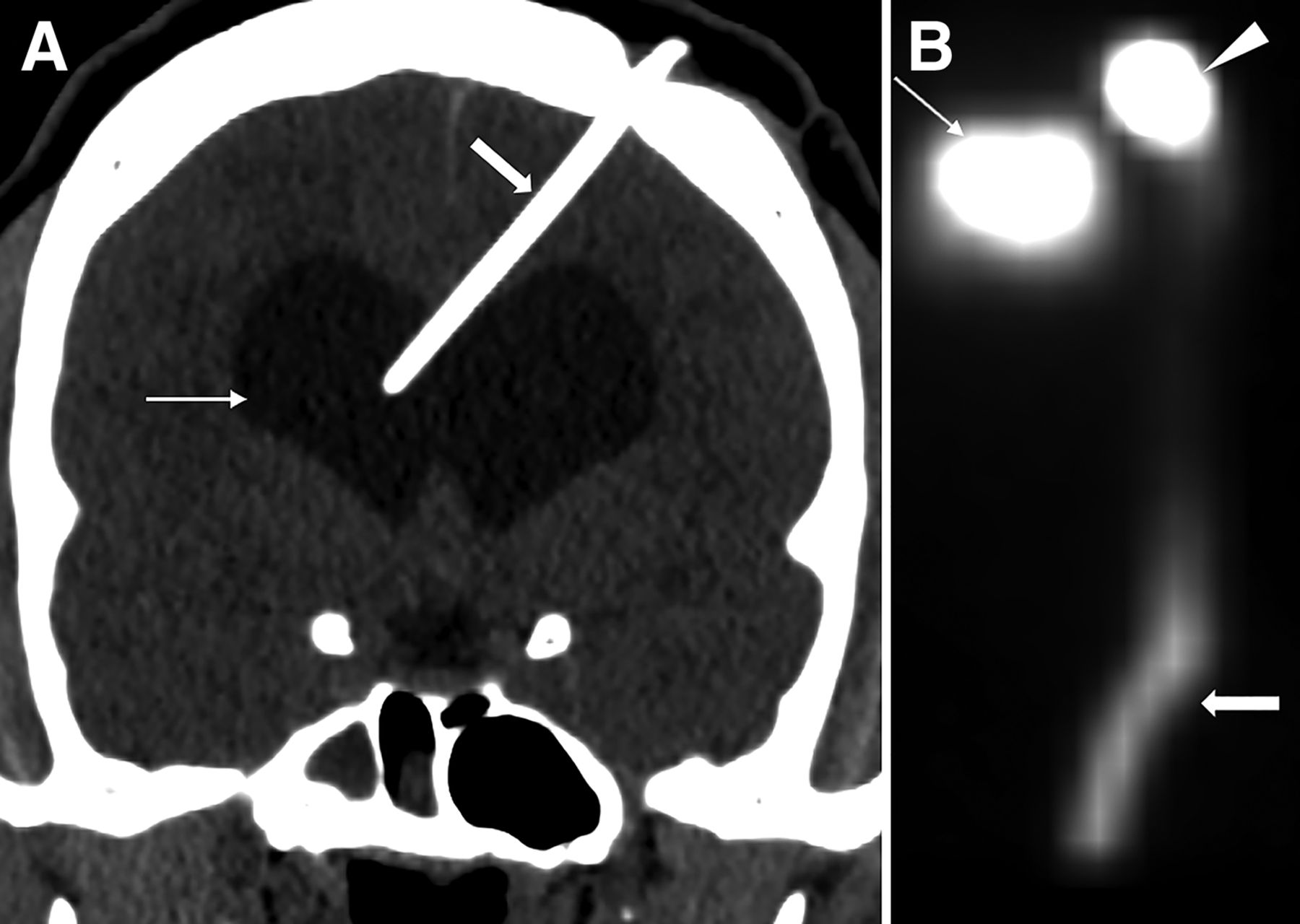
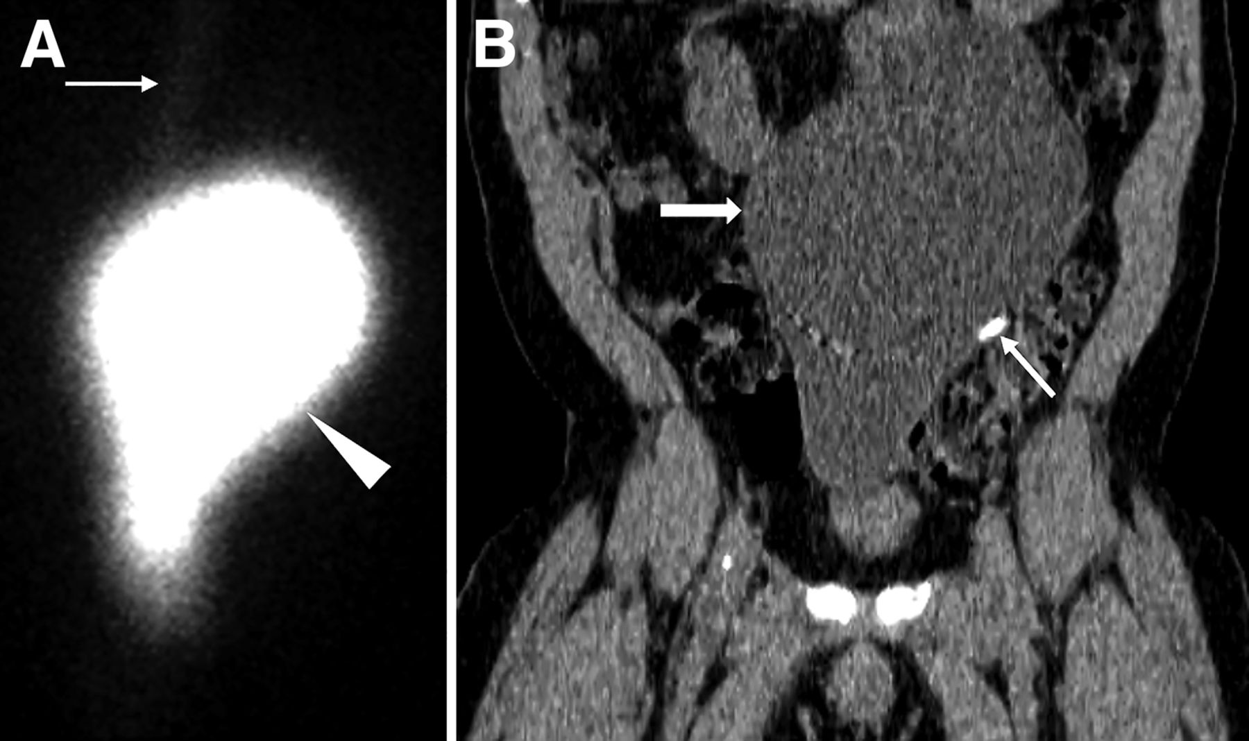
A 41-y-old man with a history of communicating hydrocephalus and ventriculoperitoneal shunt placement 11 y previously presented with headache. Shunt malfunction was suspected. A radionuclide cerebrospinal fluid shuntogram was done after injection of 106.93 MBq (2.89 mCi) of 111In-diethylenetriaminepentaacetic acid into the shunt reservoir on the left scalp. Dynamic images of the head and chest and static image of the abdomen were acquired. Injected activity was noted in the shunt reservoir. Tracer activity in the lateral ventricles suggested patent proximal tubing, and activity along the distal tubing suggested patent distal tubing (Fig. 1). However, there was a large area of well-defined loculated activity in the abdomen (Fig. 2). Further evaluation with SPECT/CT confirmed that the activity in the abdomen correlated with a large pseudocyst occupying the abdomen and pelvis. The distal tip of the shunt was seen within the pseudocyst. Excision of the pseudocyst was planned for the patient.

FIGURE 1.
  • Download figure
  • Open in new tab
  • Download powerpoint
FIGURE 1.

(A) Coronal CT of head shows hydrocephalus (thin arrow) and left frontal approach ventricular shunt catheter terminating in right ventricle (thick arrow). (B) Planar scintigraphy in anterior projection shows injected activity in shunt reservoir on left scalp (arrowhead). Activity is seen in lateral ventricles (thin arrow) suggestive of patent proximal tubing. Activity is seen tracking through distal tubing along chest and upper abdomen (thick arrow).

FIGURE 2.
  • Download figure
  • Open in new tab
  • Download powerpoint
FIGURE 2.

(A) Planar scintigraphy of abdomen demonstrates well-defined loculated activity in abdomen (arrowhead), as well as part of distal tubing in upper abdomen (arrow). (B) Coronal CT depicts correlating large pseudocyst occupying abdomen and pelvis (thick arrow) with fluid density and thin wall, as well as distal end of shunt catheter tip terminating within pseudocyst (thin arrow).

DISCUSSION

Ventriculoperitoneal shunting is the common treatment for hydrocephalus, with the cerebrospinal fluid being absorbed into the peritoneal cavity. When cerebrospinal fluid is not adequately absorbed, there can be focal peritonitis with inflammatory pseudocyst formation. Other etiologies for pseudocyst include adhesions from previous abdominal surgeries or silicone allergy (1,2). Symptoms include abdominal pain, distension, and constipation. There can be signs of increased intracranial pressure such as headache, nausea, vomiting, and fatigue due to shunt malfunction. Sometimes the pseudocyst can get infected. Ultrasonography is the initial method to diagnose a pseudocyst and can be confirmed with CT. Routine radiography can detect other shunt complications, such as kinking or disconnection. With persisting suspicion, cerebrospinal fluid shunt scintigraphy aids to further assess shunt patency. In our case, the pseudocyst was incidentally detected on the scintigram done to evaluate shunt patency. Excision of the cyst and shunt relocation is the definitive treatment. Percutaneous drainage can be therapeutic and diagnostic.

CONCLUSION

Though ultrasonography is the method of choice for the initial diagnosis, we report initial detection of a large abdominal pseudocyst incidentally on scintigraphy in a patient with a history of ventriculoperitoneal shunt placement and headache. An abdominal pseudocyst has to be suspected, especially if there are abdominal symptoms. Timely diagnosis and management can alleviate the patient’s symptoms.

DISCLOSURE

No potential conflict of interest relevant to this article was reported.

Footnotes

  • Published online Sep. 12, 2023.

REFERENCES

  1. 1.↵
    1. Hamid R,
    2. Baba AA,
    3. Bhat NA,
    4. Mufti G,
    5. Mir YA,
    6. Sajad W
    . Post ventriculoperitoneal shunt abdominal pseudocyst: challenges posed in management. Asian J Neurosurg. 2017;12:13–16.
    OpenUrl
  2. 2.↵
    1. Achufusi TGO,
    2. Chebaya P,
    3. Rawlins S
    . Abdominal cerebrospinal fluid pseudocyst as a complication of ventriculoperitoneal shunt placement. Cureus. 2020;12:e9363.
    OpenUrl
  • Received for publication May 17, 2023.
  • Revision received August 4, 2023.
PreviousNext
Back to top

In this issue

Journal of Nuclear Medicine Technology: 53 (1)
Journal of Nuclear Medicine Technology
Vol. 53, Issue 1
March 1, 2025
  • Table of Contents
  • About the Cover
  • Index by author
  • Complete Issue (PDF)
Print
Download PDF
Article Alerts
Sign In to Email Alerts with your Email Address
Email Article

Thank you for your interest in spreading the word on Journal of Nuclear Medicine Technology.

NOTE: We only request your email address so that the person you are recommending the page to knows that you wanted them to see it, and that it is not junk mail. We do not capture any email address.

Enter multiple addresses on separate lines or separate them with commas.
Scintigraphic Detection of Abdominal Pseudocyst: A Complication of Ventriculoperitoneal Shunt
(Your Name) has sent you a message from Journal of Nuclear Medicine Technology
(Your Name) thought you would like to see the Journal of Nuclear Medicine Technology web site.
Citation Tools
Scintigraphic Detection of Abdominal Pseudocyst: A Complication of Ventriculoperitoneal Shunt
Fathima Fijula Palot Manzil, Muhammad Yahya Hameed, Joshua Eichhorn
Journal of Nuclear Medicine Technology Sep 2023, jnmt.123.266042; DOI: 10.2967/jnmt.123.266042

Citation Manager Formats

  • BibTeX
  • Bookends
  • EasyBib
  • EndNote (tagged)
  • EndNote 8 (xml)
  • Medlars
  • Mendeley
  • Papers
  • RefWorks Tagged
  • Ref Manager
  • RIS
  • Zotero
Share
Scintigraphic Detection of Abdominal Pseudocyst: A Complication of Ventriculoperitoneal Shunt
Fathima Fijula Palot Manzil, Muhammad Yahya Hameed, Joshua Eichhorn
Journal of Nuclear Medicine Technology Sep 2023, jnmt.123.266042; DOI: 10.2967/jnmt.123.266042
Twitter logo Facebook logo LinkedIn logo Mendeley logo
  • Tweet Widget
  • Facebook Like
  • Google Plus One
Bookmark this article

Jump to section

  • Article
    • Abstract
    • CASE REPORT
    • DISCUSSION
    • CONCLUSION
    • DISCLOSURE
    • Footnotes
    • REFERENCES
  • Figures & Data
  • Info & Metrics
  • PDF

Related Articles

  • No related articles found.
  • PubMed
  • Google Scholar

Cited By...

  • No citing articles found.
  • Google Scholar

More in this TOC Section

  • High-Sensitivity Troponin Elevation in a Young Woman with Typical Chest Pain: The Heart of the Matter
  • Pulmonary Adenocarcinoma Revealed by Parathyroid Scintigraphy: An Incidental Case to Remember
  • Prominent Right Ventricular Tracer Uptake: A Harbinger of Multivessel Coronary Artery Disease
Show more Teaching Case Studies

Similar Articles

Keywords

  • abdominal CSF pseudocyst
  • VP shunt malfunction
  • 111In-DTPA shunt study
SNMMI

© 2025 SNMMI

Powered by HighWire