


Abstract
Incidental detection of coronavirus disease 2019 (COVID-19)–related lung changes on 18F-FDG PET/CT images of oncology patients has been increasingly reported. Most of the case reports or series have stressed the retrospective diagnosis of COVID-19 with the help of 18F-FDG PET/CT lung findings. In this case report, we introduce a different aspect of COVID-19–related lung changes on 18F-FDG PET/CT, interfering with the evaluation of metastatic lung lesions in a patient with renal cell carcinoma.
Various lung involvement patterns have been reported on 18F-FDG PET/CT scans of coronavirus disease 2019 (COVID-19) patients undergoing workup for various malignancies. The patterns range from 18F-FDG–avid diffuse ground-glass opacities to 18F-FDG–avid patchy consolidatory changes, with or without 18F-FDG–avid mediastinal lymph nodes, depending on the imaging time from the onset of infection and other unknown factors (1–4). COVID-19 infection was a retrospective diagnosis in most reported cases, after the typical findings were seen on the 18F-FDG PET/CT images (3–7). Here, we present a different aspect of COVID-19 on 18F-FDG PET/CT, in which there was interference with response assessment in a patient receiving chemotherapy for pulmonary metastasis from renal cell carcinoma.
CASE REPORT
A 45 y-old man with a known case of metastatic renal cell carcinoma underwent cytoreduction nephrectomy followed by first-line chemotherapy with pembrolizumab and axitinib because of multiple cannonball metastases in the lungs. 18F-FDG PET/CT at the end of treatment showed disease progression in the form of an increase in the number and size of lung nodules. The patient was then started on second-line chemotherapy with oral lenvatinib (18 mg daily) and everolimus (5 mg daily). His interim 18F-FDG PET/CT scan (Fig. 1) showed a favorable response (>30% reduction in size and 18F-FDG avidity compared with baseline PET/CT) to second-line therapy, and he was continued on the same treatment. He was diagnosed with COVID-19 in May 2020 on evaluation for malaise and chills. He was managed conservatively with antibiotics, antipyretics, and multivitamins in a local hospital. He had no symptoms or signs suggestive of pneumonia and never required oxygen support during the 11-d course in the hospital. He was discharged from the hospital after a negative nucleic acid test 1 wk before he was scheduled for an 18F-FDG PET/CT scan at 6 mo of chemotherapy to determine the response. The 18F-FDG PET/CT scan (Fig. 2) showed 18F-FDG–avid diffuse ground-glass opacities/patchy consolidatory changes bilaterally in the lung fields from apex to base, obscuring the metastatic lesions. The COVID-19–related lung changes obscured both the anatomic and the metabolic features of the metastatic lesions, leading to difficulty in assessing the response to treatment.
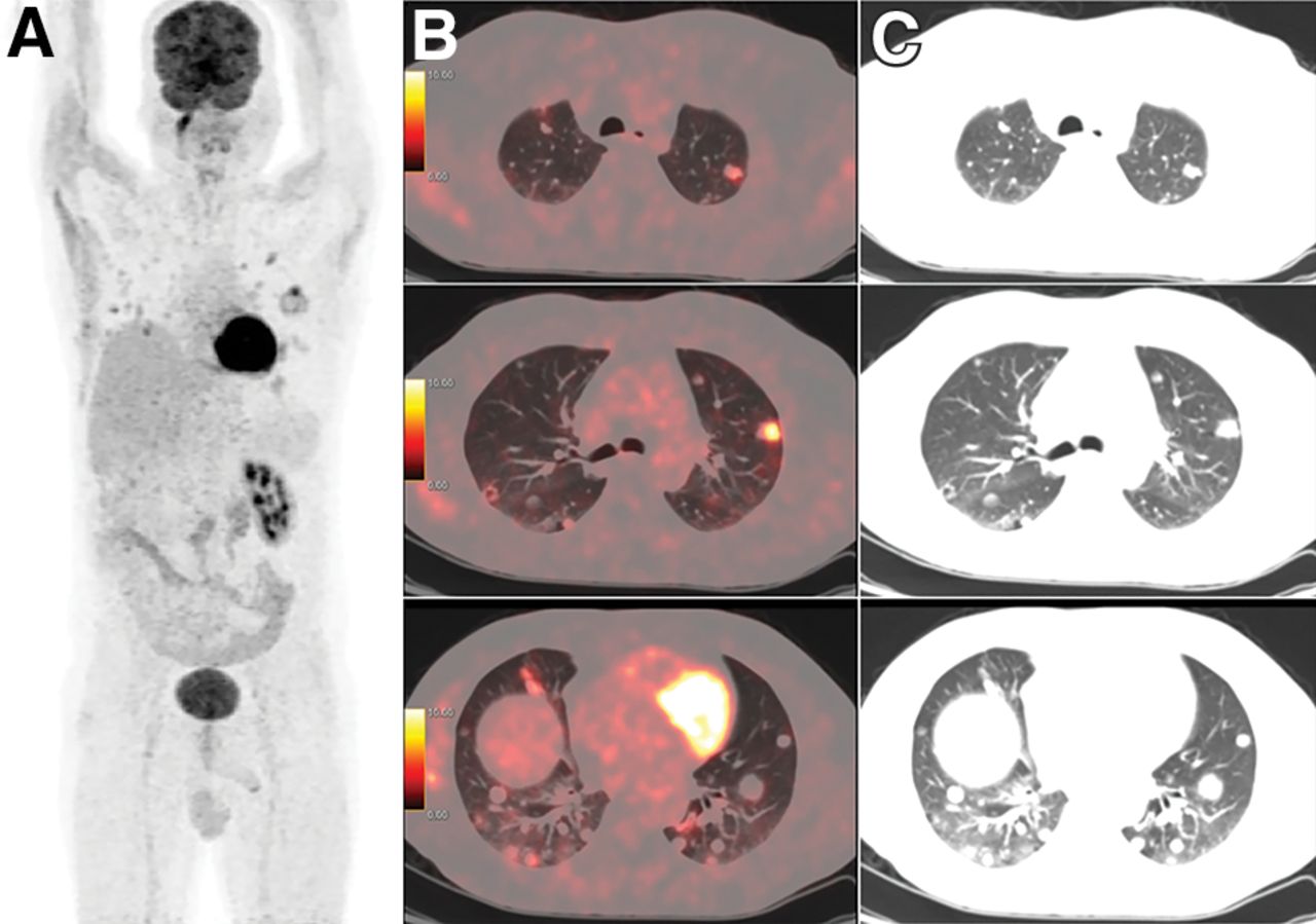
18F-FDG PET/CT whole-body maximum-intensity-projection image (A), axial PET/CT images (B), and corresponding CT images (C) showing variably 18F-FDG–avid random nodules in both lung fields (SUVmax of hottest nodule, 9.9).
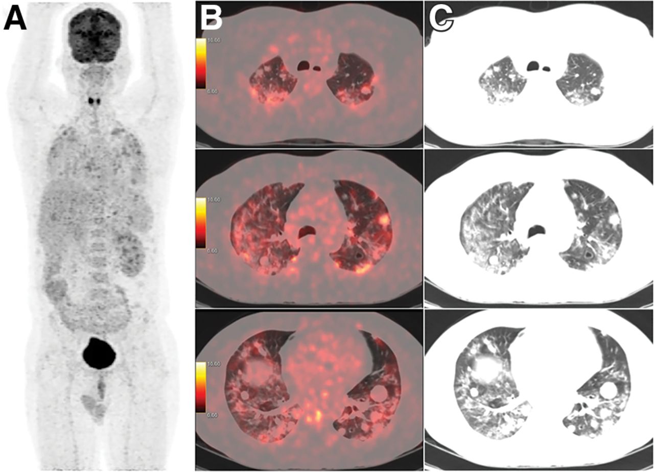
18F-FDG PET/CT whole-body maximum-intensity-projection image (A), axial PET/CT images (B), and corresponding CT images (C) showing 18F-FDG–avid diffuse ground-glass opacities/patchy consolidatory changes bilaterally in lung fields from apex to base, obscuring details of metastatic lesions (SUVmax of hottest nodule, 7.8; SUVmax of ground-glass opacities, 7.3).
DISCUSSION
18F-FDG uptake in ground-glass opacities in the background may add spill-in counts to metastatic lesions, causing a falsely high uptake in metastatic lesions (8). For this reason, an accurate assessment of the metabolic response was not possible in this patient. The patient was advised to repeat the nucleic acid test because of 18F-FDG avidity in the ground-glass opacities/consolidatory changes and was found to be positive. The patient was then advised to remain home in isolation again.
CONCLUSION
During the COVID-19 pandemic phase, we have to consider sources of possible interference such as described in this report before scheduling patients for 18F-FDG PET/CT scans for various oncologic purposes.
DISCLOSURE
No potential conflict of interest relevant to this article was reported.
Footnotes
Published online July 9, 2021.
Immediate Open Access: Creative Commons Attribution 4.0 International License (CC BY) allows users to share and adapt with attribution, excluding materials credited to previous publications. License: https://creativecommons.org/licenses/by/4.0/. Details: http://jnm.snmjournals.org/site/misc/permission.xhtml.
REFERENCES
- Received for publication February 16, 2021.
- Accepted for publication March 26, 2021.


