
Main menu

  • Home
  • Content
    • Current
      • JNMT Supplement
    • Ahead of print
    • Past Issues
    • Continuing Education
    • JNMT Podcast
    • SNMMI Annual Meeting Abstracts
  • Subscriptions
    • Subscribers
    • Rates
    • Journal Claims
    • Institutional and Non-member
  • Authors
    • Submit to JNMT
    • Information for Authors
    • Assignment of Copyright
    • AQARA Requirements
  • Info
    • Reviewers
    • Permissions
    • Advertisers
    • Corporate & Special Sales
  • About
    • About Us
    • Editorial Board
    • Contact Information
  • More
    • Alerts
    • Feedback
    • Help
    • SNMMI Journals
  • SNMMI
    • JNMT
    • JNM
    • SNMMI Journals
    • SNMMI

User menu

  • Subscribe
  • My alerts
  • Log in
  • Log out
  • My Cart

Search

  • Advanced search
Journal of Nuclear Medicine Technology
  • SNMMI
    • JNMT
    • JNM
    • SNMMI Journals
    • SNMMI
  • Subscribe
  • My alerts
  • Log in
  • Log out
  • My Cart
Journal of Nuclear Medicine Technology

Advanced Search

  • Home
  • Content
    • Current
    • Ahead of print
    • Past Issues
    • Continuing Education
    • JNMT Podcast
    • SNMMI Annual Meeting Abstracts
  • Subscriptions
    • Subscribers
    • Rates
    • Journal Claims
    • Institutional and Non-member
  • Authors
    • Submit to JNMT
    • Information for Authors
    • Assignment of Copyright
    • AQARA Requirements
  • Info
    • Reviewers
    • Permissions
    • Advertisers
    • Corporate & Special Sales
  • About
    • About Us
    • Editorial Board
    • Contact Information
  • More
    • Alerts
    • Feedback
    • Help
    • SNMMI Journals
  • Watch or Listen to JNMT Podcast
  • Visit SNMMI on Facebook
  • Join SNMMI on LinkedIn
  • Follow SNMMI on Twitter
  • Subscribe to JNMT RSS feeds
Review ArticleTEACHING CASE STUDIES

The Pinwheel Sign: Artifact from Head Rotation During SPECT Acquisition for Dopamine Transporter Imaging

Amy K. Janicek, Ryan J. Avery and Phillip H. Kuo
Journal of Nuclear Medicine Technology March 2014, 42 (1) 75-76; DOI: https://doi.org/10.2967/jnmt.113.126078
Amy K. Janicek
1Department of Medical Imaging, University of Arizona, Tucson, Arizona; and
  • Find this author on Google Scholar
  • Find this author on PubMed
  • Search for this author on this site
Ryan J. Avery
1Department of Medical Imaging, University of Arizona, Tucson, Arizona; and
  • Find this author on Google Scholar
  • Find this author on PubMed
  • Search for this author on this site
Phillip H. Kuo
1Department of Medical Imaging, University of Arizona, Tucson, Arizona; and
2Departments of Medicine and Biomedical Engineering, University of Arizona, Tucson, Arizona
  • Find this author on Google Scholar
  • Find this author on PubMed
  • Search for this author on this site
  • Article
  • Figures & Data
  • Info & Metrics
  • PDF
Loading

Abstract

This case study illustrates the pattern and significance of the pinwheel sign, a SPECT artifact from rotational head motion that can complicate the diagnosis of Parkinson disease. Monitoring the patient during image acquisition is crucial since rotational motion artifacts are difficult to identify on raw or processed images and may lead to incorrect interpretation of findings.

  • neurology
  • quality assurance
  • SPECT
  • DAT scan
  • Parkinson’s disease
  • artifact

Diagnosis of Parkinson disease can be complicated in patients with preexisting neurologic conditions such as essential tremor or drug-induced parkinsonism. In such situations, dopamine transporter imaging improves diagnostic accuracy and optimization of therapy (1). The accuracy of dopamine transporter imaging relies on quality control at all stages in the imaging chain, starting with initial positioning (2). Motion artifacts may lead to incorrect visual reads and may degrade the utility of quantification programs for computer-aided diagnosis (3). Here, we describe the pattern and significance of the pinwheel sign, an artifact from rotational head motion.

CASE STUDY

A 73-y-old man with Asperger syndrome developed rest and action tremors that interfered with writing and brushing teeth. Propranolol helped to control the action tremor but not the rest tremor. The patient also took valproic acid, which has a potential side effect of tremor.

The neurologist ordered dopamine transporter imaging to differentiate Parkinson disease from essential tremor or drug-induced parkinsonism. Two nuclear medicine physicians found the initial scan difficult to evaluate. Both readers inclined to interpret the scan findings as abnormal because of decreased activity in both putamina (worse on the right) (Fig. 1).

FIGURE 1.
  • Download figure
  • Open in new tab
  • Download powerpoint
FIGURE 1.

(A) Representative 123I-ioflupane scan with normal findings demonstrates properly aligned striata with comma configurations. Dual-head camera set for circular orbit with 13-cm radius acquired data at rate of 30 s/frame at 3° per stop for 30 min. Postprocessing reconstruction was performed using filtered backprojection with Butterworth filter with cutoff of 0.6 and order of 8 and no attenuation correction. Axial images have slice thickness of 2.5 mm. (B) Initial imaging was compromised by rotation of head leftward during acquisition. Before injection of 225.7 MBq (6.1 mCi) of 123I-ioflupane, patient drank 4 drops of Lugol solution. (C) Imaging was repeated without motion. Standard reconstruction algorithm was same as used for initial scan.

However, the technologist noted that the patient had turned his head to the left approximately halfway through the acquisition. The technologist and physician reviewed the sinograms and projection data in cine mode and could not detect the rotary motion. Because the patient could not tolerate a full repeated examination, the acquisition was repeated in half the time and with the head secured to prevent motion.

The 30-min initial acquisition collected 1,740,000 counts, and the 15-min repeated acquisition collected 834,000 counts. The repeated acquisition was considered adequate because more than 1,000,000 counts would have been collected if the study had been classically normal (4). Technologists carefully observed the patient to confirm no motion. Both readers found the repeated scan also difficult to interpret (Fig. 1C). It demonstrated improvement of the anteroposterior orientation of the basal ganglia and unexpected normalization of the right putamen. The first reader again inclined to interpret the findings as abnormal because of decreased activity in the left putamen, but the second reader inclined to interpret the findings as normal because of the imperfect but relatively comma-shaped configuration of the striata. The final diagnosis is being determined through continuing long-term clinical follow-up.

The repeated imaging revealed that leftward rotation of the head caused blurring of the activity in the left caudate head posteriorly, thereby falsely adding activity to the region of the left putamen. Likewise, blurring of the activity in the right putamen anteriorly gave a falsely abnormal right putamen (Fig. 2). The pinwheel analogy was selected because it conjures a mental image of rotary motion with subsequent blurring.

FIGURE 2.
  • Download figure
  • Open in new tab
  • Download powerpoint
FIGURE 2.

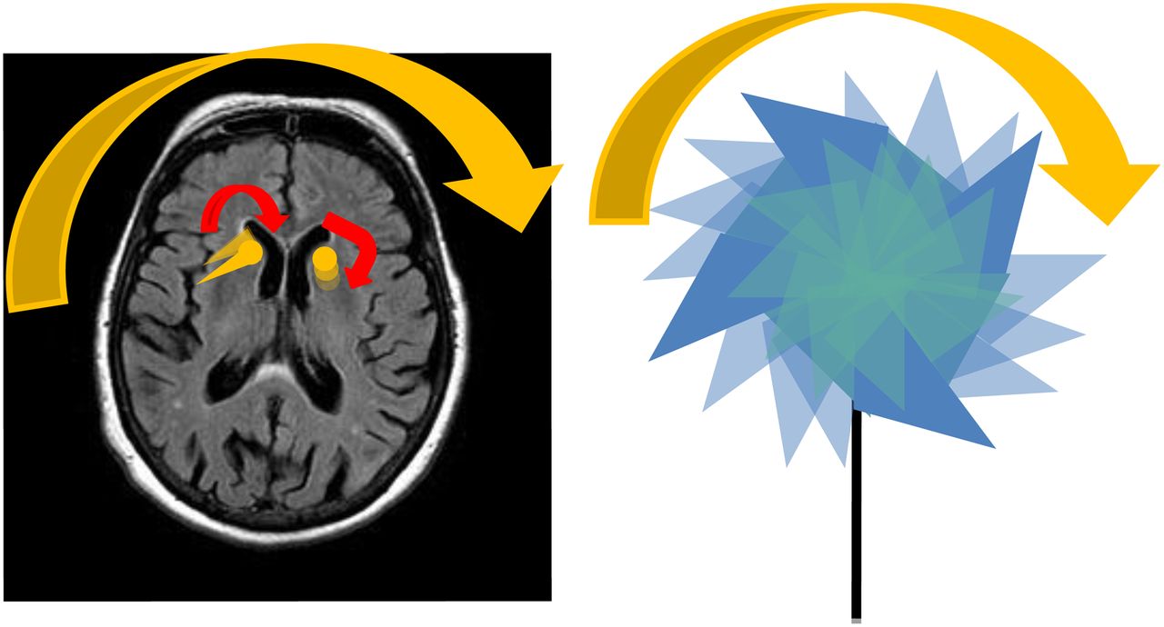
(Left) Graphic depicting leftward rotation of caudate heads and putamen (yellow) overlay on MR image of brain. (Right) Illustration of rotating pinwheel for memorable analogy for rotational motion and blurring.

DISCUSSION

Routinely, technologists assess for motion before submission of images. In myocardial perfusion imaging, technologists examine the cine raw images and sinograms. Physicians can detect motion artifacts on postprocessed images by, for example, the hurricane sign, in which short-axis slices of the heart resemble the National Weather Service symbol for a hurricane (5). Motion artifacts are surprisingly difficult to detect on raw and processed dopamine transporter images, unlike other SPECT images.

CONCLUSION

Monitoring the patient during SPECT acquisition is crucial because rotational motion artifacts are difficult to identify on raw or processed images. Clinical interpretation of dopamine transporter images relies on assessment of the morphology of small structures and is therefore particularly susceptible to motion artifacts. Recognizing the pinwheel sign may aid in detecting this critical artifact.

DISCLOSURE

Phillip H. Kuo has consulted for and received grants from GE Healthcare. No other potential conflict of interest relevant to this article was reported.

Footnotes

  • Published online Jan. 30, 2014.

REFERENCES

  1. 1.↵
    1. Kägi G,
    2. Bhatia KP,
    3. Tolosa E
    . The role of DAT-SPECT in movement disorders. J Neurol Neurosurg Psychiatry. 2010;81:5–12.
    OpenUrlAbstract/FREE Full Text
  2. 2.↵
    1. Covington MF,
    2. McMillan NA,
    3. Avery RJ,
    4. Kuo PH
    . The semicolon sign: dopamine transporter (DAT) imaging artifact from head tilt. J Nucl Med Technol. 2013;41:105–107.
    OpenUrlAbstract/FREE Full Text
  3. 3.↵
    1. Kuo PH,
    2. Avery R,
    3. Krupinski E,
    4. et al
    . Receiver-operator-characteristic analysis of an automated program for analyzing striatal uptake of 123I-ioflupane SPECT images: calibration using visual reads. J Nucl Med Technol. 2013;41:26–31.
    OpenUrlAbstract/FREE Full Text
  4. 4.↵
    1. Djang DS,
    2. Janssen M,
    3. Bohnen N,
    4. et al
    . SNM practice guideline for dopamine transporter imaging with 123I-ioflupane SPECT 1.0. J Nucl Med. 2012;53:154–163.
    OpenUrlFREE Full Text
  5. 5.↵
    1. Sorrell V,
    2. Figueroa B,
    3. Hansen CL
    . The “hurricane sign”: evidence of patient motion artifact on cardiac single-photon emission computed tomographic imaging. J Nucl Cardiol. 1996;3:86–88.
    OpenUrlCrossRefPubMed
  • Received for publication May 13, 2013.
  • Accepted for publication October 10, 2013.
PreviousNext
Back to top

In this issue

Journal of Nuclear Medicine Technology: 42 (1)
Journal of Nuclear Medicine Technology
Vol. 42, Issue 1
March 1, 2014
  • Table of Contents
  • About the Cover
  • Index by author
Print
Download PDF
Article Alerts
Sign In to Email Alerts with your Email Address
Email Article

Thank you for your interest in spreading the word on Journal of Nuclear Medicine Technology.

NOTE: We only request your email address so that the person you are recommending the page to knows that you wanted them to see it, and that it is not junk mail. We do not capture any email address.

Enter multiple addresses on separate lines or separate them with commas.
The Pinwheel Sign: Artifact from Head Rotation During SPECT Acquisition for Dopamine Transporter Imaging
(Your Name) has sent you a message from Journal of Nuclear Medicine Technology
(Your Name) thought you would like to see the Journal of Nuclear Medicine Technology web site.
Citation Tools
The Pinwheel Sign: Artifact from Head Rotation During SPECT Acquisition for Dopamine Transporter Imaging
Amy K. Janicek, Ryan J. Avery, Phillip H. Kuo
Journal of Nuclear Medicine Technology Mar 2014, 42 (1) 75-76; DOI: 10.2967/jnmt.113.126078

Citation Manager Formats

  • BibTeX
  • Bookends
  • EasyBib
  • EndNote (tagged)
  • EndNote 8 (xml)
  • Medlars
  • Mendeley
  • Papers
  • RefWorks Tagged
  • Ref Manager
  • RIS
  • Zotero
Share
The Pinwheel Sign: Artifact from Head Rotation During SPECT Acquisition for Dopamine Transporter Imaging
Amy K. Janicek, Ryan J. Avery, Phillip H. Kuo
Journal of Nuclear Medicine Technology Mar 2014, 42 (1) 75-76; DOI: 10.2967/jnmt.113.126078
Twitter logo Facebook logo LinkedIn logo Mendeley logo
  • Tweet Widget
  • Facebook Like
  • Google Plus One
Bookmark this article

Jump to section

  • Article
    • Abstract
    • CASE STUDY
    • DISCUSSION
    • CONCLUSION
    • DISCLOSURE
    • Footnotes
    • REFERENCES
  • Figures & Data
  • Info & Metrics
  • PDF

Related Articles

  • No related articles found.
  • PubMed
  • Google Scholar

Cited By...

  • Pitfalls and Artifacts of 123I-Ioflupane SPECT in Parkinsonian Syndromes: A Quality Improvement Teaching Tool
  • False-Positive Findings on Dopamine Transporter SPECT Due to Therapeutic Dextroamphetamine and Amphetamine
  • What do patients with scans without evidence of dopaminergic deficit (SWEDD) have? New evidence and continuing controversies
  • Google Scholar

More in this TOC Section

  • High-Sensitivity Troponin Elevation in a Young Woman with Typical Chest Pain: The Heart of the Matter
  • Pulmonary Adenocarcinoma Revealed by Parathyroid Scintigraphy: An Incidental Case to Remember
  • Prominent Right Ventricular Tracer Uptake: A Harbinger of Multivessel Coronary Artery Disease
Show more Teaching Case Studies

Similar Articles

Keywords

  • Neurology
  • quality assurance
  • SPECT
  • DAT scan
  • Parkinson’s disease
  • artifact
SNMMI

© 2025 SNMMI

Powered by HighWire