
Main menu

  • Home
  • Content
    • Current
      • JNMT Supplement
    • Ahead of print
    • Past Issues
    • Continuing Education
    • JNMT Podcast
    • SNMMI Annual Meeting Abstracts
  • Subscriptions
    • Subscribers
    • Rates
    • Journal Claims
    • Institutional and Non-member
  • Authors
    • Submit to JNMT
    • Information for Authors
    • Assignment of Copyright
    • AQARA Requirements
  • Info
    • Reviewers
    • Permissions
    • Advertisers
    • Corporate & Special Sales
  • About
    • About Us
    • Editorial Board
    • Contact Information
  • More
    • Alerts
    • Feedback
    • Help
    • SNMMI Journals
  • SNMMI
    • JNMT
    • JNM
    • SNMMI Journals
    • SNMMI

User menu

  • Subscribe
  • My alerts
  • Log in
  • My Cart

Search

  • Advanced search
Journal of Nuclear Medicine Technology
  • SNMMI
    • JNMT
    • JNM
    • SNMMI Journals
    • SNMMI
  • Subscribe
  • My alerts
  • Log in
  • My Cart
Journal of Nuclear Medicine Technology

Advanced Search

  • Home
  • Content
    • Current
    • Ahead of print
    • Past Issues
    • Continuing Education
    • JNMT Podcast
    • SNMMI Annual Meeting Abstracts
  • Subscriptions
    • Subscribers
    • Rates
    • Journal Claims
    • Institutional and Non-member
  • Authors
    • Submit to JNMT
    • Information for Authors
    • Assignment of Copyright
    • AQARA Requirements
  • Info
    • Reviewers
    • Permissions
    • Advertisers
    • Corporate & Special Sales
  • About
    • About Us
    • Editorial Board
    • Contact Information
  • More
    • Alerts
    • Feedback
    • Help
    • SNMMI Journals
  • Watch or Listen to JNMT Podcast
  • Visit SNMMI on Facebook
  • Join SNMMI on LinkedIn
  • Follow SNMMI on Twitter
  • Subscribe to JNMT RSS feeds
Research ArticleContinuing Education

PET/CT Case Series: Unmasking the Mystery of Cardiac Sarcoidosis

Diane K. Soulek
Journal of Nuclear Medicine Technology April 2025, jnmt.125.269732; DOI: https://doi.org/10.2967/jnmt.125.269732
Diane K. Soulek
Department of Radiology, University of Iowa, Iowa City, Iowa
  • Find this author on Google Scholar
  • Find this author on PubMed
  • Search for this author on this site
  • Article
  • Figures & Data
  • Info & Metrics
  • PDF
Loading

Article Figures & Data

Figures

  • Figure
    • Download figure
    • Open in new tab
    • Download powerpoint
  • FIGURE 1.
    • Download figure
    • Open in new tab
    • Download powerpoint
    FIGURE 1.

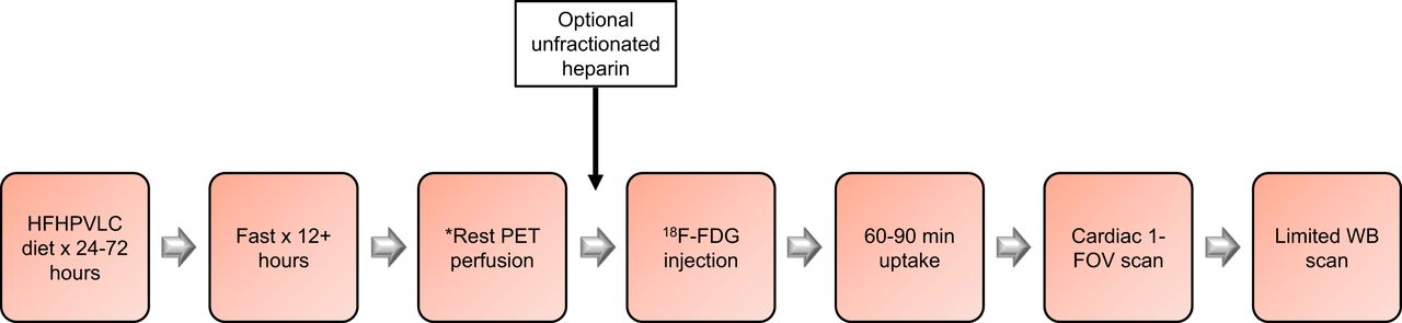
    Workflow example for 1-d cardiac sarcoid PET/CT imaging. *One-day PET/CT protocols currently use 82Rb-chloride or 13N-ammonia for rest PET perfusion imaging. Use of 18F perfusion radiopharmaceuticals may require increased time between rest PET perfusion and 18F-FDG scans.

  • FIGURE 2.
    • Download figure
    • Open in new tab
    • Download powerpoint
    FIGURE 2.

    Scan 1 82Rb-chloride rest perfusion (top rows) and 18F-FDG cardiac metabolism (bottom rows). Rest perfusion images show small mid inferolateral wall defect (white arrows) and normal distribution of activity throughout remaining LV myocardium. Gated rest LVEF is 32%. 18F-FDG cardiac slices show suboptimal display intensity for 18F-FDG cardiac sarcoidosis imaging, as neither 18F-FDG myocardial blood-pool activity nor activity in LV myocardial wall can be appreciated in current display.
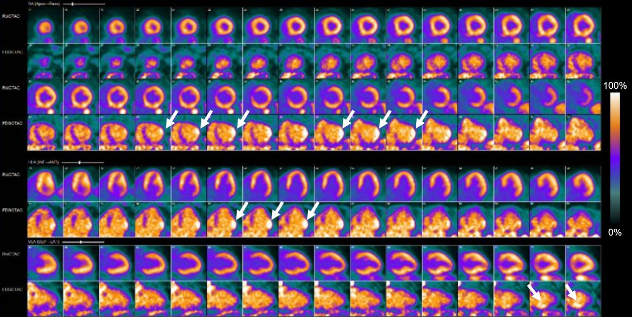
  • FIGURE 3.
    • Download figure
    • Open in new tab
    • Download powerpoint
    FIGURE 3.

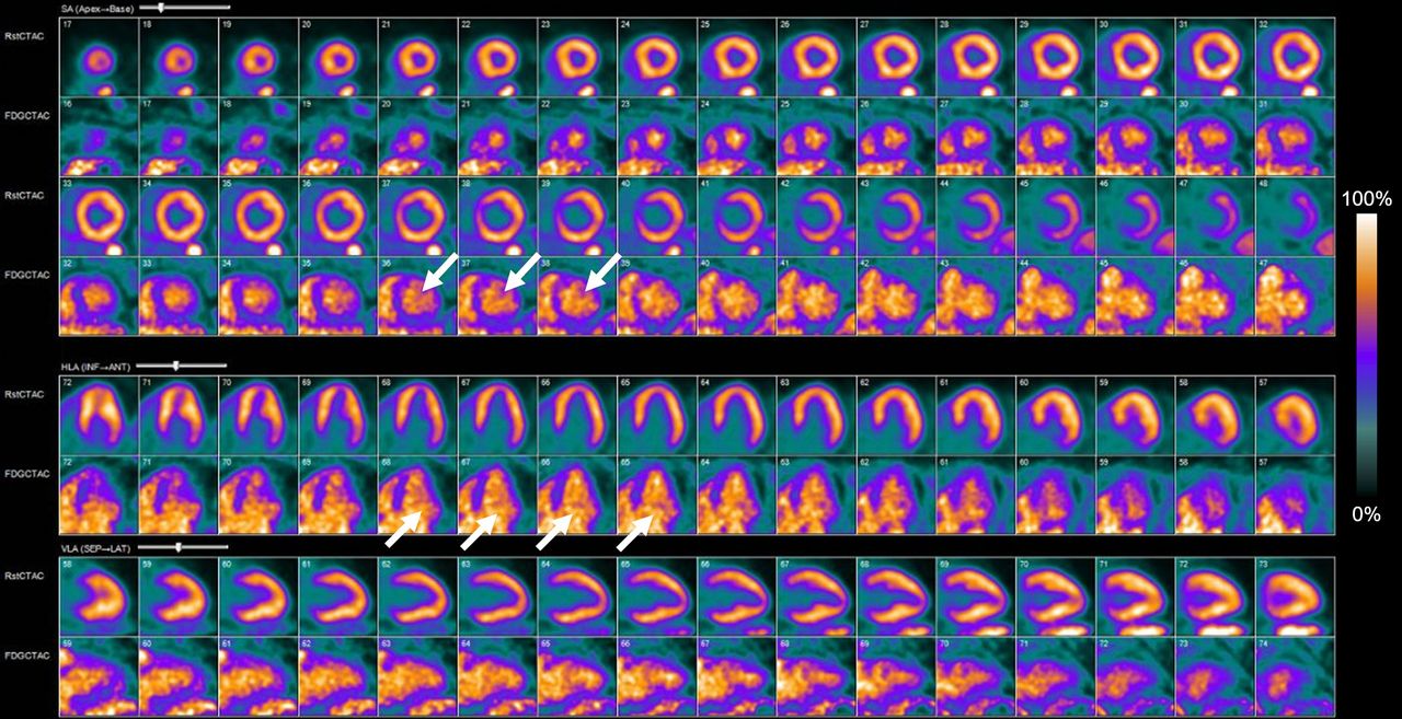
    Scan 1 82Rb-chloride rest perfusion (top rows) and rescaled 18F-FDG cardiac metabolism (bottom rows). Images show appropriate 18F-FDG display intensity with blood-pool activity (white arrows) brighter than LV wall, indicating normal FDG uptake pattern. No evidence of any focally increased 18F-FDG uptake in LV myocardial wall is seen, which indicates negative examination for active cardiac sarcoidosis.

  • FIGURE 4.
    • Download figure
    • Open in new tab
    • Download powerpoint
    FIGURE 4.

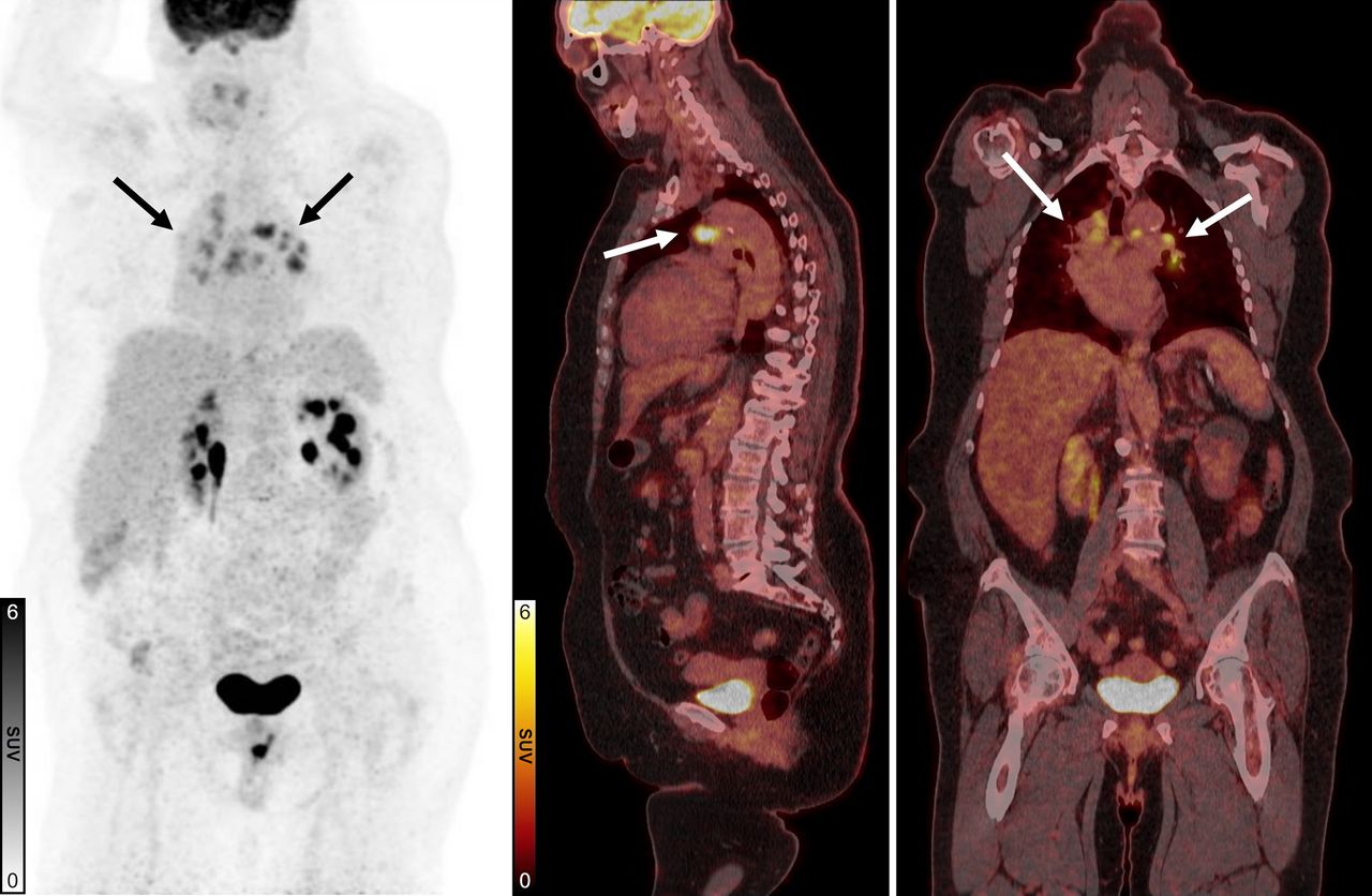
    Scan 1 18F-FDG maximum-intensity projection (left), fused 18F-FDG PET/CT sagittal (middle), and fused 18F-FDG PET/CT coronal (right) views. Images show diffuse hypermetabolic uptake (arrows) in mediastinal and hilar lymphadenopathy biopsy-proven noncaseating granulomatous disease. 18F-FDG PET/CT scan 1 is negative for cardiac sarcoidosis but positive for extracardiac sarcoidosis.

  • FIGURE 5.
    • Download figure
    • Open in new tab
    • Download powerpoint
    FIGURE 5.

    Scan 2 82Rb-chloride rest perfusion (top rows) and 18F-FDG cardiac metabolism (bottom rows). Images show diagnostic evaluation with 18F-FDG blood-pool activity indicating adequate dietary preparation. Rest images show borderline hypoperfusion in basal to mid inferolateral wall and moderate LV enlargement. Rest gated images showed 31% LVEF. 18F-FDG images show suspected early-to-progressive stable cardiac sarcoidosis with moderate inflammatory 18F-FDG uptake in basal to mid inferolateral wall (arrows).
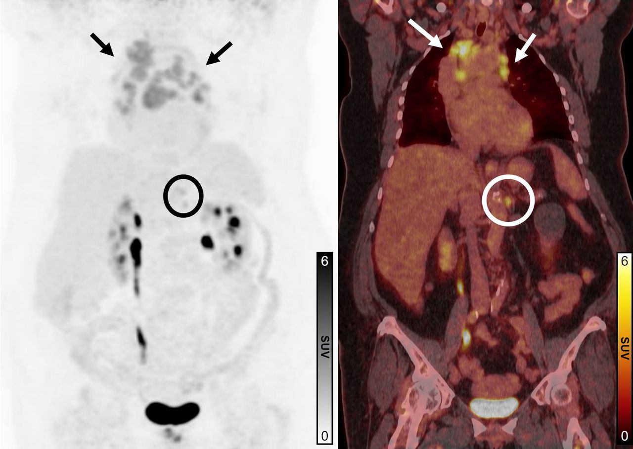
  • FIGURE 6.
    • Download figure
    • Open in new tab
    • Download powerpoint
    FIGURE 6.

    Scan 2 18F-FDG maximum-intensity projection (left) and 18F-FDG PET/CT fused coronal (right) images. Persistent 18F-FDG–avid bilateral mediastinal and hilar/perihilar adenopathy is visualized from known extracardiac sarcoidosis (arrows). Solitary 18F-FDG–avid left upper abdominal node is suspicious for additional sarcoid involvement (circles).
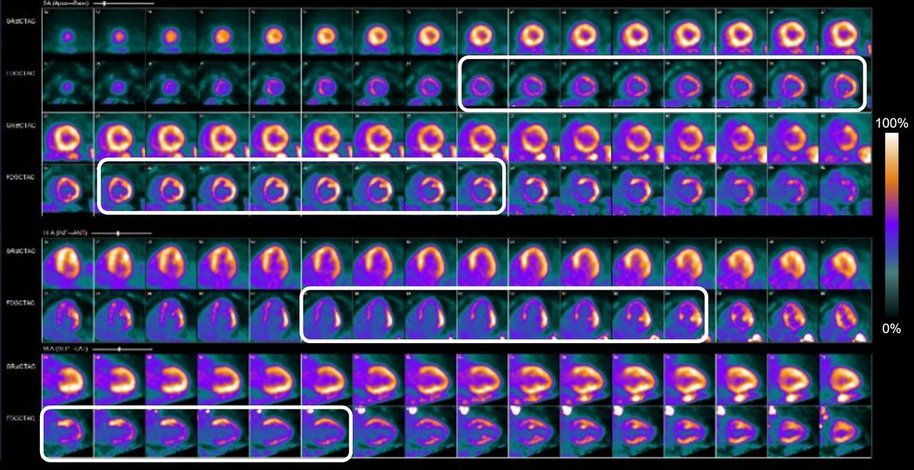
  • FIGURE 7.
    • Download figure
    • Open in new tab
    • Download powerpoint
    FIGURE 7.

    Scan 3 82Rb-chloride rest perfusion (top rows) and 18F-FDG cardiac metabolism (bottom rows). Rest MPI shows no significant perfusion defects in LV. 18F-FDG PET images show diffusely increased myocardial uptake (rectangles), more prominent in lateral wall. This pattern of diffusely increased 18F-FDG cardiac uptake indicates hyperinsulinemic state, suggesting possible inadequate dietary compliance, and limits evaluation of 18FDG-PET images for cardiac sarcoidosis. Gated rest images show estimated LVEF of 24%.
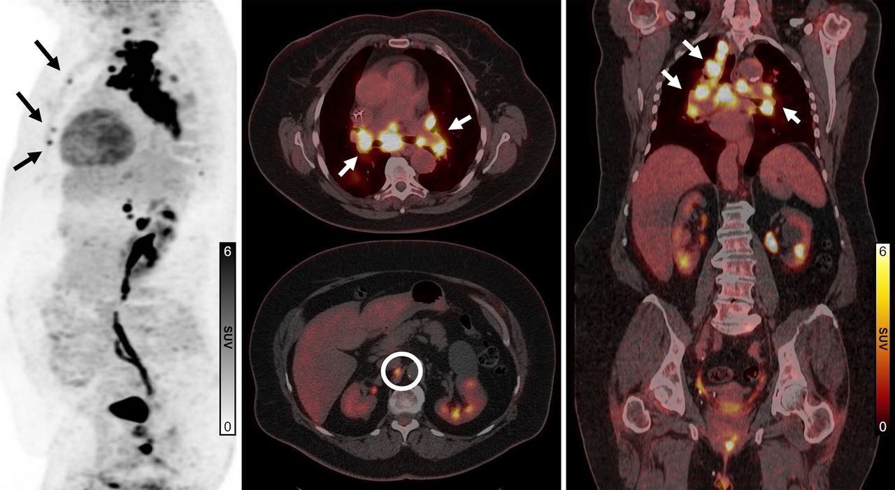
  • FIGURE 8.
    • Download figure
    • Open in new tab
    • Download powerpoint
    FIGURE 8.

    Scan 3 18F-FDG sagittal maximum-intensity projection (left), 18F-FDG PET/CT fused axial (middle), and 18F-FDG PET/CT fused coronal (right) views. Images show mediastinal, supraclavicular (white arrows), and retroperitoneal (circle) hypermetabolic lymphadenopathy with new or enlarged lymph nodes since scan 1, consistent with sarcoidosis with possible involvement of bilateral ribs (black arrows).
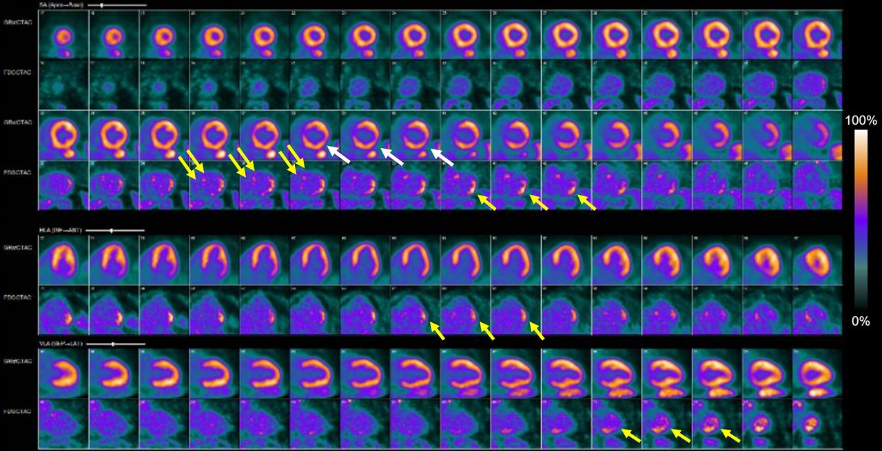
  • FIGURE 9.
    • Download figure
    • Open in new tab
    • Download powerpoint
    FIGURE 9.

    Scan 4 82Rb-chloride rest perfusion (top rows) and 18F-FDG cardiac metabolism (bottom rows). Rest perfusion images show medium-sized mild defect in basal to mid lateral and inferolateral wall (white arrows). Gated rest LVEF was 31%. 18F-FDG images show diffusely suppressed myocardial uptake, indicating adequate dietary preparation. Medium-sized area of increased 18F-FDG uptake is visualized in basal to mid lateral and inferolateral wall (single yellow arrows) similar to scan 2. New small-sized area of mildly increased 18F-FDG uptake is visualized in basal anteroseptal wall (double yellow arrows).
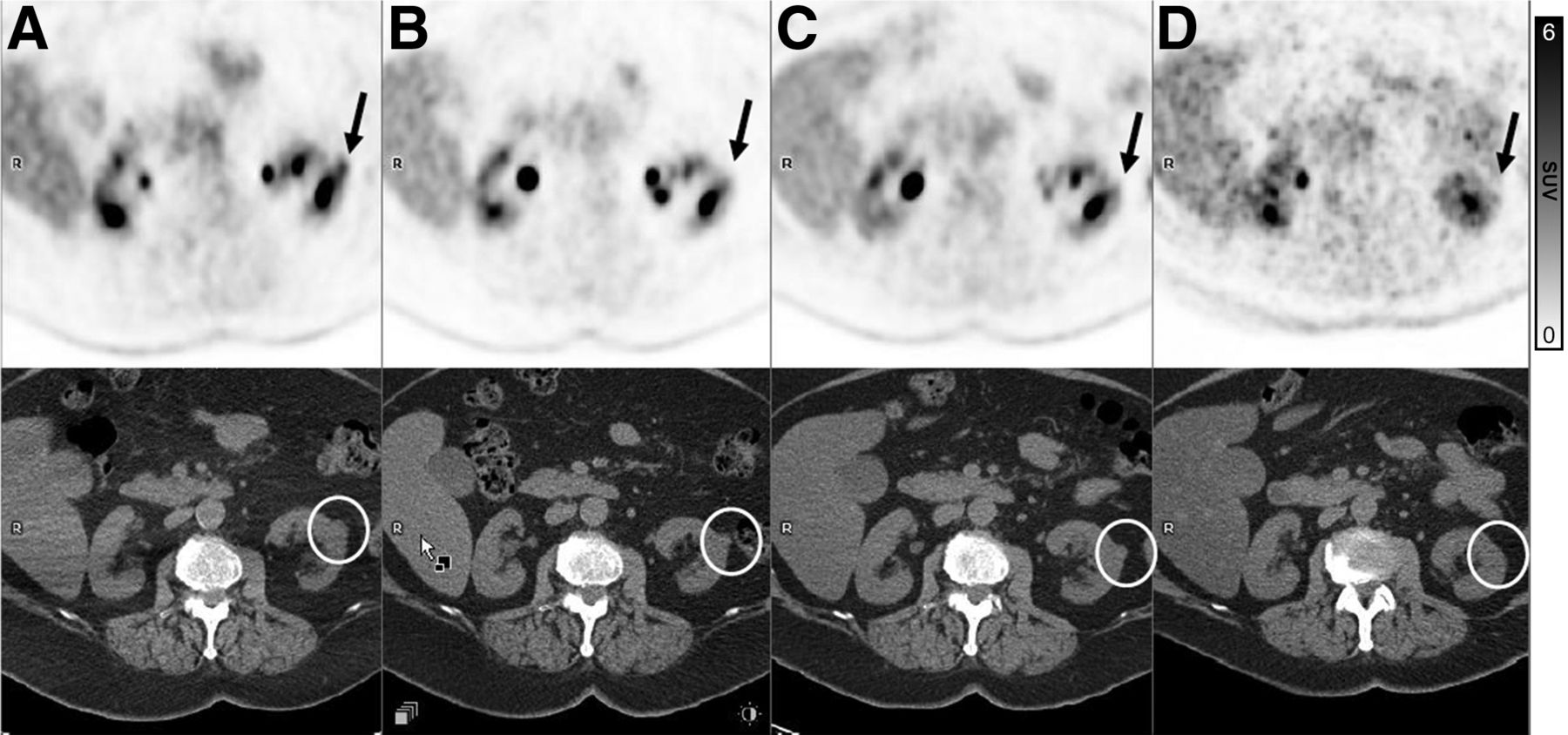
  • FIGURE 10.
    • Download figure
    • Open in new tab
    • Download powerpoint
    FIGURE 10.

    18F-FDG PET axial (top row) and CT axial (bottom row) images from scan 4 (A), scan 3 (B), scan 2 (C), and scan 1 (D). CT axial images from limited WB 18F-FDG PET/CT scan show incidental 18F-FDG–avid exophytic 1.7-cm isodense nodule in mid left renal pole (circles). This lesion is new since scan 1, has slowly enlarged since most recent examination, and displays increased 18F-FDG activity on scan 4 (arrows).

PreviousNext
Back to top

In this issue

Journal of Nuclear Medicine Technology: 53 (1)
Journal of Nuclear Medicine Technology
Vol. 53, Issue 1
March 1, 2025
  • Table of Contents
  • About the Cover
  • Index by author
  • Complete Issue (PDF)
Print
Download PDF
Article Alerts
Sign In to Email Alerts with your Email Address
Email Article

Thank you for your interest in spreading the word on Journal of Nuclear Medicine Technology.

NOTE: We only request your email address so that the person you are recommending the page to knows that you wanted them to see it, and that it is not junk mail. We do not capture any email address.

Enter multiple addresses on separate lines or separate them with commas.
PET/CT Case Series: Unmasking the Mystery of Cardiac Sarcoidosis
(Your Name) has sent you a message from Journal of Nuclear Medicine Technology
(Your Name) thought you would like to see the Journal of Nuclear Medicine Technology web site.
Citation Tools
PET/CT Case Series: Unmasking the Mystery of Cardiac Sarcoidosis
Diane K. Soulek
Journal of Nuclear Medicine Technology Apr 2025, jnmt.125.269732; DOI: 10.2967/jnmt.125.269732

Citation Manager Formats

  • BibTeX
  • Bookends
  • EasyBib
  • EndNote (tagged)
  • EndNote 8 (xml)
  • Medlars
  • Mendeley
  • Papers
  • RefWorks Tagged
  • Ref Manager
  • RIS
  • Zotero
Share
PET/CT Case Series: Unmasking the Mystery of Cardiac Sarcoidosis
Diane K. Soulek
Journal of Nuclear Medicine Technology Apr 2025, jnmt.125.269732; DOI: 10.2967/jnmt.125.269732
Twitter logo Facebook logo LinkedIn logo Mendeley logo
  • Tweet Widget
  • Facebook Like
  • Google Plus One
Bookmark this article

Jump to section

  • Article
    • Visual Abstract
    • Abstract
    • BACKGROUND
    • CASE SERIES JOURNEY
    • CONCLUSION
    • DISCLOSURE
    • ACKNOWLEDGMENTS
    • Footnotes
    • REFERENCES
  • Figures & Data
  • Info & Metrics
  • PDF

Related Articles

  • No related articles found.
  • PubMed
  • Google Scholar

Cited By...

  • No citing articles found.
  • Google Scholar

More in this TOC Section

  • Illuminating the Hidden: Standardizing Cardiac MIBG Imaging for Sympathetic Dysfunction
  • Delivery Methods of Radiopharmaceuticals: Exploring Global Strategies to Minimize Occupational Radiation Exposure
Show more Continuing Education

Similar Articles

Keywords

  • cardiac sarcoidosis
  • immunosuppressive
  • metabolic shift
  • inflammation
  • HFHPVLC diet
SNMMI

© 2025 SNMMI

Powered by HighWire