


Abstract
In a 32-y-old man with neurofibromatosis type 1, 18F-FDG PET/CT incidentally revealed a vesicourachal diverticulum, a rare anatomic variant. The PET/CT, performed for staging a malignant peripheral nerve sheath tumor, highlighted a distinctive 18F-FDG–avid pattern crucial for accurate diagnosis. Recognizing such features enhances disease assessment and clarifies distinctions between benign urogenital anomalies and malignancies in 18F-FDG PET/CT staging.
The urachus connects the fetal bladder to the umbilicus, aiding waste removal. Second-trimester descent obliterates it, forming the median umbilical ligament. Occasionally, incomplete closure leads to urachal anomalies, affecting 1 in 5,000 adults (1). A vesicourachal diverticulum, a rare variant, manifests as an outpouching from the bladder to the umbilicus and is often asymptomatic. Identified uniquely on 18F-FDG PET/CT because of tracer excretion, it appears as an 18F-FDG–avid, fluid-filled lumen. Notably, this finding, usually associated with malignant transformation, stands out on imaging (2). In contrast, neurofibromatosis type 1, a genetic disorder causing neurologic tumors, rarely involves the bladder, with only 3 reported cases (3). In a single case, a neurofibroma originating from a urachal mass was noted, highlighting the exceptional nature of these associations (4). For the current case study, the need for informed consent was waived by the institutional review board (waiver form 118).
CASE REPORT
A 32-y-old man with neurofibromatosis type 1, who had a prior neurofibroma resection in his right lower extremity, presented with a rapidly enlarging mass in the same area. CT revealed a heterogeneous lesion in the right femur, and MRI displayed a T1-isointense, T2-weighted short-tau inversion recovery hyperintense lesion. Additionally, numerous T2-hyperintense neurofibromas were seen along the course of the sciatic nerve in this known case of neurofibromatosis (Fig. 1). Biopsy confirmed a malignant peripheral nerve sheath tumor. 18F-FDG PET/CT showed intense uptake in the tumor and a linear structure in the anterior lower abdomen, indicating physiologic tracer excretion into a vesicourachal diverticulum (Figs. 2 and 3A–C). CT confirmed a fluid-filled structure in the anterior lower abdomen, extending to the umbilicus (Fig. 3D). These findings emphasize the diagnostic significance of multimodal imaging in neurofibromatosis-related malignancies and the unique identification of a vesicourachal diverticulum on PET/CT.

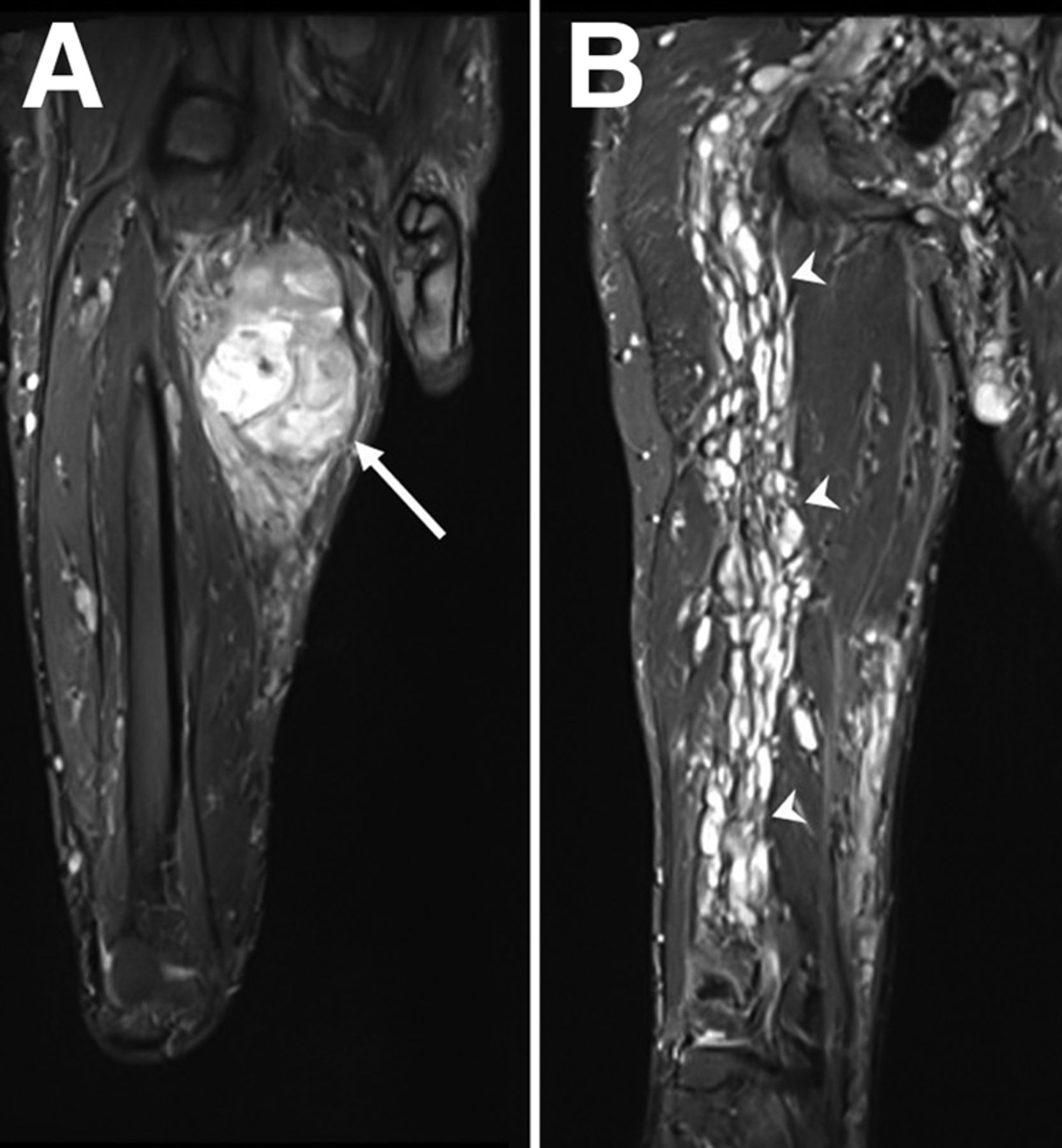
Coronal anterior (A) and posterior (B) MR images of right femur. Heterogeneous, hyperintense lesion (arrow) in right proximal thigh is seen with mild adjacent edema. Additionally, numerous T2-hyperintense neurofibromas are seen along course of sciatic nerve (arrowheads).

Initial 18F-FDG PET/CT images: maximum-intensity projection (A), axial PET (B), axial CT (C), and axial PET/CT (D). Intense tracer activity (white arrows) is seen in linear structure in anterior lower abdomen, without clear bladder morphology. Mild 18F-FDG uptake (red arrows) appears in lower-left-quadrant soft-tissue mass, and intense 18F-FDG uptake (blue arrow) in right proximal thigh corresponds to anterior thigh mass on MRI, indicating malignant transformation of neuromas. Numerous subcutaneous neurofibromas appear in posterior abdominal wall without increased 18F-FDG uptake (arrowheads).

(A–C) Initial staging 18F-FDG PET/CT images: PET (A), CT (B), and PET/CT (C) (coronal images at top and sagittal images at bottom). Intense tracer avidity (arrows) in fluid-density linear structure is seen extending from pelvis to umbilicus, suggesting physiologic tracer excretion into vesicourachal diverticulum. Heterogeneous intense 18F-FDG uptake in right proximal thigh corresponds to soft-tissue mass on MRI, indicating malignant transformation of neuromas. Numerous subcutaneous and intraabdominal neurofibromas are seen without increased 18F-FDG uptake. (D) Postcontrast CT images (coronal images at top and sagittal images at bottom). Nonenhancing fluid-filled structure (arrows) extending from pelvis to umbilicus correlates with intense 18F-FDG avidity on PET/CT, suggesting tracer excretion into vesicourachal diverticulum. Minimal contrast is seen in hypoplastic urinary bladder. Numerous subcutaneous and intraabdominal neurofibromas (arrowheads) are seen.
DISCUSSION
A vesicourachal diverticulum, an outpouching from the bladder to the umbilicus, is often asymptomatic but may present complications such as malignancies or infections. On 18F-FDG PET/CT, it appears as an avid, fluid-filled tract. The association of neurofibromatosis type 1 with the bladder, particularly originating from a urachal mass, is rare (3,4).
Physiologic 18F-FDG excretion in the urinary system results in visualized activity in the kidneys, renal pelvis, and bladder. A patent urachus or vesicourachal diverticulum, continuous with the bladder lumen, can appear 18F-FDG–avid. Distinguishing features from malignancy include the location of the tracer uptake, a concurrent hypoplastic bladder, and the absence of a focal mass. This case highlights the unique appearance of vesicourachal diverticulum on 18F-FDG PET/CT.
CONCLUSION
In 18F-FDG PET/CT disease staging, recognizing metabolically active tissue is crucial for assessing severity. This case emphasizes discerning urogenital anomalies during interpretation, enhancing the distinction between benign anatomic variants and malignancies.
DISCLOSURE
No potential conflict of interest relevant to this article was reported.
Footnotes
Published online Jan. 9, 2024.
- Received for publication August 22, 2023.
- Revision received November 18, 2023.
In this issue


Jump to section
Related Articles
Cited By...
- No citing articles found.