


Abstract
99mTc-sestamibi scintigraphy localizes parathyroid adenoma as a persistent focus of uptake on delayed images, whereas thyroid glands in normal or ectopic locations are seen on only early images and wash out on delayed images. We report a case of absence of eutopic neck thyroid activity and synchronous ectopic lingual thyroid and mediastinal parathyroid adenoma on scintigraphy confirmed with CT.
Though it is not uncommon to have a lingual thyroid or mediastinal parathyroid, it is rare to have a concomitant ectopic thyroid and parathyroid.
CASE REPORT
A 61-y-old euthyroid woman with hyperparathyroidism (parathyroid hormone level, 115 pg/mL) underwent 99mTc-sestamibi scintigraphy after receiving a 1,010.1-MBq (27.3 mCi) tracer injection. The images showed a superior mediastinal focus on SPECT/CT images at 15 min and persisting at 2 h (Figs. 1A and 1B), suggestive of an ectopic parathyroid adenoma and confirmed on 4-dimensional CT (Fig. 1C). Normal neck thyroid uptake was absent, with posterior tongue uptake on early images showing washout on delayed images suggestive of lingual thyroid (Fig. 1A). The ectopic lingual thyroid and absence of native thyroid were confirmed on CT (Fig. 2). The intraoperative parathyroid hormone level dropped to 27.8 pg/mL after transcervical mediastinal parathyroidectomy.

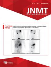
(A) 99mTc-sestamibi SPECT image at 15 min (left) shows uptake at posterior tongue (thick arrow) and mediastinal focus (horizontal thin arrow). SPECT image at 2 h (right) shows posterior tongue tracer washout (thick arrow) suggesting ectopic lingual thyroid, with persistent uptake in mediastinal focus (horizontal thin arrow) consistent with ectopic parathyroid adenoma. Physiologic uptake is seen in salivary glands (oblique arrows). Uptake at anterior tongue region (arrowhead) is likely salivary activity. (B) 99mTc-sestamibi SPECT at 2 h (left) shows focal uptake in anterior superior mediastinum (arrow), with axial CT component of SPECT/CT (right) showing correlating 1.6-cm lesion (arrow) consistent with ectopic parathyroid adenoma. Other peripheral uptake is artifactual activity in muscles. (C) Axial 4-dimensional parathyroid CT scan shows 39 Hounsfield units (HU) within lesion on precontrast phase (left), 161 HU hyperenhancement on arterial phase (middle), and 89 HU washout on delayed phase (right), rendering high confidence for parathyroid adenoma.

(A) Postcontrast axial CT scan shows enhancing lingual thyroid (arrow). (B) Sagittal reformatted postcontrast CT depicts both ectopic lingual thyroid (thin arrow) and ectopic parathyroid adenoma (thick arrow), without any eutopic neck thyroid gland.
DISCUSSION
The thyroid gland originates as an endodermal diverticulum between the first and second pharyngeal pouches, whereas the parathyroid glands develop from the third and fourth pouches (inferior and superior parathyroid, respectively). Ectopic thyroid tissue may rest anywhere along the thyroglossal duct when there is aberrant embryogenesis, as the thyroid descends from the posterior tongue foramen cecum to the lower anterior neck, with the lingual thyroid being most frequent (1,2). Before any excision, thyroid tissue elsewhere should be confirmed to prevent extreme postoperative hypothyroidism.
Superior parathyroid glands are usually located along the posterior margin of the thyroid gland or, less commonly, posteroinferior to the thyroid as a dropped gland and ectopically in retropharyngeal or retroesophageal locations. Inferior parathyroid glands lie anteriorly, in front of the trachea and behind the strap muscles in the lower neck. Inferior parathyroid glands follow the embryologic course of the thymopharyngeal duct, with one fourth lying ectopically in the superior or anterior mediastinum along the thyrothymic ligament, and extremely rarely may lie undescended in the upper neck near the submandibular salivary gland (3).
Combined CT and 99mTc-sestamibi scintigraphy have around 100% sensitivity and a 97% positive predictive value for ectopic parathyroid adenomas. The sensitivity of scintigraphy increases with larger adenomas, likely because of more mitochondria causing high uptake of 99mTc-sestamibi.
CONCLUSION
We report an extremely rare synchronous ectopic thyroid and parathyroid diagnosed with 99mTc-sestamibi scintigraphy and correlative imaging.
DISCLOSURE
No potential conflict of interest relevant to this article was reported.
Footnotes
Published online Jun. 14, 2023.
- Received for publication November 23, 2022.
- Revision received January 31, 2023.
In this issue


Jump to section
Related Articles
Cited By...
- No citing articles found.