
Main menu

  • Home
  • Content
    • Current
      • JNMT Supplement
    • Ahead of print
    • Past Issues
    • Continuing Education
    • JNMT Podcast
    • SNMMI Annual Meeting Abstracts
  • Subscriptions
    • Subscribers
    • Rates
    • Journal Claims
    • Institutional and Non-member
  • Authors
    • Submit to JNMT
    • Information for Authors
    • Assignment of Copyright
    • AQARA Requirements
  • Info
    • Reviewers
    • Permissions
    • Advertisers
    • Corporate & Special Sales
  • About
    • About Us
    • Editorial Board
    • Contact Information
  • More
    • Alerts
    • Feedback
    • Help
    • SNMMI Journals
  • SNMMI
    • JNMT
    • JNM
    • SNMMI Journals
    • SNMMI

User menu

  • Subscribe
  • My alerts
  • Log in
  • My Cart

Search

  • Advanced search
Journal of Nuclear Medicine Technology
  • SNMMI
    • JNMT
    • JNM
    • SNMMI Journals
    • SNMMI
  • Subscribe
  • My alerts
  • Log in
  • My Cart
Journal of Nuclear Medicine Technology

Advanced Search

  • Home
  • Content
    • Current
    • Ahead of print
    • Past Issues
    • Continuing Education
    • JNMT Podcast
    • SNMMI Annual Meeting Abstracts
  • Subscriptions
    • Subscribers
    • Rates
    • Journal Claims
    • Institutional and Non-member
  • Authors
    • Submit to JNMT
    • Information for Authors
    • Assignment of Copyright
    • AQARA Requirements
  • Info
    • Reviewers
    • Permissions
    • Advertisers
    • Corporate & Special Sales
  • About
    • About Us
    • Editorial Board
    • Contact Information
  • More
    • Alerts
    • Feedback
    • Help
    • SNMMI Journals
  • Watch or Listen to JNMT Podcast
  • Visit SNMMI on Facebook
  • Join SNMMI on LinkedIn
  • Follow SNMMI on Twitter
  • Subscribe to JNMT RSS feeds
Research ArticleContinuing Education

Cardiac Amyloidosis Imaging, Part 1: Amyloidosis Etiology and Image Acquisition

Monica Embry-Dierson, Mary Beth Farrell, Eric Schockling, Jaime Warren and Scott Jerome
Journal of Nuclear Medicine Technology June 2023, 51 (2) 83-89; DOI: https://doi.org/10.2967/jnmt.123.265415
Monica Embry-Dierson
1Noninvasive Cardiology, Norton Audubon Hospital, Louisville, Kentucky;
  • Find this author on Google Scholar
  • Find this author on PubMed
  • Search for this author on this site
Mary Beth Farrell
2Intersocietal Accreditation Commission, Ellicott City, Maryland;
  • Find this author on Google Scholar
  • Find this author on PubMed
  • Search for this author on this site
Eric Schockling
3Outpatient Cardiovascular Diagnostics, Norton Healthcare, LLC, Louisville, Kentucky;
  • Find this author on Google Scholar
  • Find this author on PubMed
  • Search for this author on this site
Jaime Warren
4MedAxiom, Neptune Beach, Florida; and
  • Find this author on Google Scholar
  • Find this author on PubMed
  • Search for this author on this site
Scott Jerome
5University of Maryland School of Medicine, Westminster, Maryland
  • Find this author on Google Scholar
  • Find this author on PubMed
  • Search for this author on this site
  • Article
  • Figures & Data
  • Info & Metrics
  • PDF
Loading

Article Figures & Data

Figures

  • Tables
  • FIGURE 1.
    • Download figure
    • Open in new tab
    • Download powerpoint
    FIGURE 1.

    Amyloid molecular mechanisms and imaging characteristics. Source, protein, misfolding, fibril formation, and deposition are depicted for cardiac ATTR and AL. In ATTR (both wild-type and variant), transthyretin proteins are secreted by liver, fold abnormally, and form fibrils that are deposited in myocardium. In AL, immunoglobulin light-chain proteins misfold and form fibrils that are also deposited in myocardium. Echocardiography, CMR, and PET can detect both types of cardiac amyloidosis. However, nuclear imaging with bone-seeking tracers can differentiate between ATTR and AL, although there is evidence to suggest that a percentage of AL will be positive on nuclear imaging. TTR = transthyretin; Wt = wild-type. (Adapted from (2).)

  • FIGURE 2.
    • Download figure
    • Open in new tab
    • Download powerpoint
    FIGURE 2.

    Common types of cardiac amyloidosis. Two kinds of amyloidosis account for approximately 95% of all cardiac amyloidosis cases: AL and ATTR.
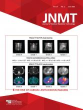
  • FIGURE 3.
    • Download figure
    • Open in new tab
    • Download powerpoint
    FIGURE 3.

    Abnormal planar and SPECT 99mTc-pyrophosphate cardiac amyloidosis scans. (A) 99mTc-pyrophosphate planar anterior image showing avid myocardial uptake. (B) SPECT short-axis (top), vertical long-axis (middle), and horizontal long-axis (bottom) images of same patient showing diffuse myocardial uptake. This scan is considered diagnostic of ATTR cardiomyopathy if serum and urine studies for AL are negative. (Reprinted from (2).)

Tables

  • Figures
    • View popup
    TABLE 1.

    Cardiac Amyloidosis Subtypes and Clinical Characteristics

    SubtypeCauseProteinAge range (y)Sex frequencyFrequency of cardiac involvementOther organ involvement or conditionsAssociated conditions
    ALPlasma cell dyscrasiaImmunoglobulin light chain40–80Men = women70%Heart, kidney, gastrointestinal, tongue, nerves, liver, soft tissueMultiple myeloma
    Wild-type ATTRAgingTransthyretin>70Men > women100%Heart, peripheral nervesBilateral carpal tunnel syndrome, lumbar spinal stenosis, atrial fibrillation, biceps tendon rupture
    Variant ATTRInherited genetic mutationTransthyretin55–75Men > women30%–100% (depending on mutation)Heart, nervesBilateral carpal tunnel syndrome, polyneuropathy
    • View popup
    TABLE 2.

    99mTc-Pyrophosphate Cardiac Amyloidosis Imaging Parameters

    ParameterCharacteristicsStandard/optional/preferred
    Camera typeLarge-field-of-view γ-cameraStandard
    Cadmium zinc tellurideOptional*
    Energy peak140 keVStandard
    Energy window15%–20%Standard
    CollimatorLow-energy, all-purposeStandard
    Patient positionSupineStandard
    Field of viewHeart/chestStandard
    Injection-to-imaging time3 hStandard
    1 hOptional
    Planar
     Acquisition typeStaticStandard
    Whole-body imagingOptional†
     Detector configuration90°Standard
     ViewsAnterior and left lateralStandard
     Number of views2Standard
     Counts per view750,000Standard
     Matrix256 × 256Standard
     Magnification1.46
    SPECT or SPECT/CT*
     Acquisition typeStep and shoot or continuousStandard
     Patient positionSupineStandard
    UprightOptional
     Orbit180°/90°Standard
    360°/180°Optional
     Matrix128 × 128 (minimum, 64 × 64)Standard
     Magnification1.46 (180° orbit)Standard
    1.0 (360° orbit)Optional
     Pixel size2.3–6.5 mmStandard
     Projections per detector40/32Standard
     Time per projection20 s/25 sStandard
     CT attenuation correctionHeartPreferred
    • ↵* Parameters defined for γ-cameras as parameters for cadmium-zinc-telluride cameras have not been firmly established.

    • ↵† Whole-body imaging is not useful when imaging with 99mTc-pyrophosphate. However, whole-body imaging when imaging with 99mTc-HMDP or 99mTc-DPD demonstrates soft-tissue uptake.

PreviousNext
Back to top

In this issue

Journal of Nuclear Medicine Technology: 51 (2)
Journal of Nuclear Medicine Technology
Vol. 51, Issue 2
June 1, 2023
  • Table of Contents
  • About the Cover
  • Index by author
  • Complete Issue (PDF)
Print
Download PDF
Article Alerts
Sign In to Email Alerts with your Email Address
Email Article

Thank you for your interest in spreading the word on Journal of Nuclear Medicine Technology.

NOTE: We only request your email address so that the person you are recommending the page to knows that you wanted them to see it, and that it is not junk mail. We do not capture any email address.

Enter multiple addresses on separate lines or separate them with commas.
Cardiac Amyloidosis Imaging, Part 1: Amyloidosis Etiology and Image Acquisition
(Your Name) has sent you a message from Journal of Nuclear Medicine Technology
(Your Name) thought you would like to see the Journal of Nuclear Medicine Technology web site.
Citation Tools
Cardiac Amyloidosis Imaging, Part 1: Amyloidosis Etiology and Image Acquisition
Monica Embry-Dierson, Mary Beth Farrell, Eric Schockling, Jaime Warren, Scott Jerome
Journal of Nuclear Medicine Technology Jun 2023, 51 (2) 83-89; DOI: 10.2967/jnmt.123.265415

Citation Manager Formats

  • BibTeX
  • Bookends
  • EasyBib
  • EndNote (tagged)
  • EndNote 8 (xml)
  • Medlars
  • Mendeley
  • Papers
  • RefWorks Tagged
  • Ref Manager
  • RIS
  • Zotero
Share
Cardiac Amyloidosis Imaging, Part 1: Amyloidosis Etiology and Image Acquisition
Monica Embry-Dierson, Mary Beth Farrell, Eric Schockling, Jaime Warren, Scott Jerome
Journal of Nuclear Medicine Technology Jun 2023, 51 (2) 83-89; DOI: 10.2967/jnmt.123.265415
Twitter logo Facebook logo LinkedIn logo Mendeley logo
  • Tweet Widget
  • Facebook Like
  • Google Plus One
Bookmark this article

Jump to section

  • Article
    • Abstract
    • WHAT IS AMYLOIDOSIS?
    • TYPES
    • WHAT IS CARDIAC AMYLOIDOSIS?
    • SIGNS AND SYMPTOMS OF CARDIAC AMYLOIDOSIS
    • 99MTC-PYROPHOSPHATE SCAN ACQUISITION
    • SUMMARY
    • Footnotes
    • REFERENCES
  • Figures & Data
  • Info & Metrics
  • PDF

Related Articles

  • No related articles found.
  • PubMed
  • Google Scholar

Cited By...

  • Illuminating the Hidden: Standardizing Cardiac MIBG Imaging for Sympathetic Dysfunction
  • Single- Versus Dual-Time-Point Imaging for Transthyretin Cardiac Amyloid Using 99mTc-Pyrophosphate
  • Lets Meet in the Windy City!
  • Google Scholar

More in this TOC Section

  • Illuminating the Hidden: Standardizing Cardiac MIBG Imaging for Sympathetic Dysfunction
  • PET/CT Case Series: Unmasking the Mystery of Cardiac Sarcoidosis
  • Delivery Methods of Radiopharmaceuticals: Exploring Global Strategies to Minimize Occupational Radiation Exposure
Show more Continuing Education

Similar Articles

Keywords

  • cardiac amyloid imaging
  • cardiac amyloidosis
  • transthyretin
  • light-chain
  • 99mTc-pyrophosphate
SNMMI

© 2025 SNMMI

Powered by HighWire