


Abstract
This report is of an unusual case of radioactive contamination of a γ-camera after scanning 2 individuals who had been treated 3 d beforehand with ablative doses of 131I for thyroid cancer. A combination of observed half-life and pulse-height spectroscopy was used to identify the contaminant. The source of the contamination was eventually found to be a single human hair, presumably contaminated when the individual was sucking her hair while waiting for the scan to start. This case demonstrates that hair can be contaminated by saliva and potentially other bodily fluids in the postablation setting and that using physical measurements, in this case the observed half-life and pulse-height spectroscopy, can be useful in identifying the radioactive contaminant.
The nuclear medicine department in our institution has 3 dual-detector γ-cameras and 1 PET/CT camera. Two of the γ-cameras are SPECT/CT devices. Of these, one has a thicker (16 mm) detector crystal than is standard (Symbia Intevo 6; Siemens Healthineers) and so is preferentially used when imaging higher-energy γ-photons from radionuclides such as 131I (364 keV), 177Lu (208 keV), 67Ga (93, 185, and 300 keV), 67Cu (93 and 185 keV), and 111In (171 and 245 keV). During a routine acquisition of a low-dose 131I scan on this γ-camera on a Monday (the first day of our working week), a low level of contamination was noticed on one of the detectors (Fig. 1). An intrinsic uniformity image acquired using 99mTc as part of routine quality control earlier that day did not show any evidence of contamination. Up to the time that the contamination was noticed, only patients who had been administered 67Ga and 131I had been scanned.
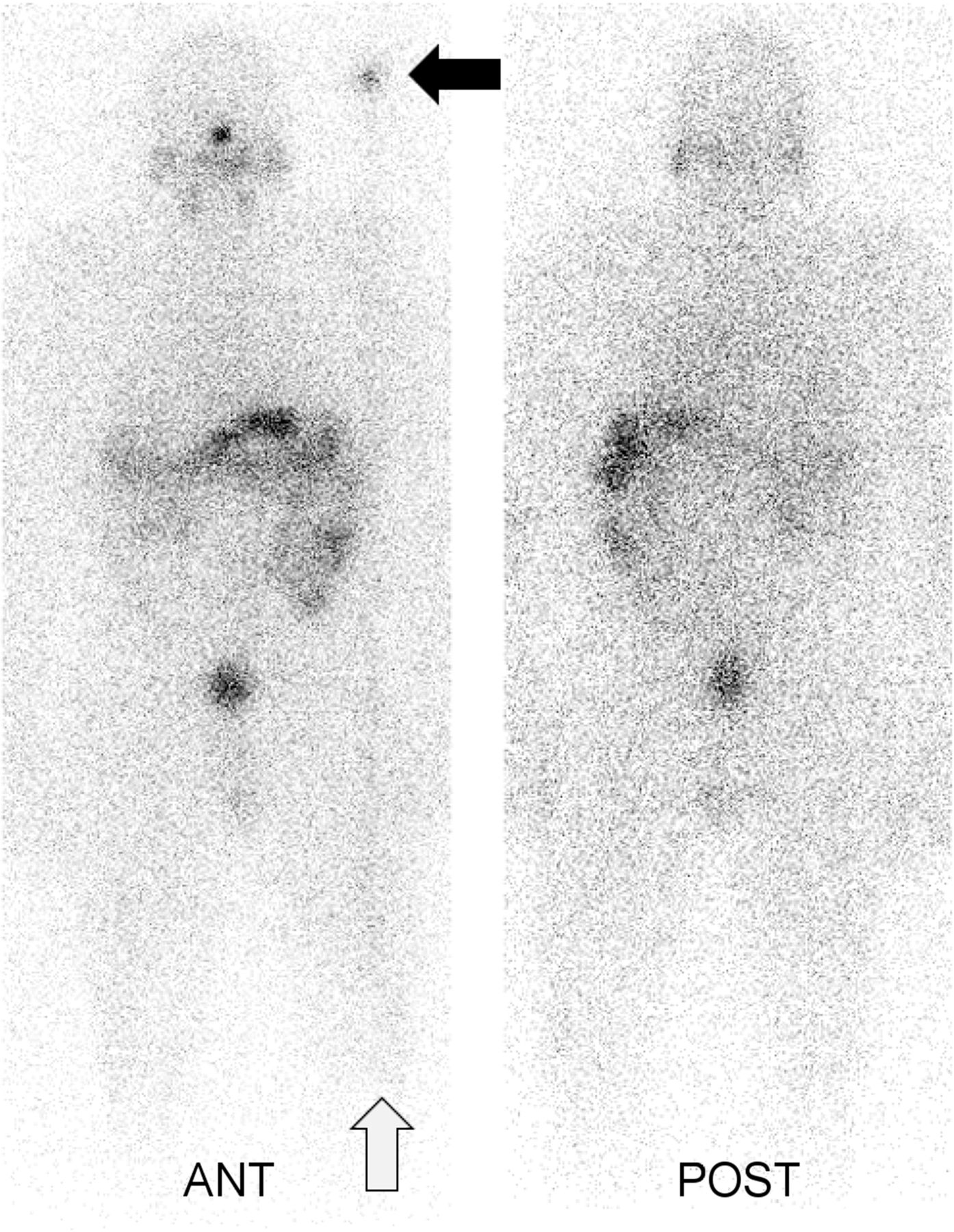
Whole-body planar post–recombinant thyroid-stimulating hormone 131I (80 MBq) scan demonstrating contamination (solid arrow) on anterior projection (detector 1). Effect of fixed site of contamination in whole-body scan where body moves continuously under detector’s z-direction is to introduce streak down image in line with site of contamination (open arrow).
QUALITY ANALYSIS
The system was inspected, and possible sources of the contamination were checked. The contamination remained fixed in location with respect to the detector when the detector heads were rotated to different angular positions, suggesting that the contamination was on the collimator or detector rather than on the scanning bed, the floor, or other nondetector location. The external face of the collimator was thoroughly cleaned with a decontamination solution, but the contamination persisted. The collimators were removed and inspected, but no obvious source of the contamination was identified. The Symbia Intevo 6 SPECT/CT system has a combination of automated collimator exchanger for low- and medium-energy collimators plus an additional manual cart exchanger for high-energy collimators. Because of the persisting contamination, the system was taken out of service for the rest of the day.
The following day, the contamination was still present, suggesting that it was not from a short-lived radionuclide such as 99mTc. Images using 67Ga energy window settings were acquired on both detectors, and the contamination was clearly seen (Fig. 2, top row). Count rates from the previous day’s images were determined and compared with the current ones in an attempt to assess the rate of decay of the contamination. A pulse-height energy spectrum was also acquired with the medium-energy collimators fitted.

Images on both detectors without any source of radioactivity present and collimators in place, demonstrating contamination on detector 1. Images are shown in both summed triple-window pulse-height analyzer settings for 67Ga (top row) and single window for 131I (bottom row).
A region of interest drawn over the area of contamination was corrected for background from an identical region of interest on the same detector in a mirrored location on the images acquired on the successive days. The decline in the count rate suggested a half-life of around 7–8 d. Using the system’s pulse-height analyzer, we found that the energy spectrum demonstrated a slight peak above background at around 360 keV. This finding suggested that the contamination was from 131I. Further images were acquired using an 131I window (Fig. 2, bottom row), which showed the contamination with better definition and a higher count rate.
On the day the contamination was first noticed, the subjects had been scanned after treatment with ablative oral doses of 131I (by capsule) for primary thyroid cancer or had been scanned using 67Ga-citrate because of suspected infection. It is our institution’s practice to hospitalize all subjects treated with at least 1 GBq of 131I. Those subjects who are treated with the largest doses that we deliver (6 GBq) are preferentially admitted to the hospital, treated Friday, and then scanned before discharge Monday morning (∼64 h later). This practice allows for extended decay and elimination of the 131I from the body, compared with our normal practice, which is to scan and discharge the subjects treated with 1–4 GBq of 131I approximately 40 h after their treatment (admitted Monday afternoon for treatment and discharged Wednesday morning, or admitted Wednesday afternoon and discharged Friday morning). On the day in question, postablation 131I scans had been acquired on 2 female subjects, each treated with 6 GBq of 131I on the previous Friday afternoon. No contamination was visible in either of these subjects’ scans, however, presumably because the amount of 131I contained within the body in these individuals remained much higher than in the diagnostic scan, which demonstrated the artifact. However, although the radionuclide had been identified, the source of the contamination was still not identified.
The collimators were removed to allow further inspection. On closer inspection of the detector surface, a single dark human hair was found on the detector (Fig. 3). The hair was removed and placed on the bench, and a handheld radiation survey meter was used to test whether the hair was contaminated. This, in fact, turned out to be the case, as it registered an increase in event rate on the meter and contamination was no longer evident on the γ-camera detector. The contamination therefore had to be from 1 of the 2 131I ablation subjects who had been treated and hospitalized over the weekend. In retrospect, it was noted that the younger of the 2 subjects who had been treated had long, dark hair and was particularly anxious. One of our staff members recalled that while waiting for her scan, she had been sucking the ends of her hair. We therefore presume that the contamination came from saliva from her mouth.

Hair that was found to be source of contamination.
This unusual source of contamination illustrates that saliva on a single human hair was able to contaminate the scanner. It is not clear how the hair came to be lodged between the underside of the collimator and the detector crystal surface.
CORRECTIVE ACTION
There were 3 observations that led to identification of this contamination. First, the fixed relationship between the contamination and the location on the detector—independent of the orientation of the γ-camera detectors—indicated that the contamination was intrinsic to the detector and not from an external source such as the scanning bed or the floor. Second, the observed count rate over the first 2 d (in a 67Ga pulse-height analyzer window) appeared to decrease, with a half-life of around 7 d, which excluded some short-lived tracers such as 99mTc. This count rate also suggested that the contaminant was not 67Ga (half-time, 78 h) and was more likely to be 131I (half time, 8 d). Finally, pulse-height spectroscopy using the NaI(Tl) detector and pulse-height analyzer of the γ-camera showed a faint peak at around 360 keV, again supporting the case for the contaminant to be 131I. Once the identified hair was found and removed, the γ-camera did not display any further contamination.
VERIFICATION OF EFFECTIVENESS
The routine practice in our institution is to instruct all subjects treated with 131I for thyroid cancer to take a shower and wash their hair thoroughly on the morning of the day that they are due to be discharged from the hospital and before they have their postablation 131I scan, as it is known that human hair can become contaminated with 131I (1,2). Our staff considers a possible site of contamination to be a subject’s hair, as well as any site that a subject’s saliva, sebum, or other bodily fluids (e.g., nasal secretions, mucus, and gastric reflux) might be able to contaminate. Using the physical characteristics (half-life, positional orientation, pulse-height analyzer of energy of emissions) of the contamination is also more relied upon now to quickly identify contamination and location. The ability for a contamination source to be located between the collimator and the detector surface is also considered.
The possibility of contamination persistence increases when large amounts of long-lived radiation are used for therapy (3), compared with most diagnostic procedures, which use lower amounts of radionuclides with typically shorter half-lives. This possibility is likely to become more of an issue as the number of radionuclide therapies administered in nuclear medicine departments increases.
CONCLUSION
Contamination of the γ-camera can arise from several causes both external to the scanning system and in the system itself. Early assessment using translation and rotation of the detectors should determine whether the contamination has a fixed geometry relative to the system or is external. Using physical characteristics such as the photon energy of the contamination and the half-life can help to identify the radionuclide and, therefore, the potential cause of the contamination. In this case, a single human hair that had become contaminated with 131I, presumably due to saliva, was able to become lodged between the underside of the collimator and the detector surface.
DISCLOSURE
No potential conflict of interest relevant to this article was reported.
Footnotes
Published online Jun. 14, 2022.
- Received for publication March 23, 2022.
- Revision received May 17, 2022.


