


Article Figures & Data
Figures
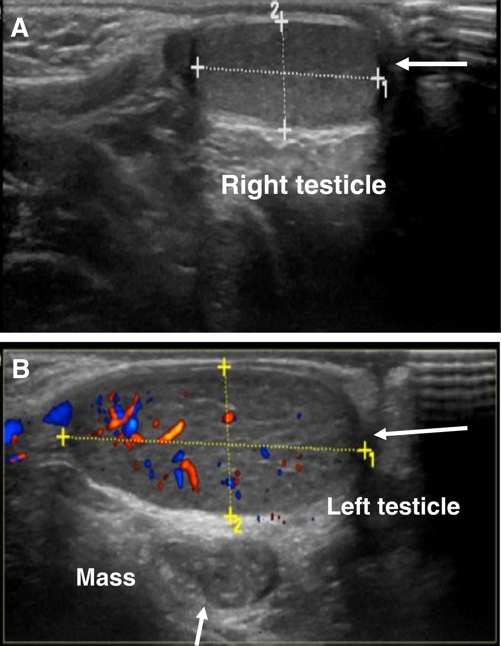
- FIGURE 1.
Ultrasound of scrotum. (A) Right testicle (arrow) measures 1.8 × 0.7 cm and shows normal echogenicity and vascularity. (B) Left testicle (horizontal arrow) measures 1.2 × 0.8 cm and shows normal echogenicity and vascularity. Well-defined, oval abnormal soft-tissue mass (vertical arrow) inseparable from left testis shows homogeneous echogenicity, measures 3 × 1.2 cm, and is adjacent to feeding vessel.
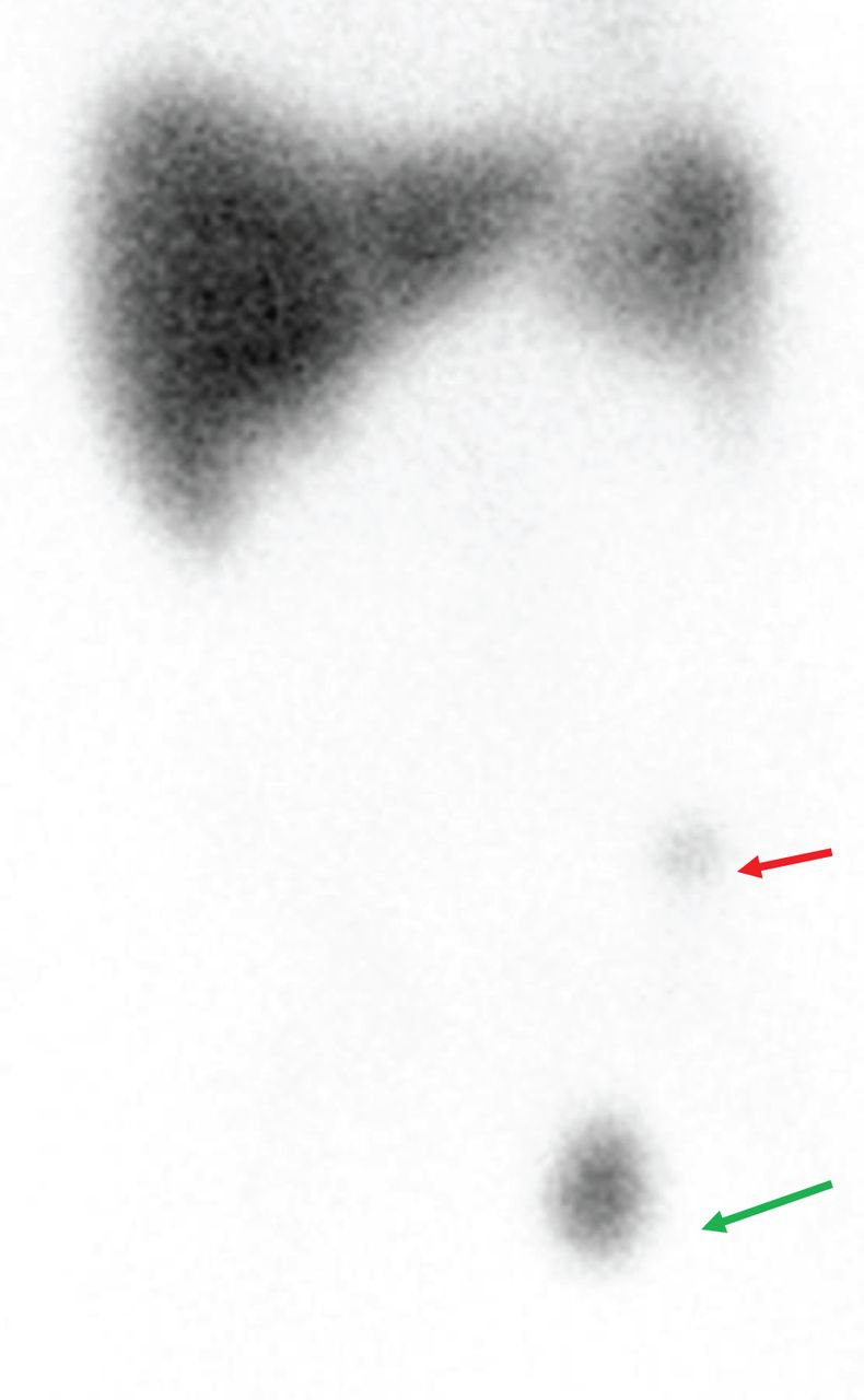
- FIGURE 2.
99mTc-SC image. Anterior planar view of abdomen and pelvis shows normal biodistribution of tracer, with 2 abnormal focal areas of uptake. One (green arrow) is more intense than the other, is in scrotum, measures about 2 × 2 cm, and matches mass noted on previous ultrasound. The other (red arrow) is in anterior part of left iliac fossa.
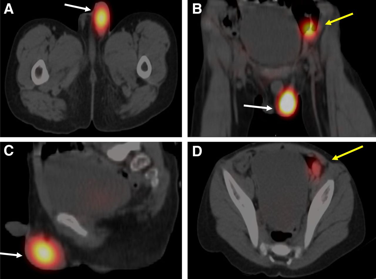
- FIGURE 3.
(A–C) 99mTc-SC SPECT/CT images showing 2 abnormal focal areas of uptake (white arrows) in axial (A), coronal (B), and sagittal (C) views and a second, less active focus (yellow arrow). (D) Axial SPECT/CT image showing that the less active focus (yellow arrow) is smaller (∼1 cm in diameter) than the other focus and is in anterior part of left iliac fossa between abdominal wall and urinary bladder; as such, it is highly suggestive of accessory spleen tissue.


In this issue


Jump to section
Related Articles
Cited By...
- No citing articles found.