


Article Figures & Data
Figures
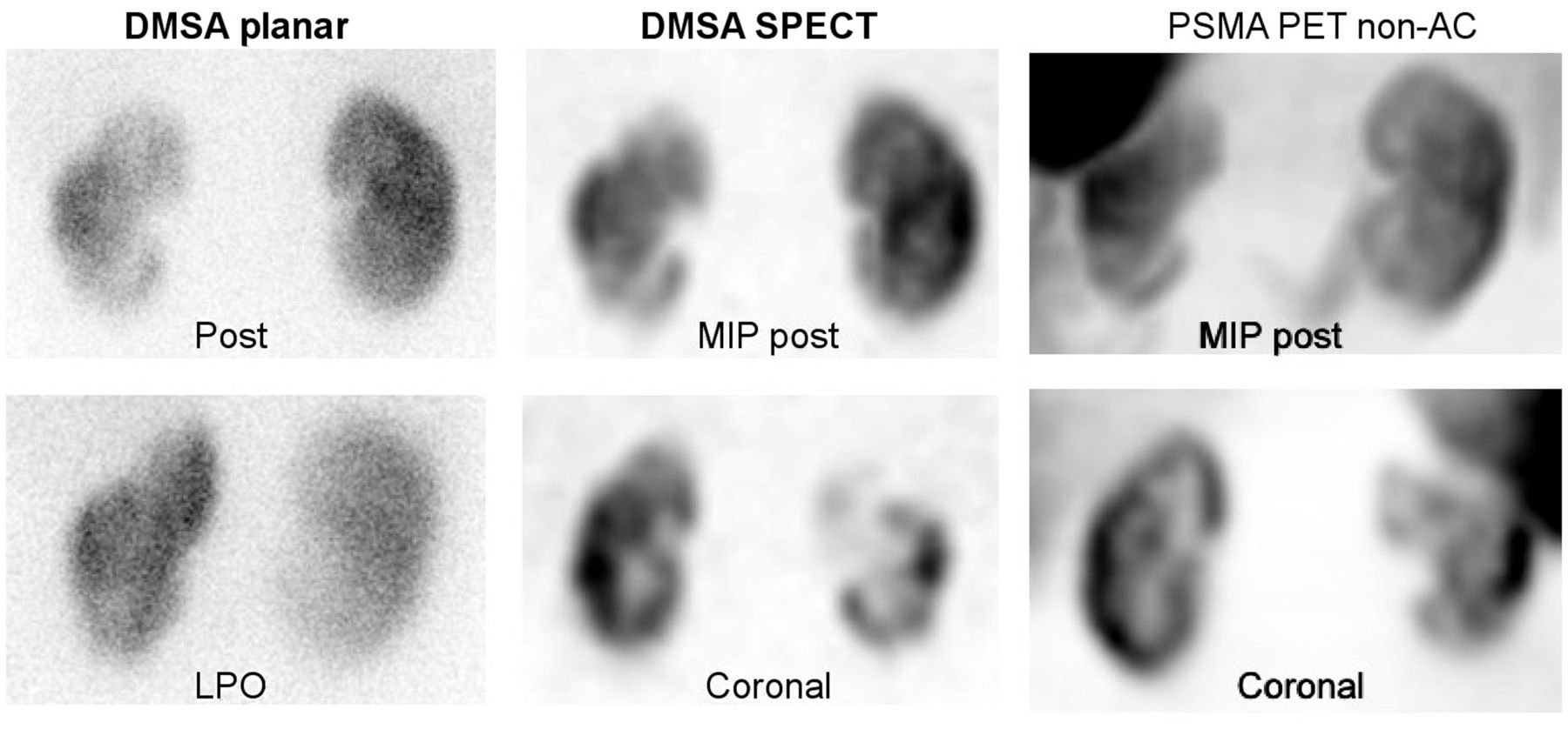
- FIGURE 1.
99mTc-DMSA planar (posterior and left posterior oblique), 99mTc-DMSA SPECT (maximum-intensity projection in posterior view and selected coronal slice), and 68Ga-PSMA-11 (non-AC maximum-intensity projection in posterior view and non-AC selected coronal slice) images demonstrating cortical defects or scars and reduced uptake in upper and lower poles of left kidney. Non-AC PET can be seen to have higher resolution than non-AC SPECT. LPO = left posterior oblique; MIP = maximum-intensity projection.
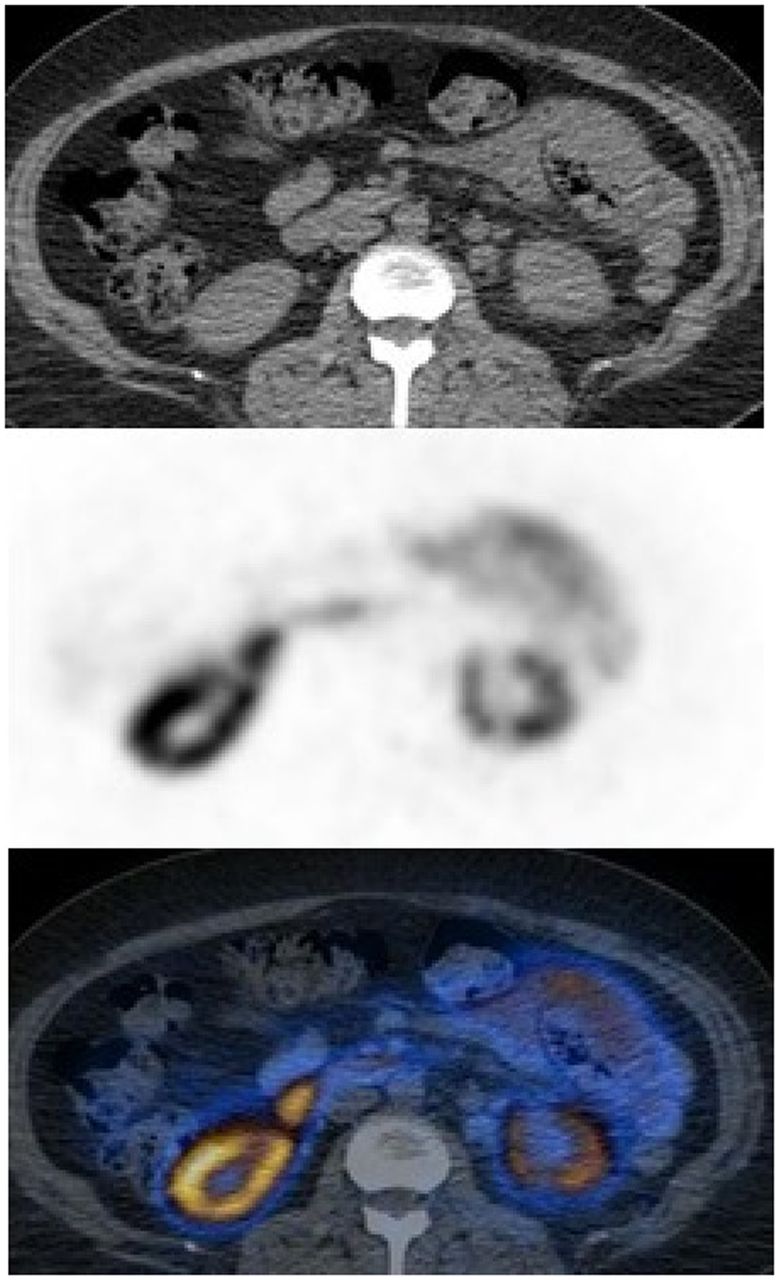
- FIGURE 2.
68Ga-PSMA-11 PET/CT (selected transaxial CT [top], PET [middle], and AC PET/CT [bottom]) images demonstrating reduced uptake in lower pole of left kidney and small cortical defect.
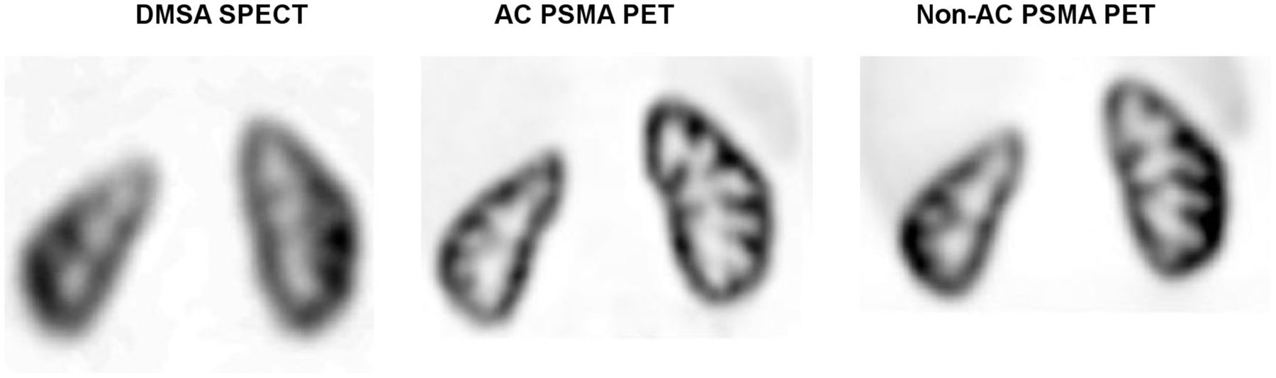
- FIGURE 3.
99mTc-DMSA SPECT (selected coronal slice) and 68Ga-PSMA-11 PET (AC and non-AC selected coronal slices) images of another patient with history of chronic recurrent pyelonephritis demonstrating mildly reduced uptake and cortical thinning in upper pole of right kidney with no parenchymal defects. Normal distribution of 68Ga-PSMA-11 is seen, with only mild activity in liver and spleen.


