
Main menu

  • Home
  • Content
    • Current
      • JNMT Supplement
    • Ahead of print
    • Past Issues
    • Continuing Education
    • JNMT Podcast
    • SNMMI Annual Meeting Abstracts
  • Subscriptions
    • Subscribers
    • Rates
    • Journal Claims
    • Institutional and Non-member
  • Authors
    • Submit to JNMT
    • Information for Authors
    • Assignment of Copyright
    • AQARA Requirements
  • Info
    • Reviewers
    • Permissions
    • Advertisers
    • Corporate & Special Sales
  • About
    • About Us
    • Editorial Board
    • Contact Information
  • More
    • Alerts
    • Feedback
    • Help
    • SNMMI Journals
  • SNMMI
    • JNMT
    • JNM
    • SNMMI Journals
    • SNMMI

User menu

  • Subscribe
  • My alerts
  • Log in
  • My Cart

Search

  • Advanced search
Journal of Nuclear Medicine Technology
  • SNMMI
    • JNMT
    • JNM
    • SNMMI Journals
    • SNMMI
  • Subscribe
  • My alerts
  • Log in
  • My Cart
Journal of Nuclear Medicine Technology

Advanced Search

  • Home
  • Content
    • Current
    • Ahead of print
    • Past Issues
    • Continuing Education
    • JNMT Podcast
    • SNMMI Annual Meeting Abstracts
  • Subscriptions
    • Subscribers
    • Rates
    • Journal Claims
    • Institutional and Non-member
  • Authors
    • Submit to JNMT
    • Information for Authors
    • Assignment of Copyright
    • AQARA Requirements
  • Info
    • Reviewers
    • Permissions
    • Advertisers
    • Corporate & Special Sales
  • About
    • About Us
    • Editorial Board
    • Contact Information
  • More
    • Alerts
    • Feedback
    • Help
    • SNMMI Journals
  • Watch or Listen to JNMT Podcast
  • Visit SNMMI on Facebook
  • Join SNMMI on LinkedIn
  • Follow SNMMI on Twitter
  • Subscribe to JNMT RSS feeds
Research ArticleContinuing Education

Brain Imaging Techniques: Improving the Quality

Rani M. Noble
Journal of Nuclear Medicine Technology September 2021, 49 (3) 209-214; DOI: https://doi.org/10.2967/jnmt.120.257592
Rani M. Noble
Nuclear Medicine Department, HonorHealth, Phoenix, Arizona
CNMT, RT(N)
  • Find this author on Google Scholar
  • Find this author on PubMed
  • Search for this author on this site
  • Article
  • Figures & Data
  • Info & Metrics
  • PDF
Loading

Article Figures & Data

Figures

  • FIGURE 1.
    • Download figure
    • Open in new tab
    • Download powerpoint
    FIGURE 1.

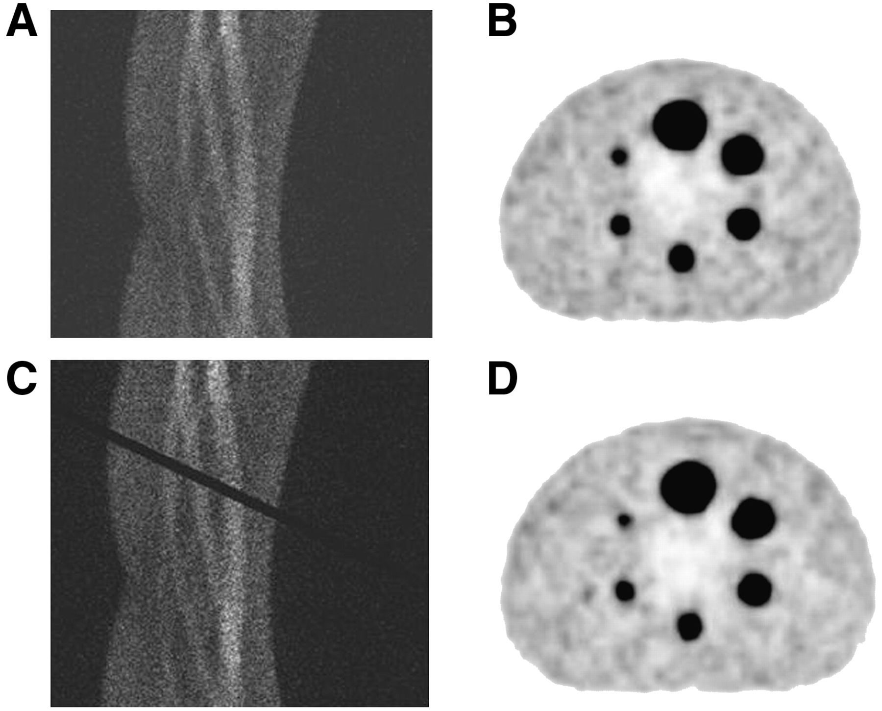
    Two-dimensional PET sinograms (A and B) and corresponding reconstructed transverse PET images (C and D, respectively) of 18F-filled phantom. Black band in C (suggestive of faulty detector) is virtually undetectable in D (11).

  • FIGURE 2.
    • Download figure
    • Open in new tab
    • Download powerpoint
    FIGURE 2.

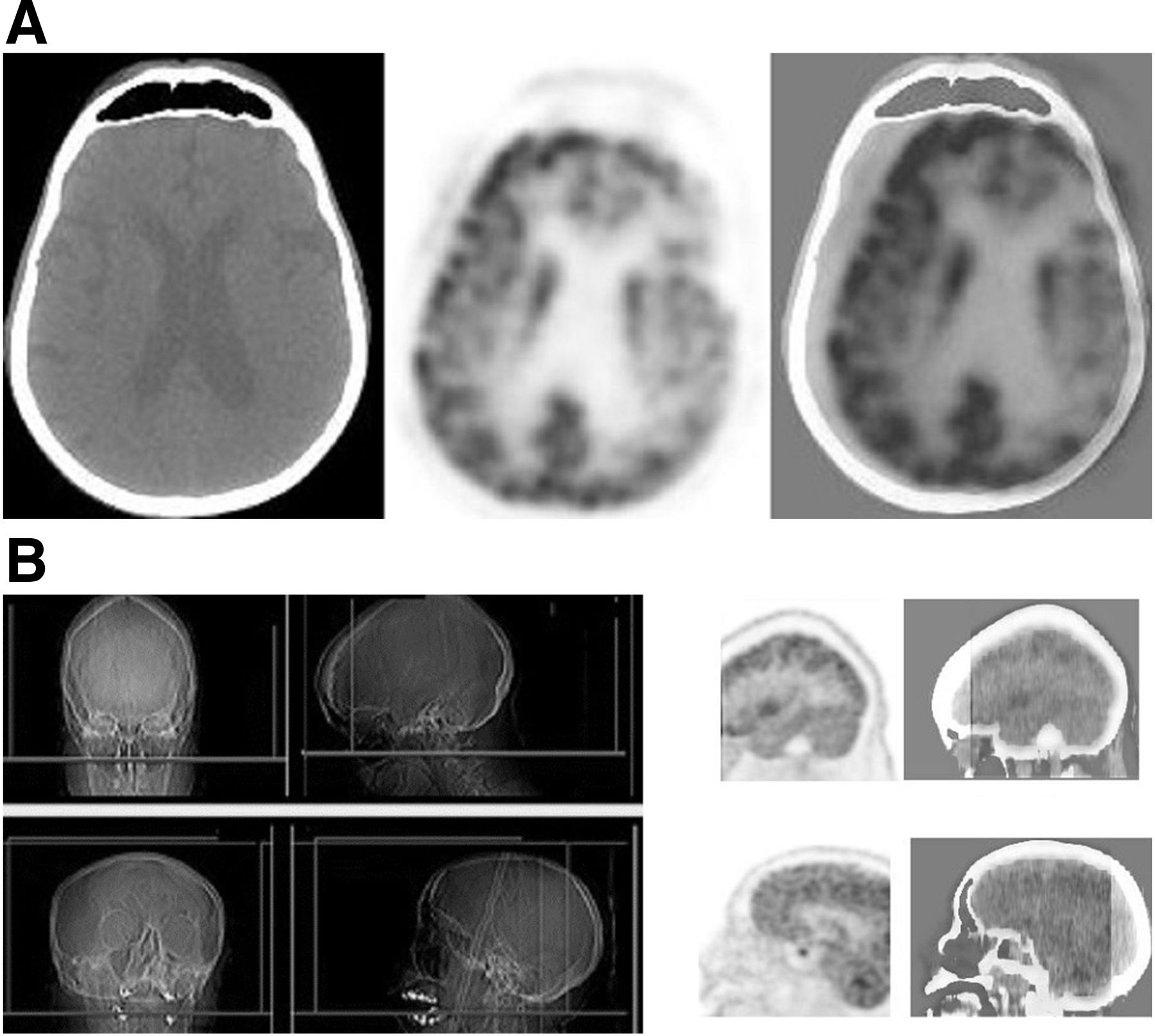
    (A) Brain 18F-FDG PET/CT images with technologist-caused motion artifact due to misplacing of region-of-interest box on scout images. (B) Transaxial and sagittal lateral radiographs (left), PET images (middle), and PET/CT images (right) of head. Artifact causes false reduction in uptake in left hemicortex and results in part of brain being cut off in final images (12).

  • FIGURE 3.
    • Download figure
    • Open in new tab
    • Download powerpoint
    FIGURE 3.

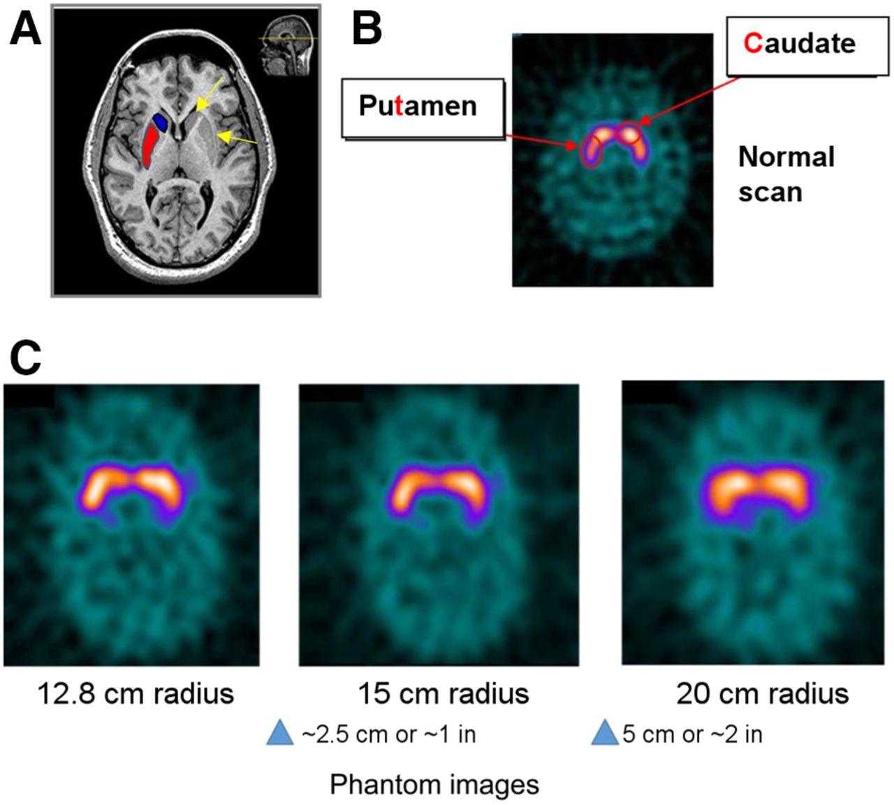
    (A and B) brain MRI (A) and PET (B) scans showing location of caudate nucleus and putamen within brain. These are main focus of 123I-ioflupane scans and need to be fully included in images. (C) PET phantom scans showing how important it is to keep detector radius within 11- to 15-cm range. More distance between patient and detector will increase blurring (9). Arrows in A point to left caudate and left putamen; blue indicates right caudate and red indicates location of right putamen. Scans in A and B are of a patient.
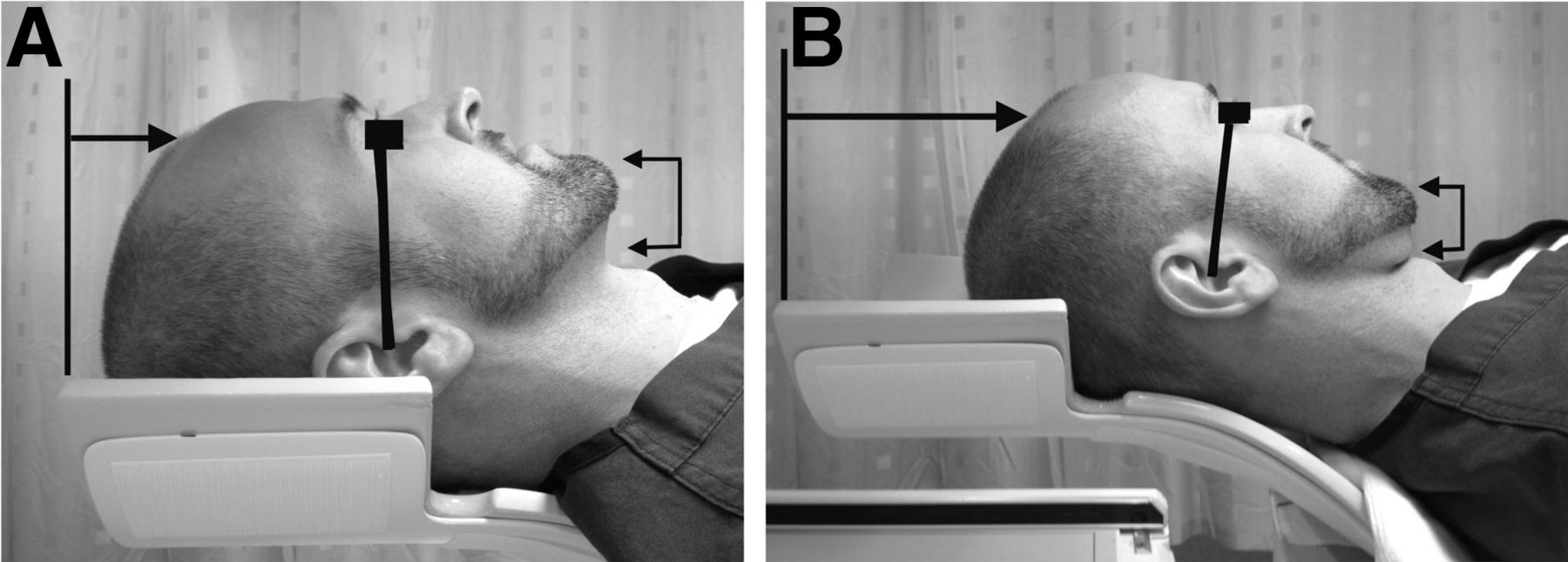
  • FIGURE 4.
    • Download figure
    • Open in new tab
    • Download powerpoint
    FIGURE 4.

    (A) Correct positioning of head during brain imaging. Canthomeatal line should be positioned as perpendicularly to imaging table as possible to eliminate head tilt and resulting artifacts. (B) Incorrect head positioning. Patient’s head is tilted and not placed fully in head holder (13).
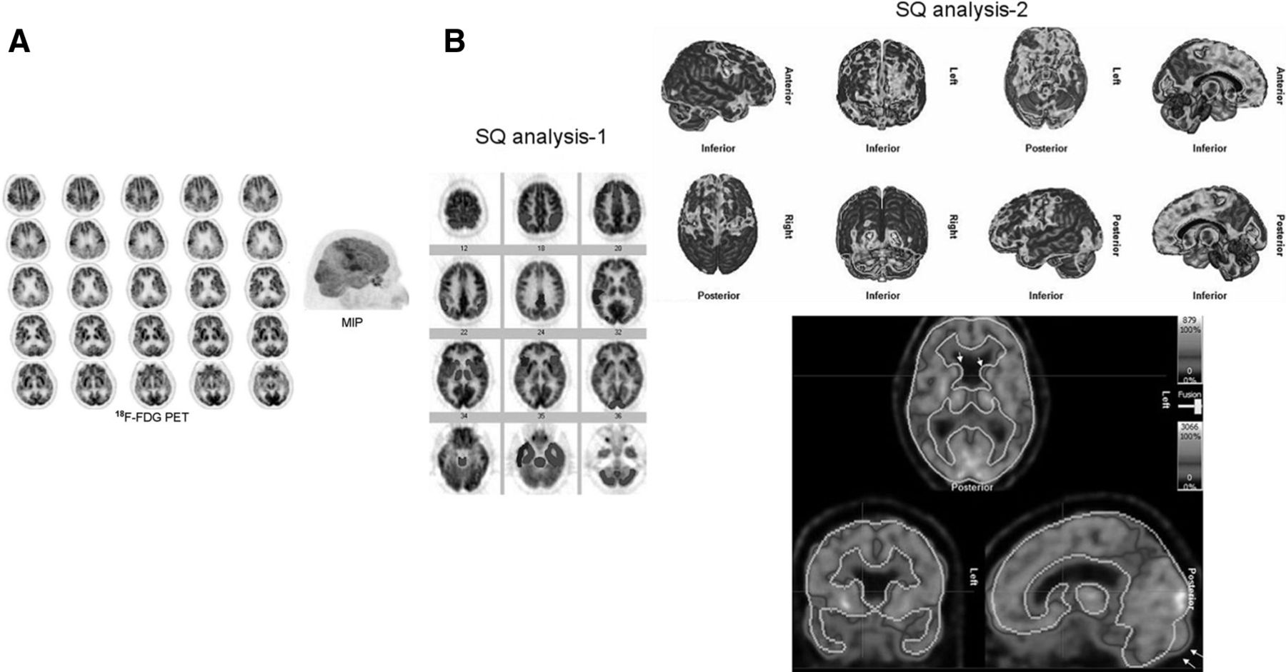
  • FIGURE 5.
    • Download figure
    • Open in new tab
    • Download powerpoint
    FIGURE 5.

    18F-FDG PET brain images showing bilateral hypometabolism in temporal, parietal, and frontal lobes, and maximum-intensity projection (MIP) showing cerebral cortical hypometabolism localization. (B) Semiquantitative (SQ) analysis showing reduction in metabolism in patient’s frontal and temporal lobes and in posterior cingulate cortices and temporoparietal junctions. Semiquantitative analysis, however, does not show hypometabolism in patient’s parietal lobes and undervalues reduction of metabolism in left temporal lobe (12). In B, SQ1 uses NeuroQ software; it presents reduced metabolism in bilateral frontal lobes, posterior cingulate cortices, temporoparietal junctions, bilateral temporal lobes with right being greater than left, and mildly in left basal ganglia. SQ2 uses Hermes BRASS software. This software furnishes greater matching results to visual analysis in cerebral cortex. This does not apply to basal ganglia, however. This program exhibits inferior registration of various parts of brain, which includes caudate heads.
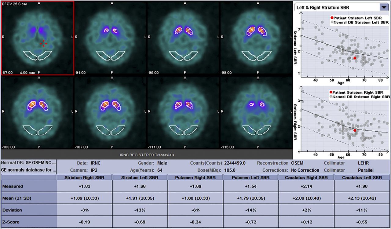
  • FIGURE 6.
    • Download figure
    • Open in new tab
    • Download powerpoint
    FIGURE 6.

    Screenshot showing results of DaTQUANT, semiquantitative analysis software used for 123I-ioflupane scans. Scans show how results would be displayed with ROIs placed (14). A = anterior; DB = database; IP2 = type of operating system in Siemens cameras, computer software; IRNC = iterative reconstruction without attenuation correction; LEHR = low-energy high-resolution; NC = normal control; OFOV = operational field of view; OSEM = ordered-subsets expectation maximization; P = posterior; SBR = striatal binding ratio.

PreviousNext
Back to top

In this issue

Journal of Nuclear Medicine Technology: 49 (3)
Journal of Nuclear Medicine Technology
Vol. 49, Issue 3
September 1, 2021
  • Table of Contents
  • About the Cover
  • Index by author
  • Complete Issue (PDF)
Print
Download PDF
Article Alerts
Sign In to Email Alerts with your Email Address
Email Article

Thank you for your interest in spreading the word on Journal of Nuclear Medicine Technology.

NOTE: We only request your email address so that the person you are recommending the page to knows that you wanted them to see it, and that it is not junk mail. We do not capture any email address.

Enter multiple addresses on separate lines or separate them with commas.
Brain Imaging Techniques: Improving the Quality
(Your Name) has sent you a message from Journal of Nuclear Medicine Technology
(Your Name) thought you would like to see the Journal of Nuclear Medicine Technology web site.
Citation Tools
Brain Imaging Techniques: Improving the Quality
Rani M. Noble
Journal of Nuclear Medicine Technology Sep 2021, 49 (3) 209-214; DOI: 10.2967/jnmt.120.257592

Citation Manager Formats

  • BibTeX
  • Bookends
  • EasyBib
  • EndNote (tagged)
  • EndNote 8 (xml)
  • Medlars
  • Mendeley
  • Papers
  • RefWorks Tagged
  • Ref Manager
  • RIS
  • Zotero
Share
Brain Imaging Techniques: Improving the Quality
Rani M. Noble
Journal of Nuclear Medicine Technology Sep 2021, 49 (3) 209-214; DOI: 10.2967/jnmt.120.257592
Twitter logo Facebook logo LinkedIn logo Mendeley logo
  • Tweet Widget
  • Facebook Like
  • Google Plus One
Bookmark this article

Jump to section

  • Article
    • Abstract
    • DEFINITION OF A QUALITY IMAGE
    • PATIENT PREPARATION
    • MECHANICAL ARTIFACTS
    • TECHNOLOGIST-TRIGGERED ARTIFACTS
    • PATIENT POSITIONING
    • PRECAUTIONS
    • IMAGE INTERPRETATION
    • CONCLUSION
    • ACKNOWLEDGMENT
    • Footnotes
    • REFERENCES
  • Figures & Data
  • Info & Metrics
  • PDF

Related Articles

  • No related articles found.
  • PubMed
  • Google Scholar

Cited By...

  • Once Again--The SNMMI "Nailed It"!
  • Google Scholar

More in this TOC Section

  • Illuminating the Hidden: Standardizing Cardiac MIBG Imaging for Sympathetic Dysfunction
  • PET/CT Case Series: Unmasking the Mystery of Cardiac Sarcoidosis
  • Delivery Methods of Radiopharmaceuticals: Exploring Global Strategies to Minimize Occupational Radiation Exposure
Show more Continuing Education

Similar Articles

Keywords

  • quality
  • brain
  • techniques
  • artifacts
SNMMI

© 2025 SNMMI

Powered by HighWire