


Article Figures & Data
Figures
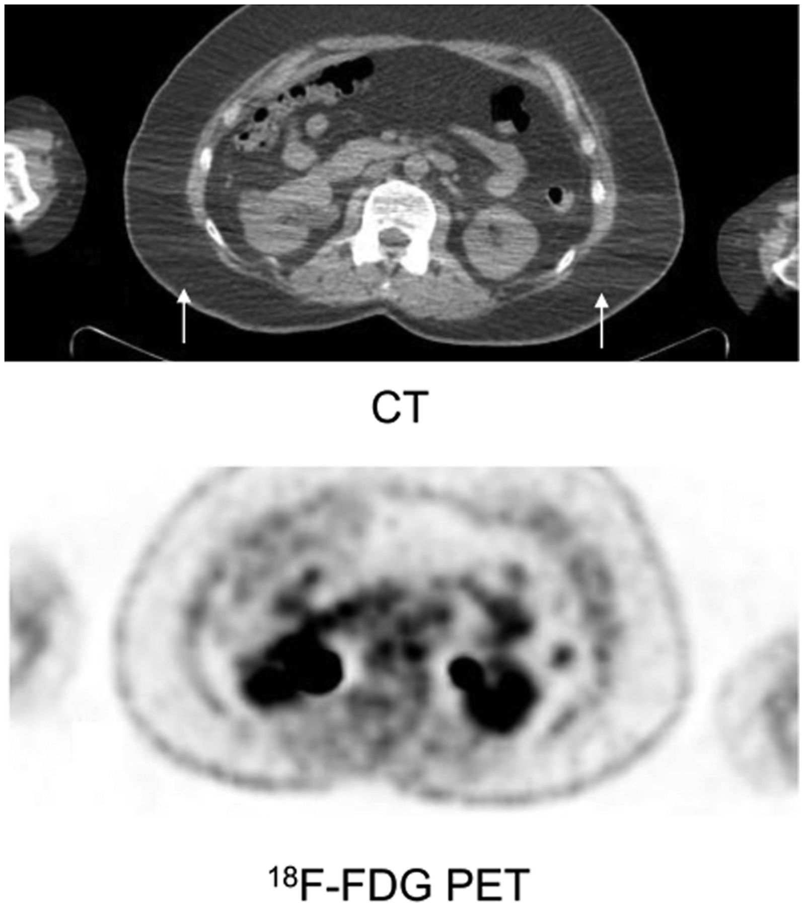
- FIGURE 1.
Selected transaxial CT image shows beam-hardening artifact between arms (arrows), with no artifact seen on PET image.
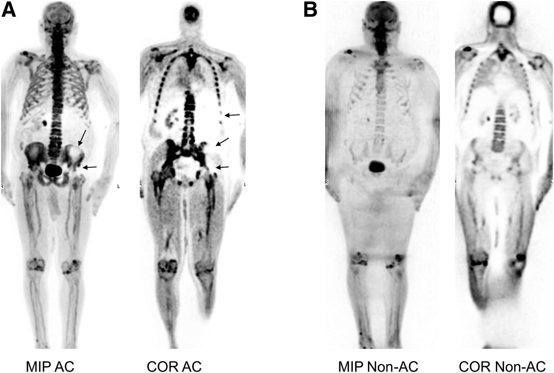
- FIGURE 2.
Whole-body coronal (COR) and maximum-intensity projection (MIP) AC 18F-NaF PET/CT images of large patient who was imaged with arms down. (A) AC PET images demonstrate large cold area (cold artifact) on left side of abdomen and pelvis, with reduced uptake in bones (left lower ribs, hemipelvis, and hip) and soft tissues (left kidney). Left arm is farther from body than right arm. (B) This cold artifact is not seen on non-AC PET images.
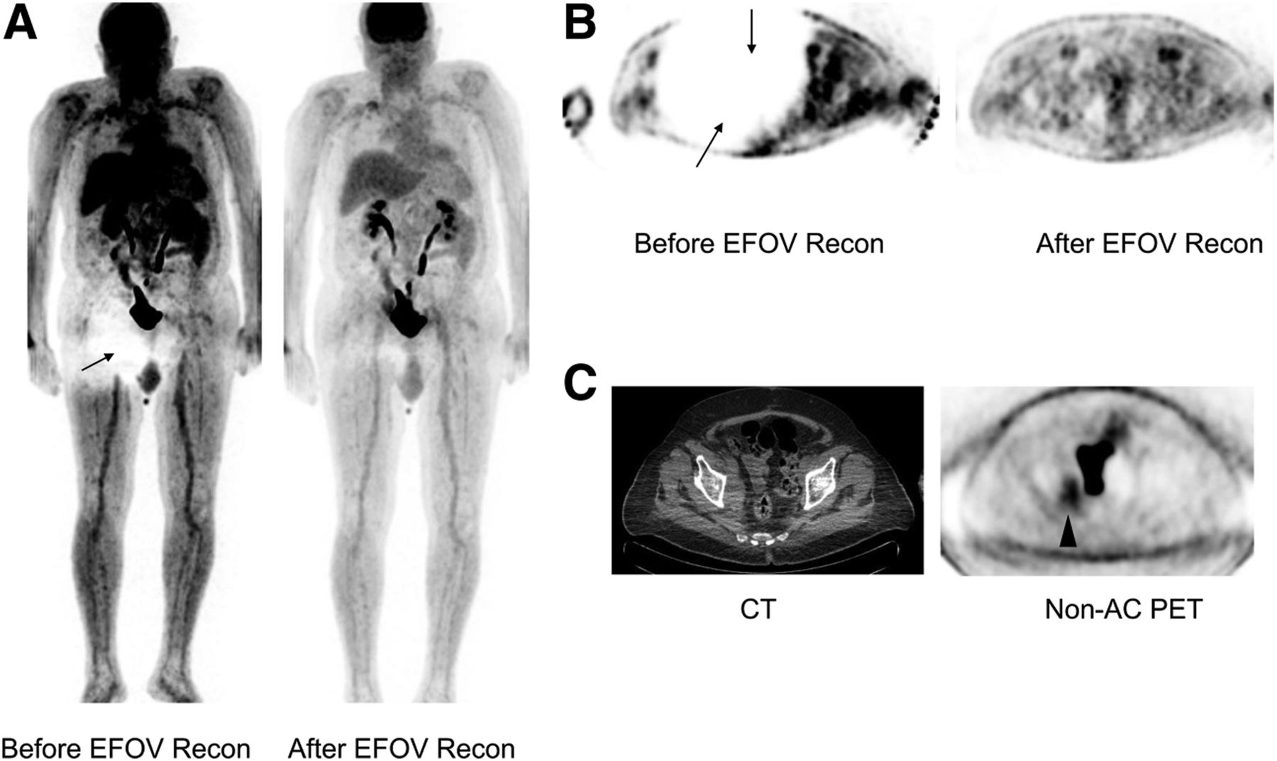
- FIGURE 3.
(A and B) Whole-body maximum-intensity projection (A) and transaxial pelvic (B) 18F-FDG PET AC images before and after extended-FOV reconstruction (EFOV recon). Cold artifact in right hip and pelvis (arrows) disappears after extended FOV reconstruction. Right arm is farther from body than left arm. (C) Cold artifact is also not present on non-AC PET image. Right pelvic hypermetabolic mass (arrowhead) on transaxial CT and non-AC PET images was missed on AC PET image because of cold artifact. (Note that images in B and C are not from the same level.)
- FIGURE 4.
Whole-body coronal CT (left and top) and AC 18F-NaF PET (right and bottom) images of large patient. Cold artifact (arrows) is seen in hips (more significant on right side) on PET image obtained with arms down. Right arm is farther from body than left arm. This cold defect is not seen after repeating PET/CT with arms up. Note the enlarged prostate elevating bladder (arrowhead).
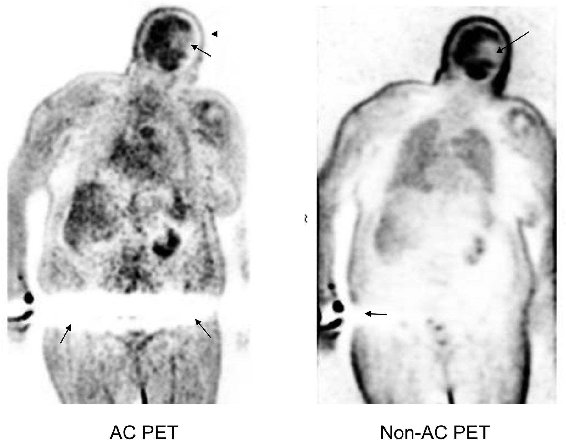
- FIGURE 5.
Whole-body coronal AC 18F-FDG PET image demonstrates linear cold artifact (bottom arrows) in pelvis of large patient imaged with arms down. This artifact is not present on non-AC PET image. Small cold artifact (bottom arrow) in skin adjacent to right arm on non-AC PET image is scatter-correction artifact due to activity extravasation at injection site. Defect on left side of brain (arrows) on both AC and non-AC images was caused by cerebral infarct (seen on CT image; not shown). Reduced uptake in scalp (arrowhead), seen on AC image only, was caused by head motion after acquiring CT scan.
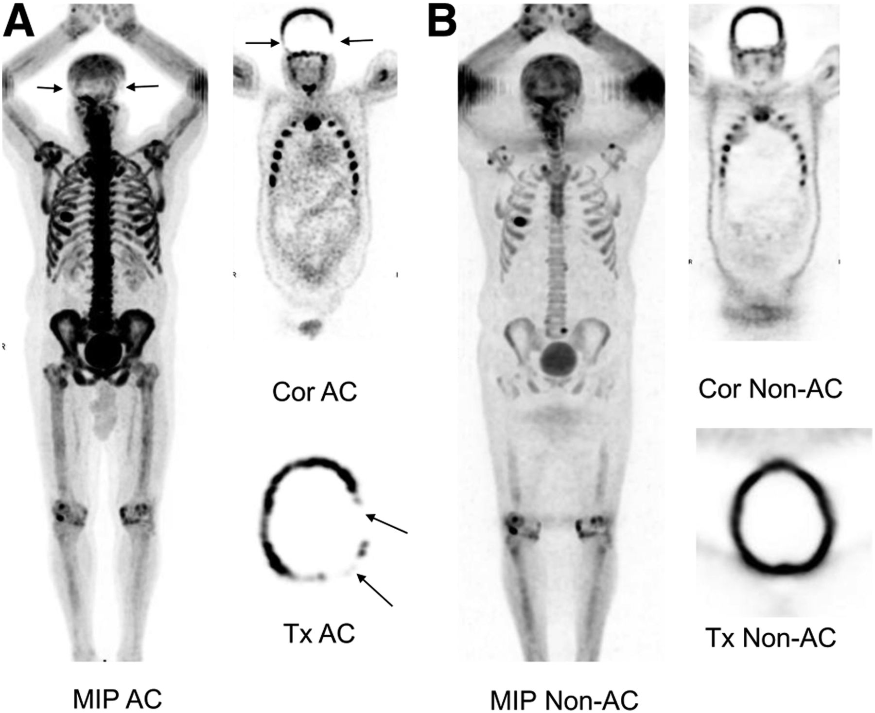
- FIGURE 6.
(A) Whole-body maximum-intensity projection (MIP) and coronal (Cor) and transaxial (Tx) AC 18F-NaF PET images demonstrate linear cold artifact in skull, more prominent on left (arrows). (B) Artifact is not present on non-AC images.
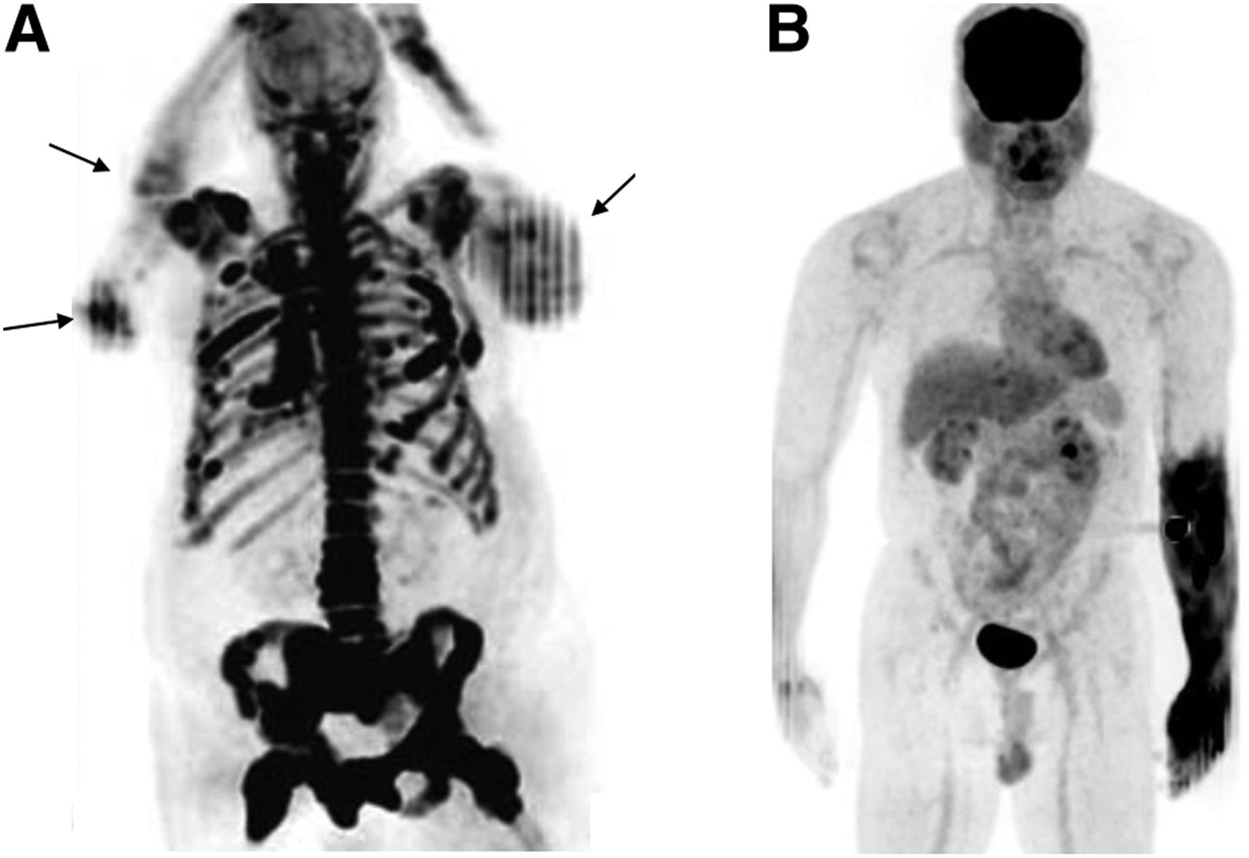
- FIGURE 7.
(A) 18F-NaF PET maximum-intensity projection (MIP) image demonstrates arm motion artifacts bilaterally (arrows). (B) Accidental intraarterial injection of radiotracer caused diffusely increased uptake in tissues of left forearm and hand seen on 18F-FDG PET maximum-intensity projection image.


