


Article Figures & Data
Figures
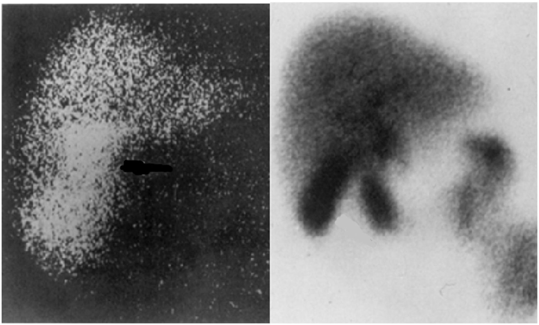
- FIGURE 1.
Early 123I-rose bengal images (left) compared to 99mTc-HIDA images (right). This image was originally published in JNMT. Ziessman. Hepatobiliary Scintigraphy. J Nucl Med Technol. 2014;42:250. © SNMMI.
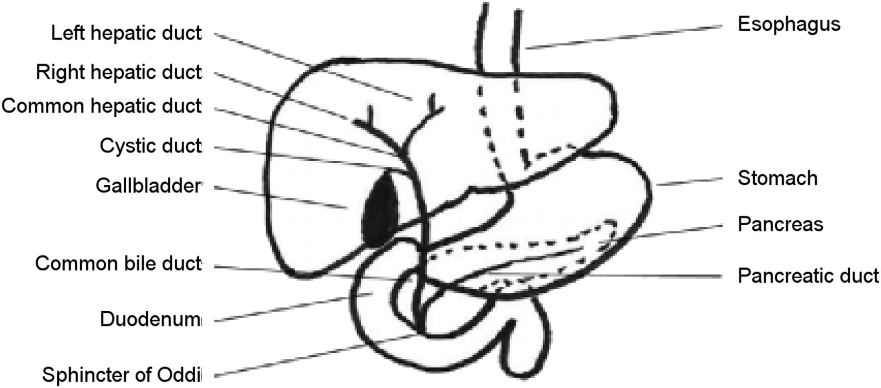
- FIGURE 2.
Diagram of the hepatobiliary system.
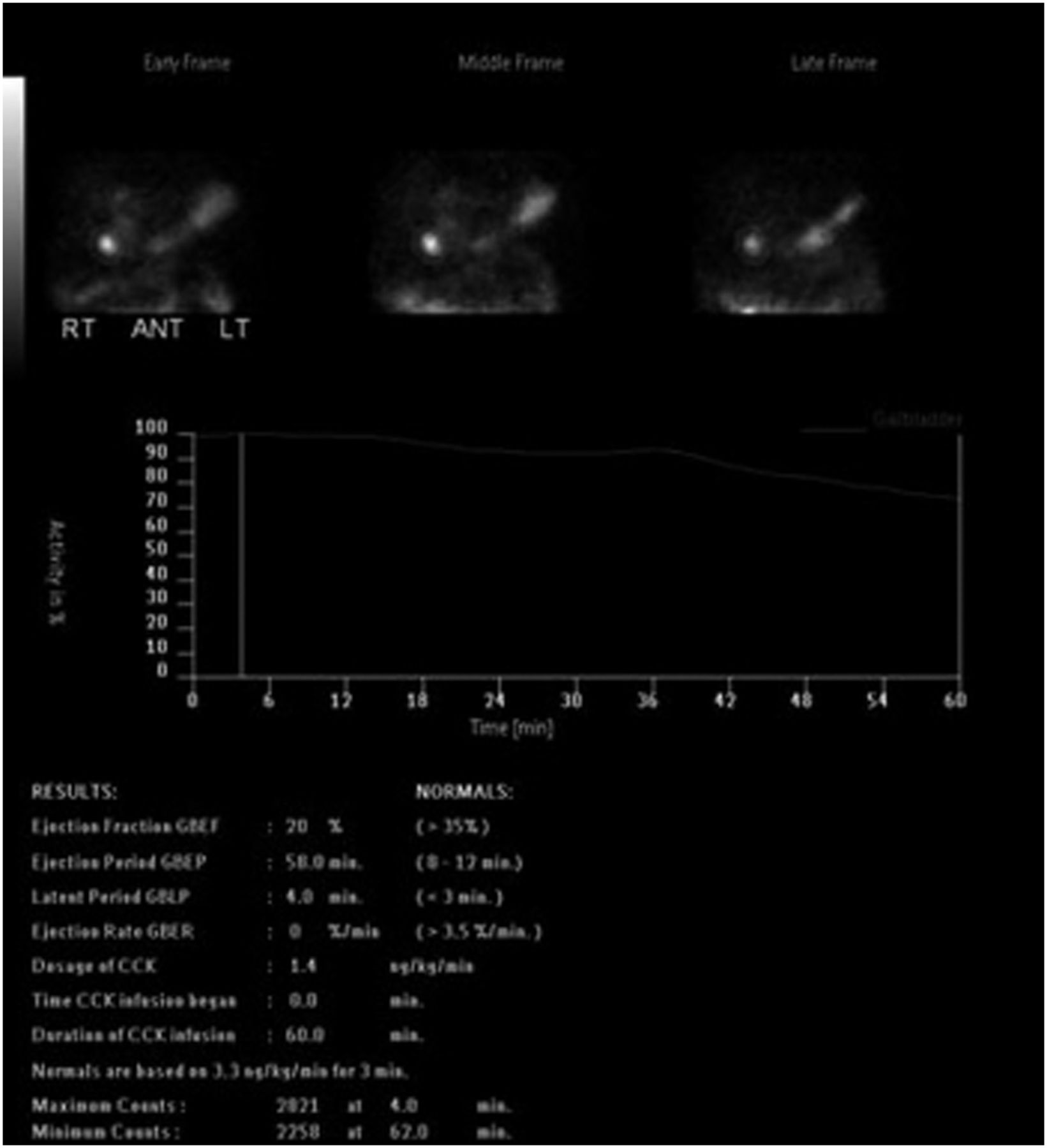
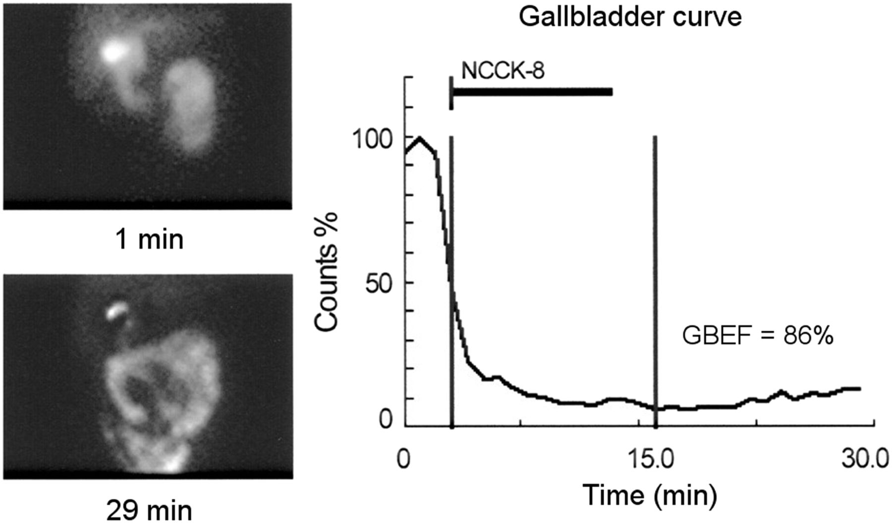
- FIGURE 3.
Normal gallbladder ejection fraction greater than 35% following the administration of CCK. This image was originally published in JNM. Krishnamurthy S. Comparison of Gallbladder Function Obtained with Regular CCK-8 and Pharmacy-Compounded CCK-8. J Nucl Med. 2008;44(4);499–504. © SNMMI.
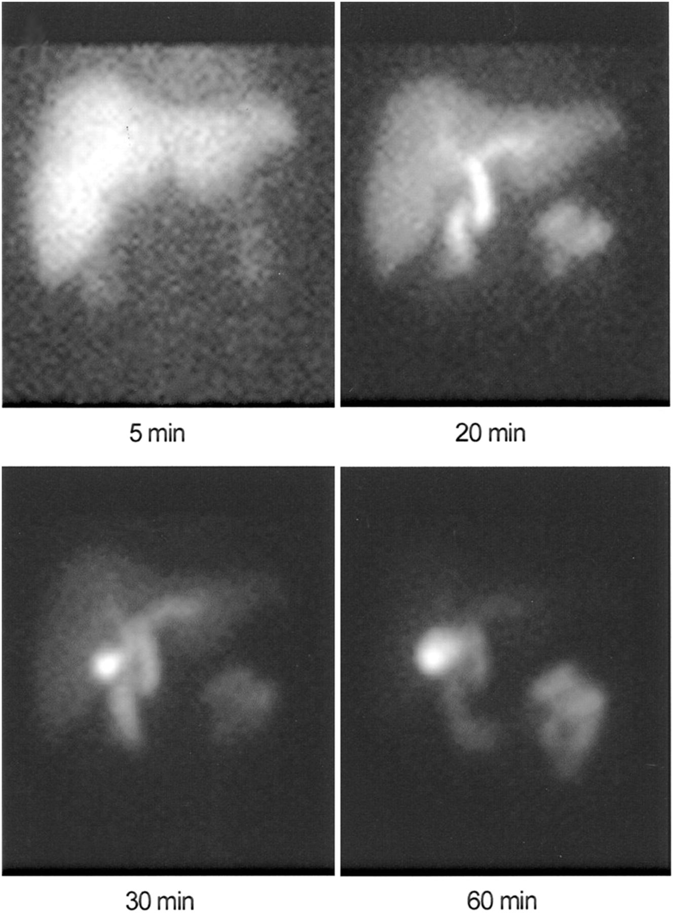
- FIGURE 4.
Normal hepatobiliary imaging demonstrating immediate uptake of the tracer within the liver and appearance of the gallbladder by 30 minutes. The right and left hepatic ducts, common hepatic duct, and common bile duct are visualized. There is visualization of tracer within the gastrointestinal tract within 30 minutes. This image was originally published in JNM. Krishnamurthy S. Comparison of Gallbladder Function Obtained with Regular CCK-8 and Pharmacy-Compounded CCK-8. J Nucl Med. 2008;44(4);499–504. © SNMMI.
- FIGURE 5.
Acute cholecystitis.
- FIGURE 6.
Chronic cholecystitis.
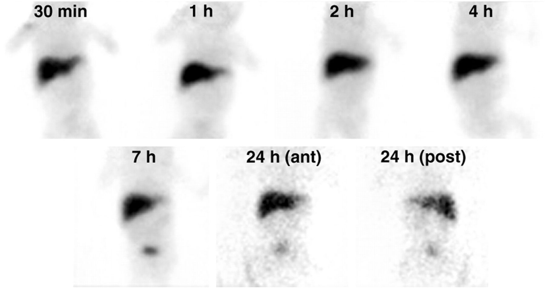
- FIGURE 7.
Hepatobiliary imaging of a 45-day-old infant with jaundice using 99mTc-mebrofenin. The images demonstrate good uptake of the tracer in the liver and bowel and nonvisualization of the biliary tree and intestines. This image was originally published in JNM. Poddar U. Ursodeoxycholic Acid—Augmented Hepatobiliary Scintigraphy in the Evaluation of Neonatal Jaundice. J Nucl Med. 2004;45(9):1488. © SNMMI.
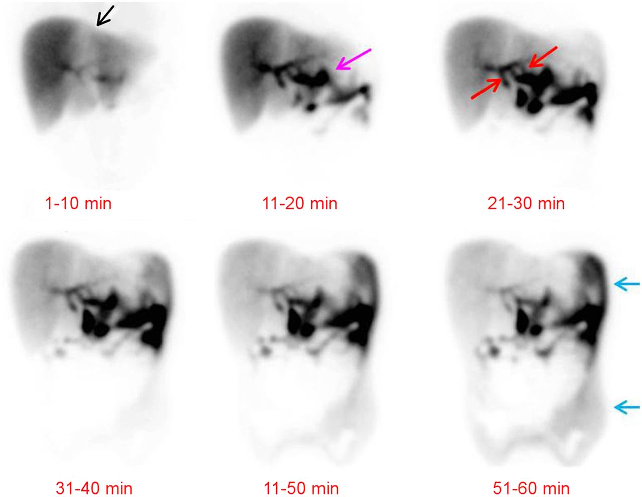
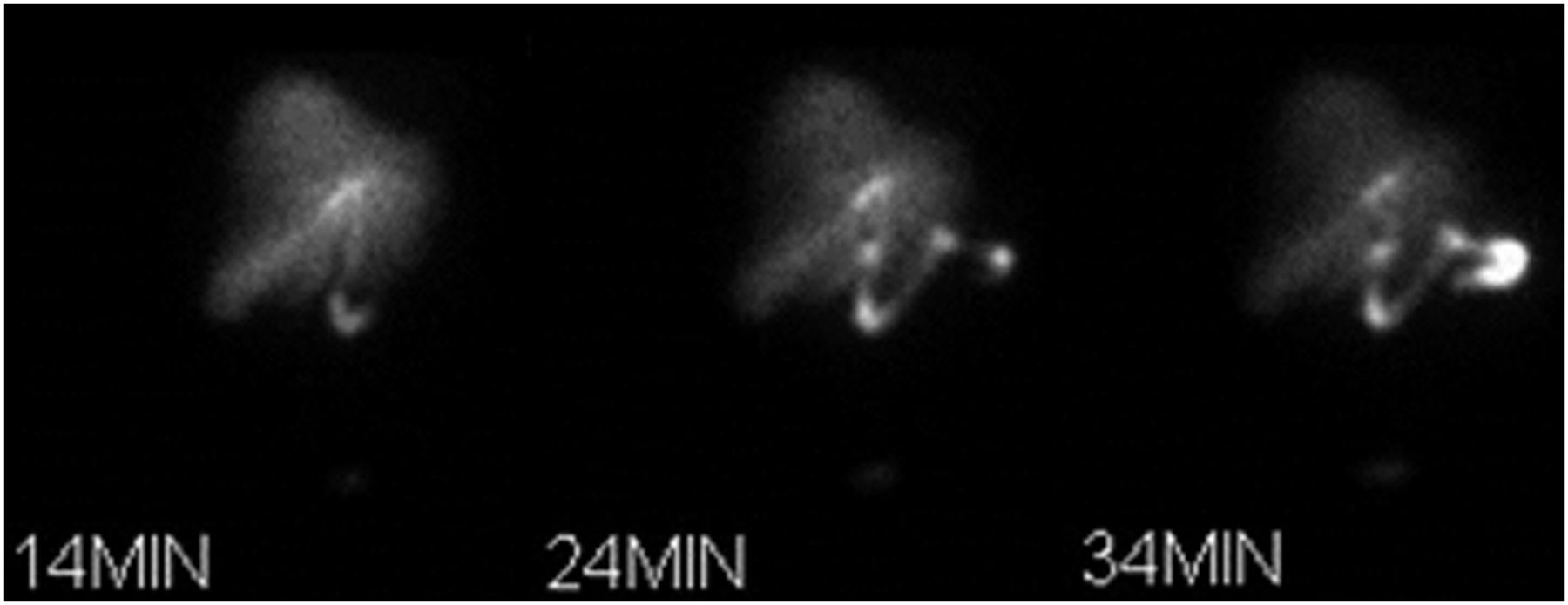
- FIGURE 8.
Hepatobiliary imaging over 60 minutes demonstrating a patient with a known hepatic laceration between the two hepatic lobes that was diagnosed by CT scan (black arrow). The images demonstrate increasing tracer accumulating outside of the biliary system (pink arrow). The red arrows show lines of tracer uptake that connect to an abnormal focus of tracer to the left of the intrahepatic duct. Over time, the abnormal tracer spreads in the paracolic gutters and sometimes throughout the abdomen, as noted by the blue arrows. These findings are indicative of a bile leak at left intrahepatic duct. This image was originally published in JNMT. Naeem S. Precise Localization of a Bile Leak with Hepatobiliary Scintigraphy. J Nucl Med Technol. 2015;44:44. © SNMMI.
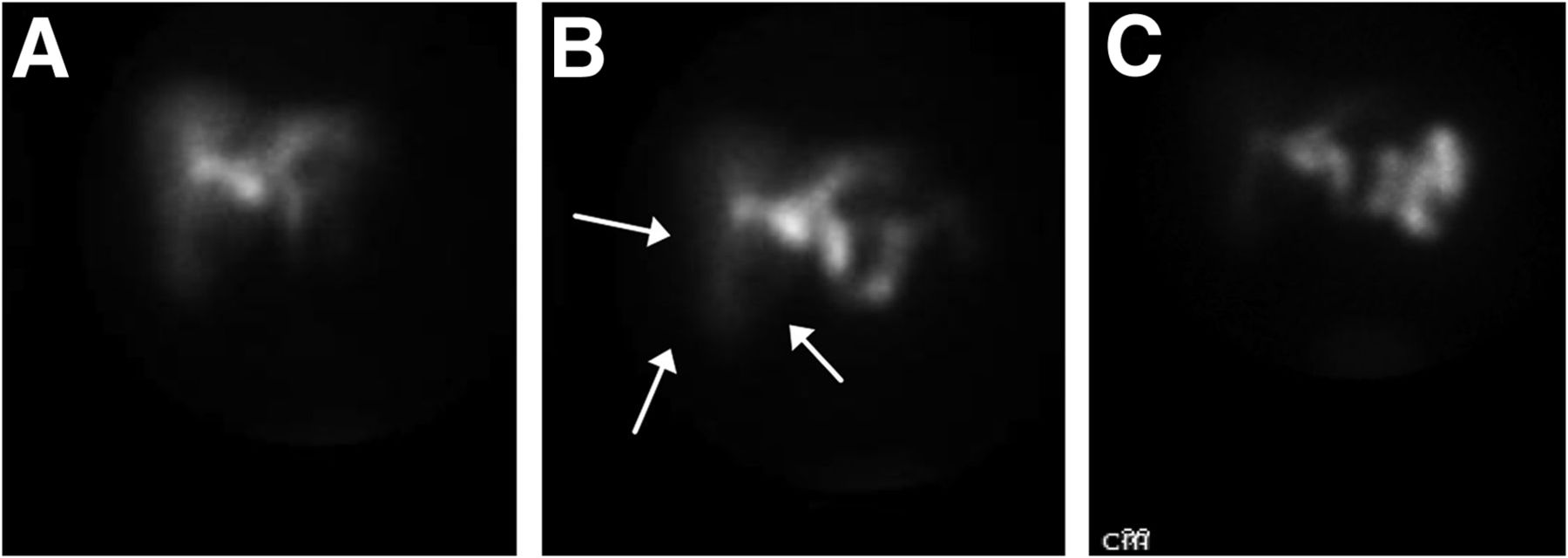
- FIGURE 9.
Hepatobiliary imaging of the “Rim Sign” (arrows) at (a) 20 minutes, (b) 30 minutes, and (c) 60 minutes. This image was originally published in JNM. Rodrique P. Gallbladder Fossa Abscess Masquerading as Cholecystitis After Cholecystectomy. J Nucl Med Technol. 2015;43. © SNMMI.
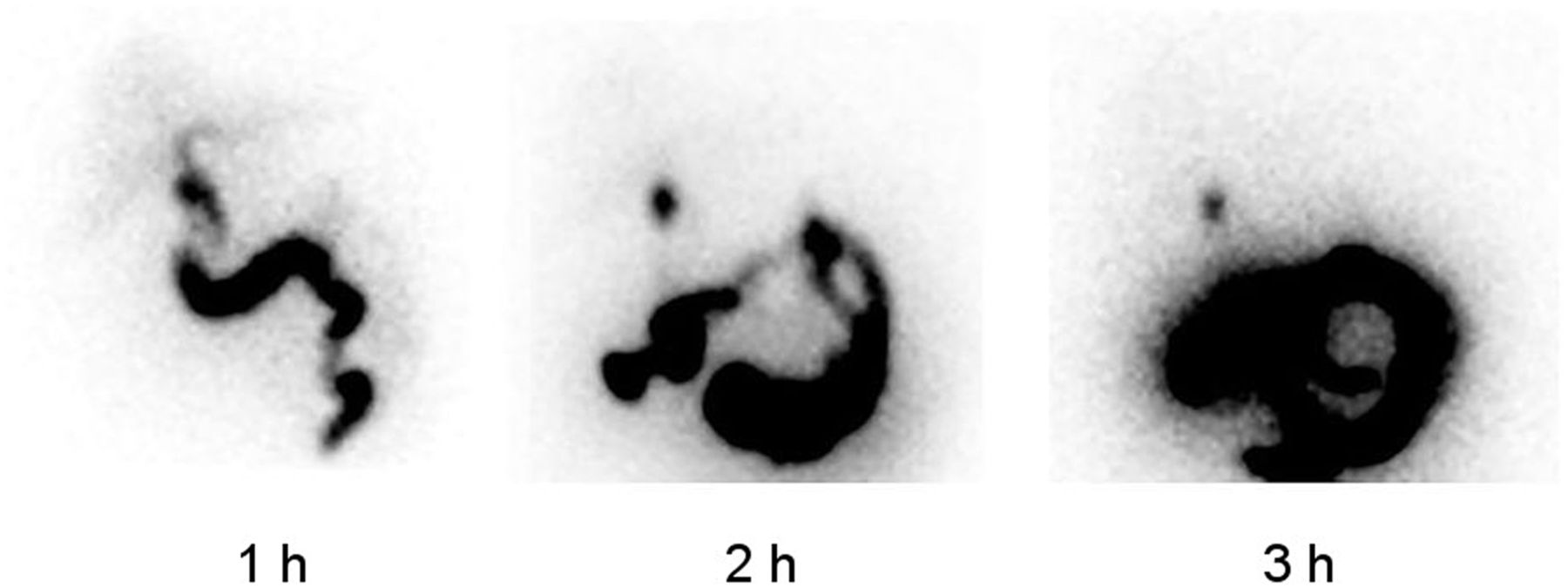
- FIGURE 10.
Hepatobiliary imaging demonstrating tracer accumulation in the cystic duct that remains unchanged over time consistent with cystic duct sign. This image was originally published in JNM. Ziessman HA. Hepatobiliary Scintigraphy in 2014. J Nucl Med. 2014;55:970. © SNMMI.
- FIGURE 11.
Retention of tracer within the duodenum mimicking the gallbladder. This image was originally published in JNMT. Bhambhvani P. Cover Image. J Nucl Med Technol. 2012;40(2):Cover Image. © SNMMI.


