


Article Figures & Data
Figures
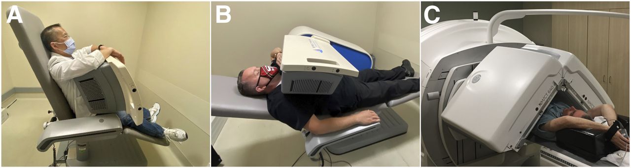
- FIGURE 1.
Multiple imaging positions can help to compensate for lack of attenuation correction. (A) Upright positioning on D-SPECT at approximately 70° chair angle, with detector at its closest position to patient. Patient can rest both arms on top of detector at about eye level, or right arm may be placed on arm rest. (B) Supine positioning on D-SPECT at approximately 30° chair angle. Right arm may be placed on arm rest, with left arm on top of camera or over patient’s head. (C) Supine positioning on conventional SPECT camera, with both detectors at 90° and arms over head.
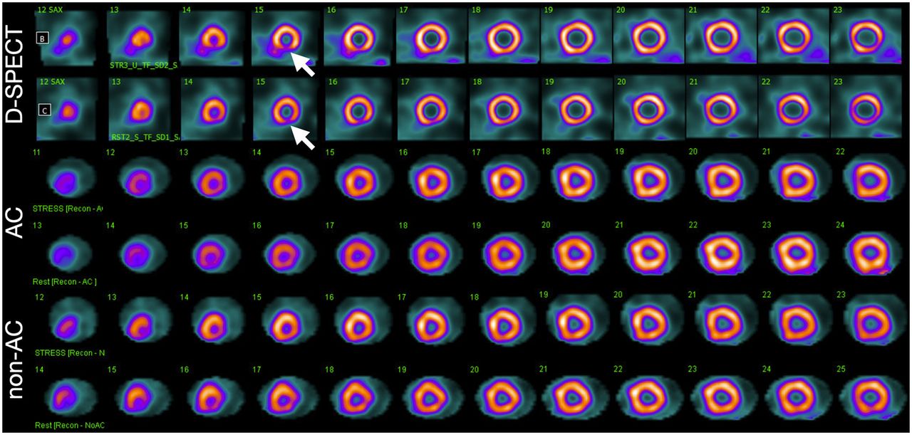
- FIGURE 2.
Example of distal inferior wall attenuation artifact (decreased uptake, arrows) seen occasionally on both rest and stress D-SPECT images. Myocardium is delineated more sharply with D-SPECT than with attenuation-corrected (AC) or non-AC SPECT/CT.
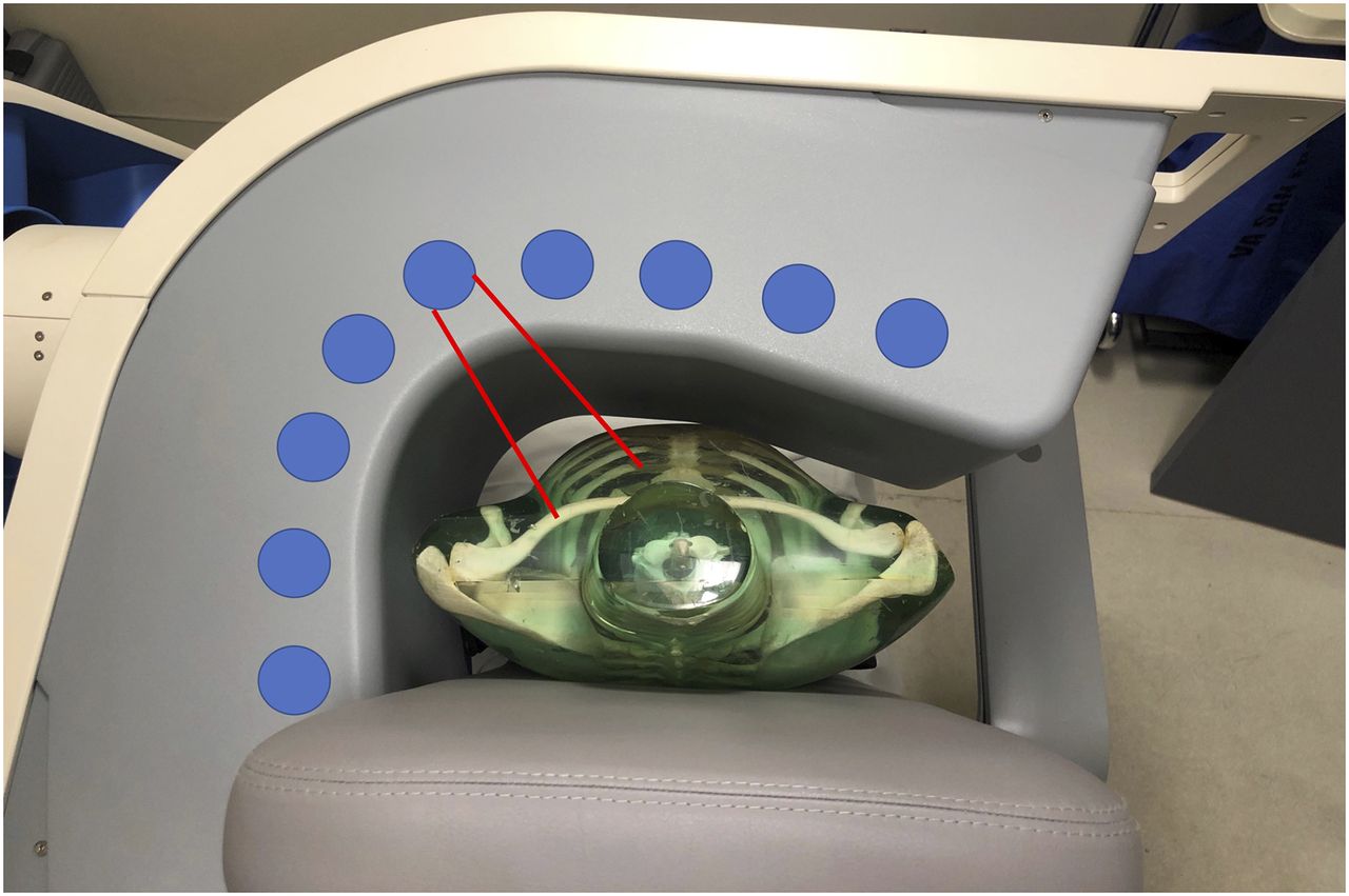
- FIGURE 3.
Aerial view of camera heads arranged inside gantry, showing approximate angles taken to achieve images. Red lines indicate heart-centric FOV of 1 of 9 detectors.
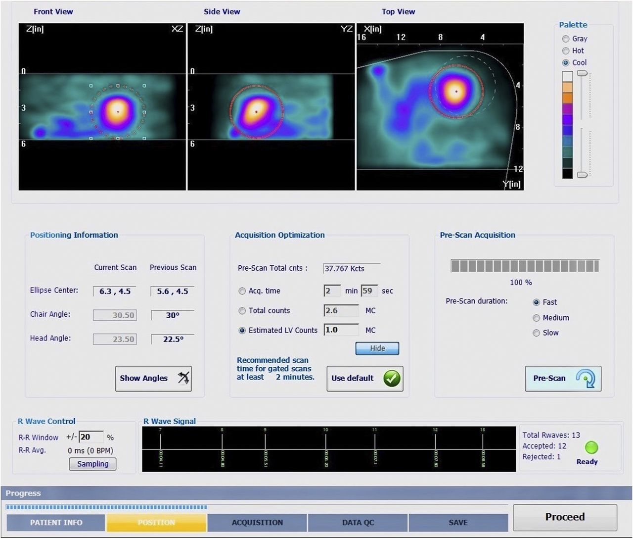
- FIGURE 4.
Prescan ensures that heart is accurately centered within FOV. Front and side views indicate superior and inferior limits. Dashed white circle in top view is D-SPECT indicator for lateral left side heart alignment; for appropriate imaging, heart must touch this circle, at minimum. Red circle needs to be adjusted to fit around myocardium on all 3 views for optimum count statistics.
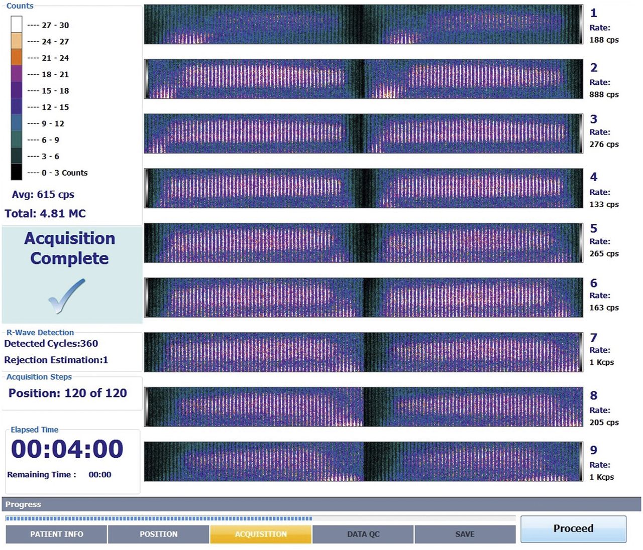
- FIGURE 5.
D-SPECT postscan. If prescan centering was accurate, each of 9 camera FOVs will be centered as in this image. Two separate vertical columns, left and right, represent first and second sweeps of detector. Scanning by the 9 detectors is based on regions of interest drawn by technologist, and then all detectors shift on linear motor and scan again. Afterward, these 2 sweeps are zipped together. Vertical lines are positions (equal to stops on traditional scanner), of which there are 60 for each detector per scan. Dark space above and below vertical lines indicates accurate prescan FOV alignment.
- FIGURE 6.
Example of poor positioning on D-SPECT prescan interface. Though frontal and side views are aligned within superior and inferior limits, top-view target area (dashed white circle) shows that heart is outside FOV. This can occur when patient is obese or when patient or camera is not adequately left-aligned.

Tables
Detector crystal Thickness (mm) Energy resolution (%) Intrinsic spatial resolution (mm) Count rate (kcps) NaI 9.5 10 4 ≥250 CZT 5 6 2.5 ≥600 Protocol Rest study Stress study Rest/stress (4,9) 296–444 MBq (8–12 mCi) (conventional SPECT) 888–1,332 MBq (24–36 mCi) (conventional SPECT) Rest/stress (12) 185 MBq (5.0 mCi) for <91 kg 463 MBq (12.5 mCi) for <91 kg 370 MBq (10.0 mCi) for >91 kg 925–1,110 MBq (25.0–30.0 mCi) for >91 kg Stress/rest (10) 359 MBq (9.7 mCi) for 75 kg 118 MBq (3.2 mCi) for 75 kg 814 MBq (22 mCi) for >110 kg 259 MBq (7.0 mCi) for >110 kg Stress/rest (11) 648–1,147 MBq (17.5–31.0 mCi) based on BMI 207–929 MBq (5.6–25.1 mCi) based on BMI Doses are based on weight and body mass index (BMI).
Recommended doses from ASNC guidelines (4,9) are weight-based and about 50% greater than doses from the other guidelines (10–12).


