


Article Figures & Data
Figures
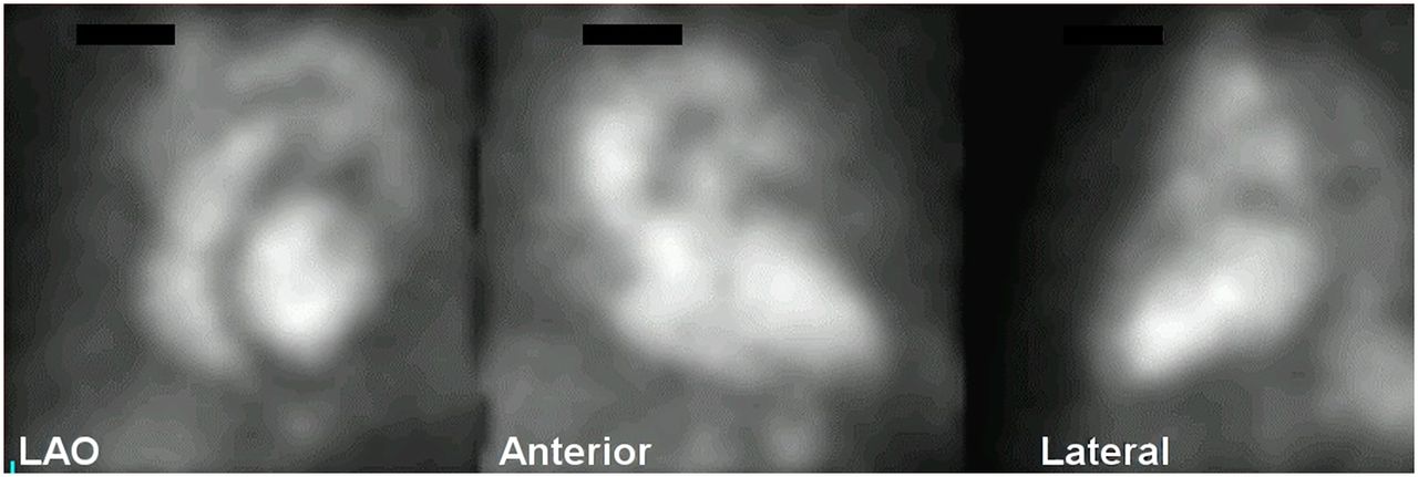
- FIGURE 1.
Standard views for ERNA.
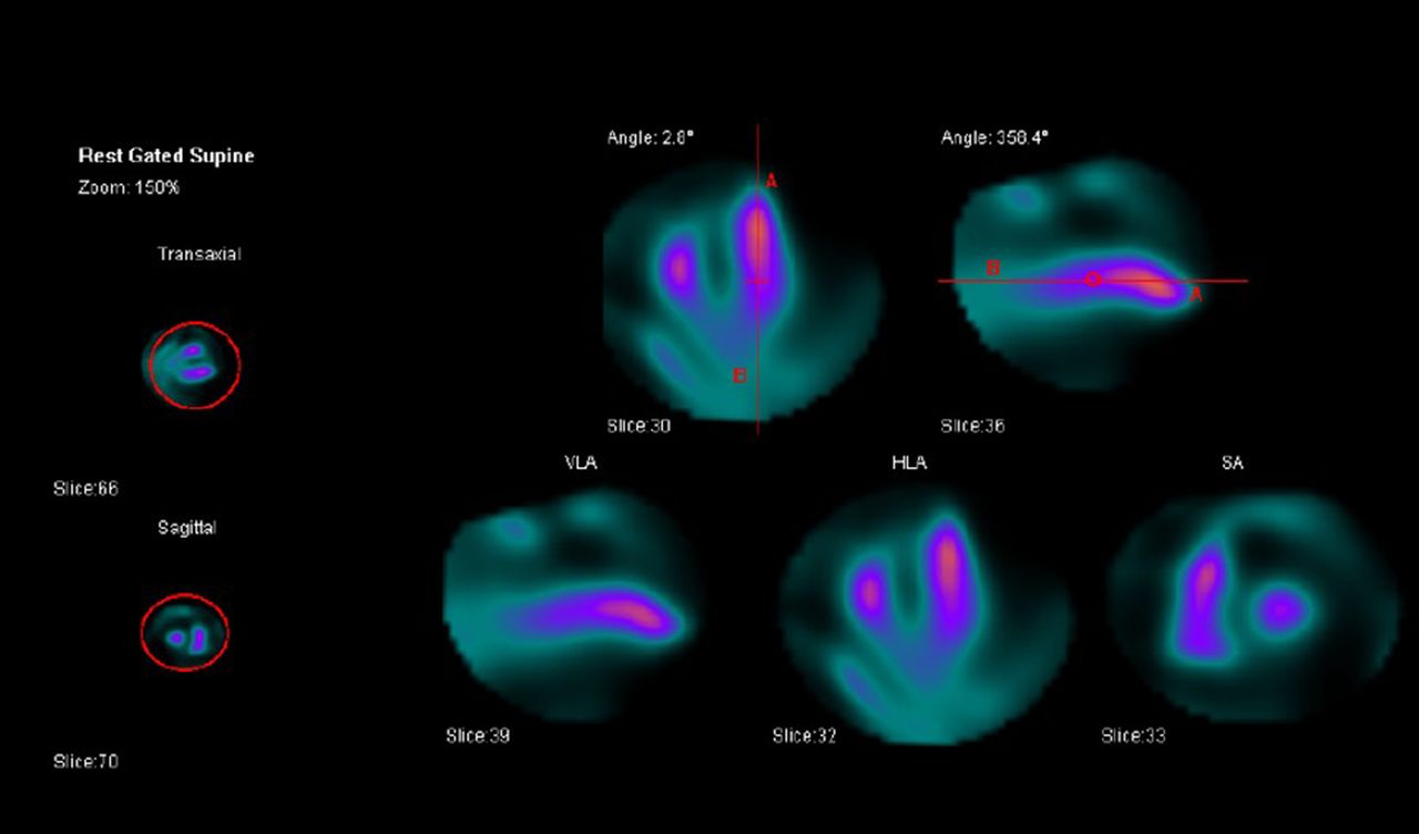
- FIGURE 2.
Example showing placement of ROIs and orientation of the ventricles before processing of SPECT ERNA data. (Image courtesy of Ronald G. Schwartz, MD, University of Rochester Medical Center, Rochester, NY. Images acquired on a cadmium-zinc-telluride (CZT) camera.)
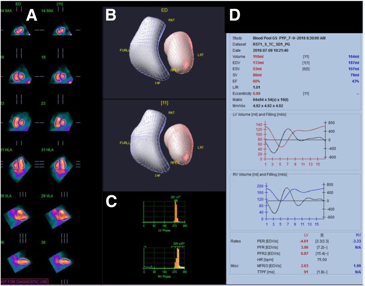
- FIGURE 3.
Ventricular function data from SPECT ERNA. SPECT ERNA data depicting end-diastolic and end-systolic contours (A), 3-dimensional rendering of LV and RV walls and morphology (B), phase data (C), and volumetric data for both ventricles (left in red and right in blue) (D). (Image courtesy of Ronald G. Schwartz, MD, University of Rochester Medical Center, Rochester, NY. Images acquired on a cadmium-zinc-telluride (CZT) camera.)
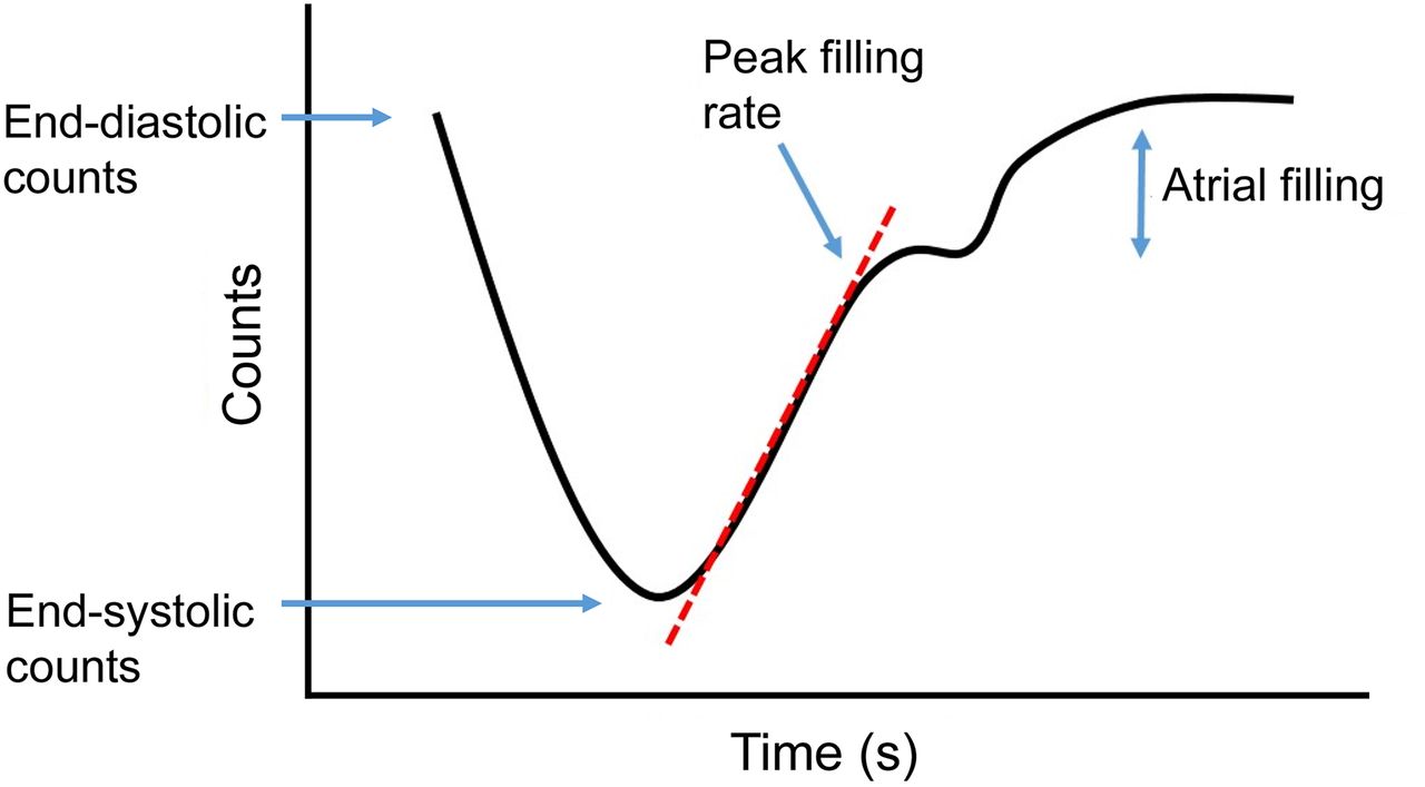
- FIGURE 4.
Example of a time–activity curve on ERNA.
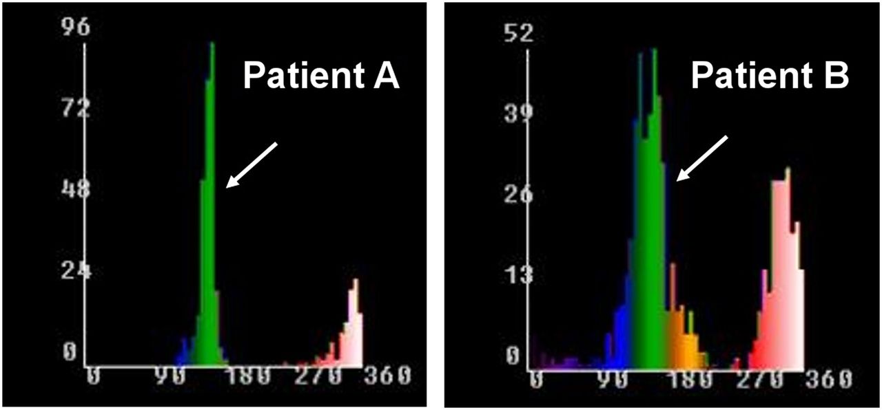
- FIGURE 5.
Assessment of LV dyssynchrony by phase analysis of ERNA. LV phase histograms (arrows) on phase analysis of ERNA from 2 patients depicting the timing of myocardial contraction (x-axis in degrees) and the frequency of the pixels achieving contraction at a particular phase (y-axis). Patient A has a normal ejection of 70%, with a uniform phase distribution and narrow phase histogram, depicting lack of dyssynchrony. Patient B has a severely reduced ejection fraction of 30%, with wide phase histogram, highlighting dyssynchronous LV contraction.
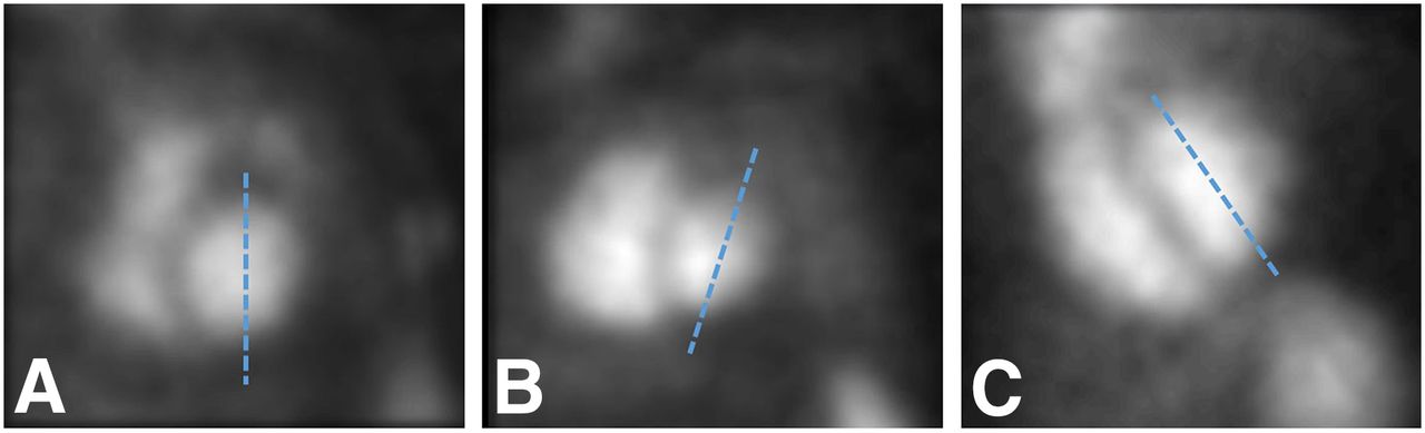
- FIGURE 6.
Correct orientation of the left ventricle in the LAO view. (A) Optimal LAO orientation, with a vertical LV axis (dashed line). Suboptimal LV axis with rightward tilting (B) and leftward tilting (C) of the LV axis.
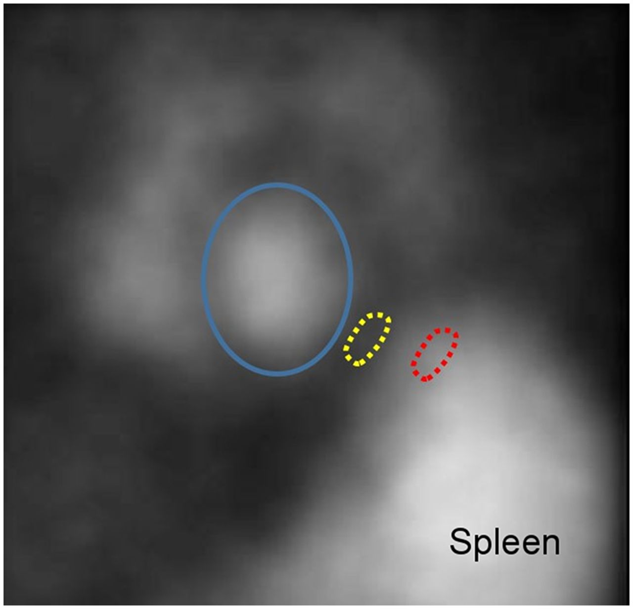
- FIGURE 7.
Location of LV and background ROI for calculation of LV function. Blue oval = LV ROI; yellow oval = correct location of background ROI; red oval = incorrect location of background ROI (over the spleen).


Tables
Valvular heart disease Timing of surgery Assessment of treatment effect Cardiomyopathy Evaluation of biventricular function Determination of type (systolic vs. diastolic) and severity Assessment of diastolic dysfunction Identification of candidates for defibrillator implantation andresynchronization therapy Evaluation of ventricular function before and after transplantation Cardio‐oncology Monitoring LV function during chemotherapy Diagnosis of cardiotoxicity from chemotherapy Guiding chemotherapy Uncommon indications Stable coronary artery disease* Diagnosis (rest/stress ERNA) Prognosis Assessment of treatment efficacy Adjunctive evaluations Evaluation of RV function Determination of ventricular dyssynchrony ↵* Performance of stress ERNA (exercise or dobutamine) in the setting of acute coronary syndromes is not recommended.
- TABLE 2
Guideline for Serial Monitoring of LVEF by ERNA for Patients Undergoing Chemotherapy with Anthracyclines (9,10)
Normal LVEF at baseline (≥50%) Baseline ERNA before initiating chemotherapy Next ERNA at 250–300 mg/m2 dose Next ERNA at 450 mg/m2 (400 mg/m2 if high risk*) Next ERNA before each dose > 450 mg/m2 Discontinue therapy if LVEF decreases ≥10% (EF units) from baseline and reaches <50% Abnormal LVEF at baseline (>30% to <50%) Baseline ERNA before initiating chemotherapy Serial ERNA before each subsequent dose Discontinue therapy if LVEF decreases ≥ 10% (EF units) from baseline or reaches ≤ 30% LVEF ≤ 30% at baseline Do not initiate therapy ↵* High-risk features include use of cyclophosphamide, heart disease, mediastinal radiation, and abnormal ECG.
99mTc-labeled RBC (17) Adult Pediatrics—10 y old Pediatrics—5 y old Urinary bladder (organ receiving the largest radiation dose) (mSv per MBq) 0.012 0.0204 0.027 Effective dose (mSv per MBq) 0.0047 0.0077 0.011 99mTc-radiolabeled human serum albumin (HSA) (17) Heart (organ receiving the largest radiation dose) (mSv per MB)q 0.0085 0.01 0.021 Effective dose (mSv per MBq) 0.0046 0.007 0.011 Cause Drug or process Oxidation of stannous ion Hydralazine Heparin Methyldopa Decreased hematocrit Inadequate reduction of 99mTc Excess stannous chloride Insufficient stannous chloride Formation of RBC antibodies Methyldopa Penicillin Quinidine Immune disorders Leukemia and lymphoma Complex formation with 99mTc Dextrose Decreased RBC labeling Prazosin Digoxin Propranolol Mechanism unknown Doxorubicin Iodinated contrast (Adapted from (4)).
Data Planar ERNA SPECT ERNA Demographic data Standard Standard Study date Standard Standard Medical record number Standard Standard Height/weight/BSA Standard Standard Acquisition information Type of study Standard Standard Radionuclide and dose Standard Standard RBC Labeling technique Standard Standard Study indication Standard Standard Study quality Recommended Recommended Study results: rest Left ventricle (LV) LV size Qualitative Standard Standard Quantitative (volume) N/A Optional LV regional wall motion Standard Standard LV hypertrophy Optional Optional Quantitative LVEF Standard Standard LV diastolic function Qualitative Standard Standard Quantitative (PFR) Preferred Preferred Right ventricle (RV) RV size Qualitative Standard Standard Quantitative (volume) N/A Optional RV regional wall motion Standard Standard RVEF Qualitative Optional Optional Quantitative* N/A Optional Atrial sizes Optional Optional Aorta and pulmonary artery size Optional Optional Pericardium Optional N/A Synchrony parameters Optional Optional Study results: stress Type of protocol: exercise/intervention Standard Standard Symptoms Standard Standard Peak BP and HR Standard Standard METs or %MPHR Standard Standard Left ventricle (LV)** LV size: change from rest Qualitative Standard — Quantitative N/A — Change in LV regional wall motion Standard — Quantitative stress LVEF Standard — Right ventricle (RV)** RV size: change from rest Standard — Change in RV regional wall motion Standard — Qualitative stress RVEF Optional — Conclusion Normal or abnormal Standard Standard Diagnostic significance of rest/stress response** Standard — Prognostic significance of rest/stress response** Optional — Comparison to prior studies Standard Standard Extracardiac findings Recommended Recommended ↵* RVEF can be obtained from SPECT ERNA, though not well validated.
↵** Pharmacologic stress SPECT ERNA can allow for assessment of these parameters, though there is little contemporary data.
BSA = body surface area; LVEF = left ventricular ejection fraction; RVEF = right ventricular ejection fraction; PFR = peak filling rate; BP = blood pressure; HR = heart rate.


