
Main menu

  • Home
  • Content
    • Current
      • JNMT Supplement
    • Ahead of print
    • Past Issues
    • Continuing Education
    • JNMT Podcast
    • SNMMI Annual Meeting Abstracts
  • Subscriptions
    • Subscribers
    • Rates
    • Journal Claims
    • Institutional and Non-member
  • Authors
    • Submit to JNMT
    • Information for Authors
    • Assignment of Copyright
    • AQARA Requirements
  • Info
    • Reviewers
    • Permissions
    • Advertisers
    • Corporate & Special Sales
  • About
    • About Us
    • Editorial Board
    • Contact Information
  • More
    • Alerts
    • Feedback
    • Help
    • SNMMI Journals
  • SNMMI
    • JNMT
    • JNM
    • SNMMI Journals
    • SNMMI

User menu

  • Subscribe
  • My alerts
  • Log in
  • My Cart

Search

  • Advanced search
Journal of Nuclear Medicine Technology
  • SNMMI
    • JNMT
    • JNM
    • SNMMI Journals
    • SNMMI
  • Subscribe
  • My alerts
  • Log in
  • My Cart
Journal of Nuclear Medicine Technology

Advanced Search

  • Home
  • Content
    • Current
    • Ahead of print
    • Past Issues
    • Continuing Education
    • JNMT Podcast
    • SNMMI Annual Meeting Abstracts
  • Subscriptions
    • Subscribers
    • Rates
    • Journal Claims
    • Institutional and Non-member
  • Authors
    • Submit to JNMT
    • Information for Authors
    • Assignment of Copyright
    • AQARA Requirements
  • Info
    • Reviewers
    • Permissions
    • Advertisers
    • Corporate & Special Sales
  • About
    • About Us
    • Editorial Board
    • Contact Information
  • More
    • Alerts
    • Feedback
    • Help
    • SNMMI Journals
  • Watch or Listen to JNMT Podcast
  • Visit SNMMI on Facebook
  • Join SNMMI on LinkedIn
  • Follow SNMMI on Twitter
  • Subscribe to JNMT RSS feeds
Review ArticleTeaching Case Studies

Primary Nasolacrimal Duct Obstruction Visualized on 123I Preablation Scan for Papillary Thyroid Carcinoma

Matthew D. Kay, Lilah F. Morris-Wiseman, Alex Beazer, Blair A. Winegar and Phillip H. Kuo
Journal of Nuclear Medicine Technology March 2020, 48 (1) 77-78; DOI: https://doi.org/10.2967/jnmt.119.235010
Matthew D. Kay
1Department of Medical Imaging, University of Arizona College of Medicine, Tucson, Arizona
  • Find this author on Google Scholar
  • Find this author on PubMed
  • Search for this author on this site
Lilah F. Morris-Wiseman
2Department of Surgery, University of Arizona College of Medicine, Tucson, Arizona
  • Find this author on Google Scholar
  • Find this author on PubMed
  • Search for this author on this site
Alex Beazer
3Department of Ophthalmology and Vision Science, University of Arizona College of Medicine, Tucson, Arizona; and
  • Find this author on Google Scholar
  • Find this author on PubMed
  • Search for this author on this site
Blair A. Winegar
1Department of Medical Imaging, University of Arizona College of Medicine, Tucson, Arizona
3Department of Ophthalmology and Vision Science, University of Arizona College of Medicine, Tucson, Arizona; and
  • Find this author on Google Scholar
  • Find this author on PubMed
  • Search for this author on this site
Phillip H. Kuo
1Department of Medical Imaging, University of Arizona College of Medicine, Tucson, Arizona
4Department of Medicine and Biomedical Engineering, University of Arizona, Tucson, Arizona
  • Find this author on Google Scholar
  • Find this author on PubMed
  • Search for this author on this site
  • Article
  • Figures & Data
  • Info & Metrics
  • PDF
Loading

Abstract

A 56-y-old man underwent total thyroidectomy and bilateral central and right lateral neck dissection for papillary thyroid carcinoma with lymph nodes metastases. Before radioiodine ablation, a 123I scan established the diagnosis of primary nasolacrimal duct obstruction (dacryostenosis).

  • nasolacrimal duct obstruction
  • 123I
  • 131I
  • papillary thyroid carcinoma

Patients with nasolacrimal duct obstruction can present with epiphora (excessive tear accumulation with subsequent overflow) or blurred vision (from an increased tear lake). Adult-onset nasolacrimal duct obstruction is classified as primary or secondary. The primary form is considered idiopathic, and the secondary causes are numerous, including inflammation (either endogenous or exogenous, the latter including radiation and radioiodine therapies), infection, neoplasm, surgery, and trauma (1,2).

CASE REPORT

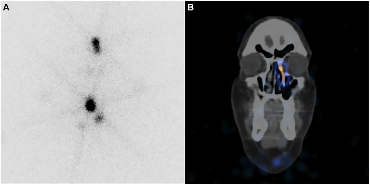
A 56-y-old man with extensive cervical lymphadenopathy due to papillary thyroid carcinoma underwent total thyroidectomy and bilateral central and right lateral neck dissection. The radioiodine ablation work-up included a pretherapy 123I scan (Fig. 1) that demonstrated left nasolacrimal duct obstruction (dacryostenosis). The nasolacrimal duct obstruction was again present on the posttherapy 131I scan (Fig. 2). On further questioning, the patient did not describe epiphora or blurred vision, suggesting that the nasolacrimal duct obstruction was incomplete. If nasolacrimal duct obstruction is present on pretherapy 123I imaging, it allows differentiation of primary nasolacrimal duct obstruction from nasolacrimal duct obstruction secondary to radioiodine iodine ablation.

FIGURE 1.
  • Download figure
  • Open in new tab
  • Download powerpoint
FIGURE 1.

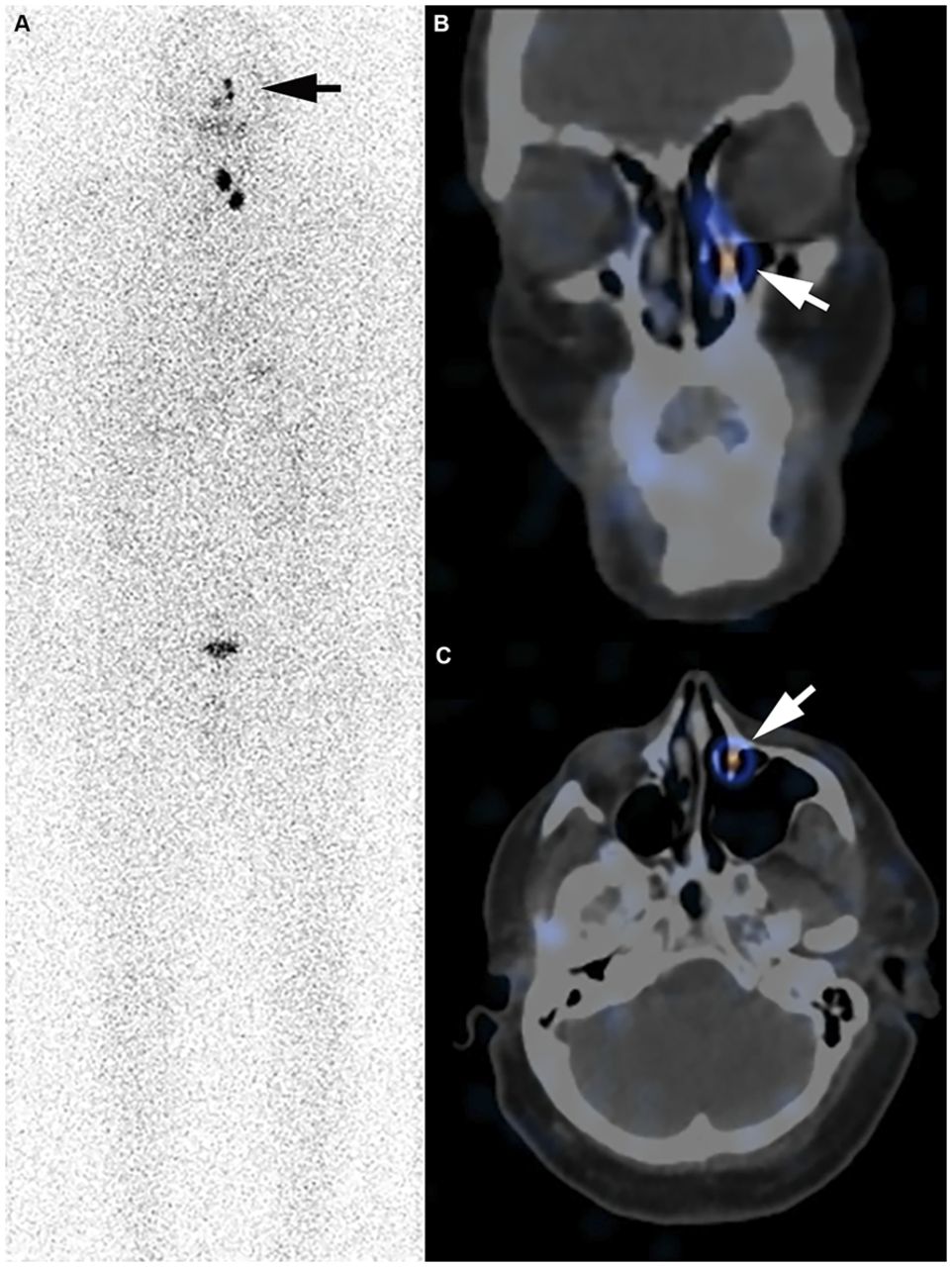
Postoperative, pretherapy 123I whole-body scan (A) and corresponding SPECT/CT of head and neck in coronal (B) and axial (C) planes demonstrate linear left nasolacrimal duct uptake (arrows) consistent with nasolacrimal duct obstruction or dacryostenosis.

FIGURE 2.
  • Download figure
  • Open in new tab
  • Download powerpoint
FIGURE 2.

Posttherapy 131I planar images of neck (A) and SPECT/CT of head and neck in coronal plane (B) reconfirm nasolacrimal duct obstruction.

DISCUSSION

Tears drain from the eyes medially via superior and inferior lacrimal canaliculi into the lacrimal sac. The lacrimal sac is seated within an osseous groove formed at the junction of the lacrimal bone and the frontal process of the maxilla. The lacrimal sac is contiguous inferiorly with the nasolacrimal duct. It functions as a reservoir for excess tears that are relayed through multiple mucous membrane folds of the nasolacrimal duct (many with eponymous names) into the inferior meatus of the nasal cavity. Nasolacrimal duct obstruction may result in epiphora or blurred vision. The incidence of nasolacrimal duct obstruction after radioiodine ablation is reported to range from 2.2% to 18% (2), but this may be an overestimation since cases without pretherapy imaging may actually be examples of primary nasolacrimal duct obstruction. Nasolacrimal duct obstruction secondary to radioiodine ablation is more likely to occur bilaterally within 6–18 mo after treatment with doses of greater than 5,550 MBq (3). The transmembrane cotransporter of sodium and iodide (sodium-iodide symporter) highly expressed by the follicular cells of the thyroid is also responsible for radioiodine secretion into the nasolacrimal drainage system, leading to localized radiotoxicity with potential for secondary nasolacrimal duct obstruction (4). Defining the etiology of nasolacrimal duct obstruction has prognostic implications, because external or endoscopic dacryocystorhinostomy (the formation of an anastomosis between the lacrimal sac and the nasal mucosa to bypass the nasolacrimal duct obstruction) is more likely to succeed in patients with primary nasolacrimal duct obstruction than in those with secondary nasolacrimal duct obstruction due to radioiodine ablation or radiotherapy (5). This case demonstrates an additional benefit of pretherapy radioiodine imaging before radioiodine ablation of differentiated thyroid cancers.

CONCLUSION

Pretherapy radioiodine imaging can differentiate primary from secondary nasolacrimal duct obstruction due to radioiodine therapy, and the primary form is more likely to improve with intervention.

DISCLOSURE

No potential conflict of interest relevant to this article was reported.

Footnotes

  • Published online Oct. 11, 2019.

REFERENCES

  1. 1.↵
    1. Ali MJ
    . Iodine-131 therapy and nasolacrimal duct obstructions: what we know and what we need to know. Ophthal Plast Reconstr Surg. 2016;32:243–248.
    OpenUrl
  2. 2.↵
    1. Bartley GB
    . Acquired lacrimal drainage obstruction: an etiologic classification system, case reports, and a review of the literature. Part 1. Ophthal Plast Reconstr Surg. 1992;8:237–242.
    OpenUrlCrossRefPubMed
  3. 3.↵
    1. Burns JA,
    2. Morgenstern KE,
    3. Cahill KV,
    4. Foster JA,
    5. Jhiang SM,
    6. Kloos RT
    . Nasolacrimal obstruction secondary to I131 therapy. Ophthal Plast Reconstr Surg. 2004;20:126–129.
    OpenUrlCrossRefPubMed
  4. 4.↵
    1. Morgenstern KE,
    2. Vadysirisack DD,
    3. Zhang Z,
    4. et al
    . Expression of sodium iodide symporter in the lacrimal drainage system: implication for the mechanism underlying nasolacrimal duct obstruction in I131-treated patients. Ophthal Plast Reconstr Surg. 2005;21:337–344.
    OpenUrlCrossRefPubMed
  5. 5.↵
    1. Sweeney AR,
    2. Davis GE,
    3. Chang SH,
    4. Amadi AJ
    . Outcomes of endoscopic dacryocystorhinostomy in secondary acquired nasolacrimal duct obstruction: a case-control study. Ophthal Plast Reconstr Surg. 2018;34:20–25.
    OpenUrl
  • Received for publication August 9, 2019.
  • Accepted for publication August 29, 2019.
PreviousNext
Back to top

In this issue

Journal of Nuclear Medicine Technology: 48 (1)
Journal of Nuclear Medicine Technology
Vol. 48, Issue 1
March 1, 2020
  • Table of Contents
  • About the Cover
  • Index by author
Print
Download PDF
Article Alerts
Sign In to Email Alerts with your Email Address
Email Article

Thank you for your interest in spreading the word on Journal of Nuclear Medicine Technology.

NOTE: We only request your email address so that the person you are recommending the page to knows that you wanted them to see it, and that it is not junk mail. We do not capture any email address.

Enter multiple addresses on separate lines or separate them with commas.
Primary Nasolacrimal Duct Obstruction Visualized on 123I Preablation Scan for Papillary Thyroid Carcinoma
(Your Name) has sent you a message from Journal of Nuclear Medicine Technology
(Your Name) thought you would like to see the Journal of Nuclear Medicine Technology web site.
Citation Tools
Primary Nasolacrimal Duct Obstruction Visualized on 123I Preablation Scan for Papillary Thyroid Carcinoma
Matthew D. Kay, Lilah F. Morris-Wiseman, Alex Beazer, Blair A. Winegar, Phillip H. Kuo
Journal of Nuclear Medicine Technology Mar 2020, 48 (1) 77-78; DOI: 10.2967/jnmt.119.235010

Citation Manager Formats

  • BibTeX
  • Bookends
  • EasyBib
  • EndNote (tagged)
  • EndNote 8 (xml)
  • Medlars
  • Mendeley
  • Papers
  • RefWorks Tagged
  • Ref Manager
  • RIS
  • Zotero
Share
Primary Nasolacrimal Duct Obstruction Visualized on 123I Preablation Scan for Papillary Thyroid Carcinoma
Matthew D. Kay, Lilah F. Morris-Wiseman, Alex Beazer, Blair A. Winegar, Phillip H. Kuo
Journal of Nuclear Medicine Technology Mar 2020, 48 (1) 77-78; DOI: 10.2967/jnmt.119.235010
Twitter logo Facebook logo LinkedIn logo Mendeley logo
  • Tweet Widget
  • Facebook Like
  • Google Plus One
Bookmark this article

Jump to section

  • Article
    • Abstract
    • CASE REPORT
    • DISCUSSION
    • CONCLUSION
    • DISCLOSURE
    • Footnotes
    • REFERENCES
  • Figures & Data
  • Info & Metrics
  • PDF

Related Articles

  • No related articles found.
  • PubMed
  • Google Scholar

Cited By...

  • No citing articles found.
  • Google Scholar

More in this TOC Section

  • High-Sensitivity Troponin Elevation in a Young Woman with Typical Chest Pain: The Heart of the Matter
  • Pulmonary Adenocarcinoma Revealed by Parathyroid Scintigraphy: An Incidental Case to Remember
  • Prominent Right Ventricular Tracer Uptake: A Harbinger of Multivessel Coronary Artery Disease
Show more Teaching Case Studies

Similar Articles

Keywords

  • Nasolacrimal duct obstruction
  • 123I
  • 131I
  • papillary thyroid carcinoma
SNMMI

© 2025 SNMMI

Powered by HighWire