


Article Figures & Data
Figures
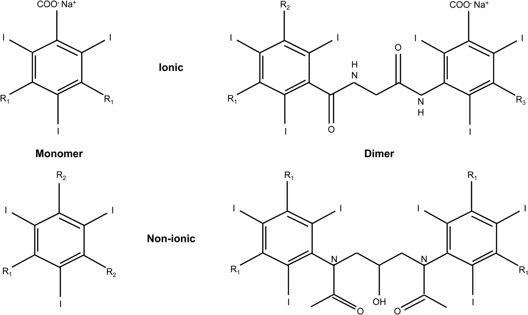
- FIGURE 1.
Chemical structure of iodinated CT contrast agents is based on 2,4,6-triiodinated benzene ring and provides 4 major classifications of iodinated CT contrast agents: ionic monomer, ionic dimer, nonionic monomer, and nonionic dimer. For ionic contrast media, carboxyl group (COOH) ionizes (COO−) with sodium or meglumine to form anion and cation pairs. Side chains (R) vary but tend to be longer for nonionic contrast media.
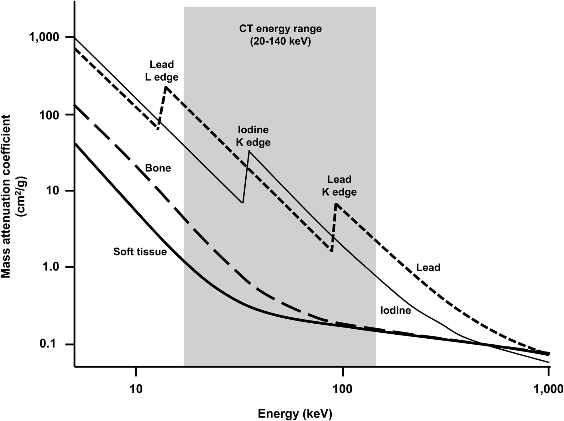
- FIGURE 2.
Schematic representation on log–log scales of mass attenuation coefficient against x-ray energy. Iodine K edge at 33 keV demonstrates abrupt increase in attenuation that produces equivalent attenuation greater than lead and several orders of magnitude greater than bone and soft tissue. Within range of 30–100 keV, attenuation coefficients for biologic tissues remain fairly uniform whereas contrast agent (iodine) varies substantially. (Adapted from (15).)
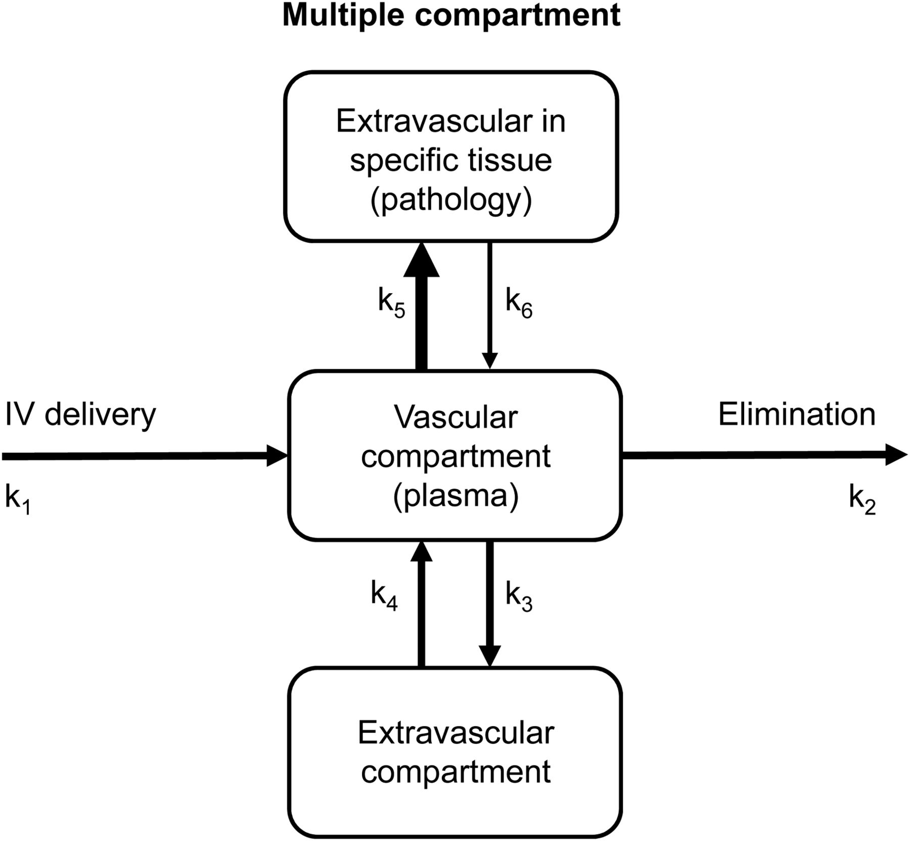
- FIGURE 3.
Modified 2-compartment model for iodinated CT or gadolinium MRI contrast media administered intravenously. Second extravascular compartment represents pathologic tissue that may enhance with contrast administration and, therefore, be differentiated by surrounding normal tissue by greater rate constant (k5 over k3 or k4 over k6). A previous article (2) in this series provides a more detailed interpretation of compartment models and rate constants.
- FIGURE 4.
Flowchart of iodinated and gadolinium contrast reaction classification.
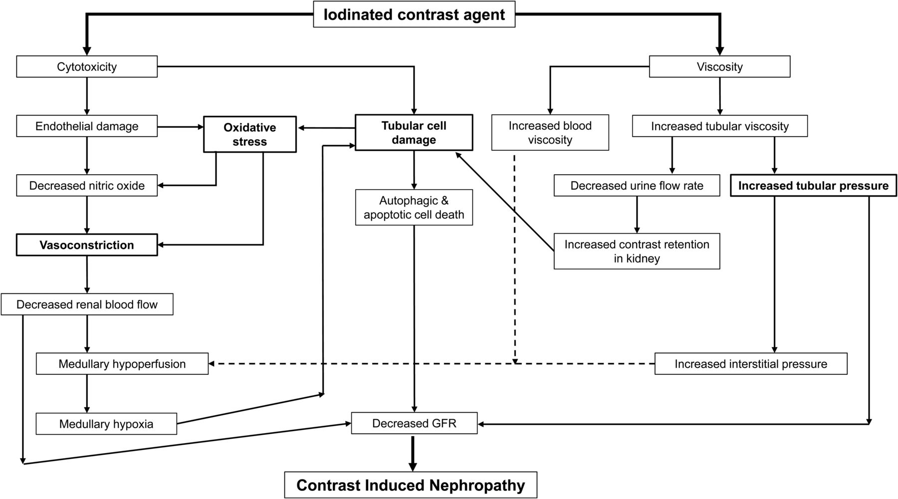
- FIGURE 5.
Flowchart outlining interplay between factors contributing to development of contrast-induced nephrotoxicity. As outlined by bold boxes, vasoconstriction, oxidative stress, tubular cell damage, and increased tubular pressure are key drivers associated with cytotoxicity and viscosity as mediators.
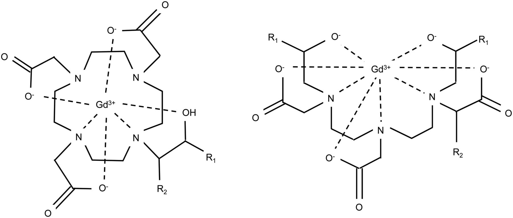
- FIGURE 6.
Chemical structure of gadolinium contrast agents adopts either macrocyclic base (left) or linear base (right), with major differences between each agent being changes to R groups.
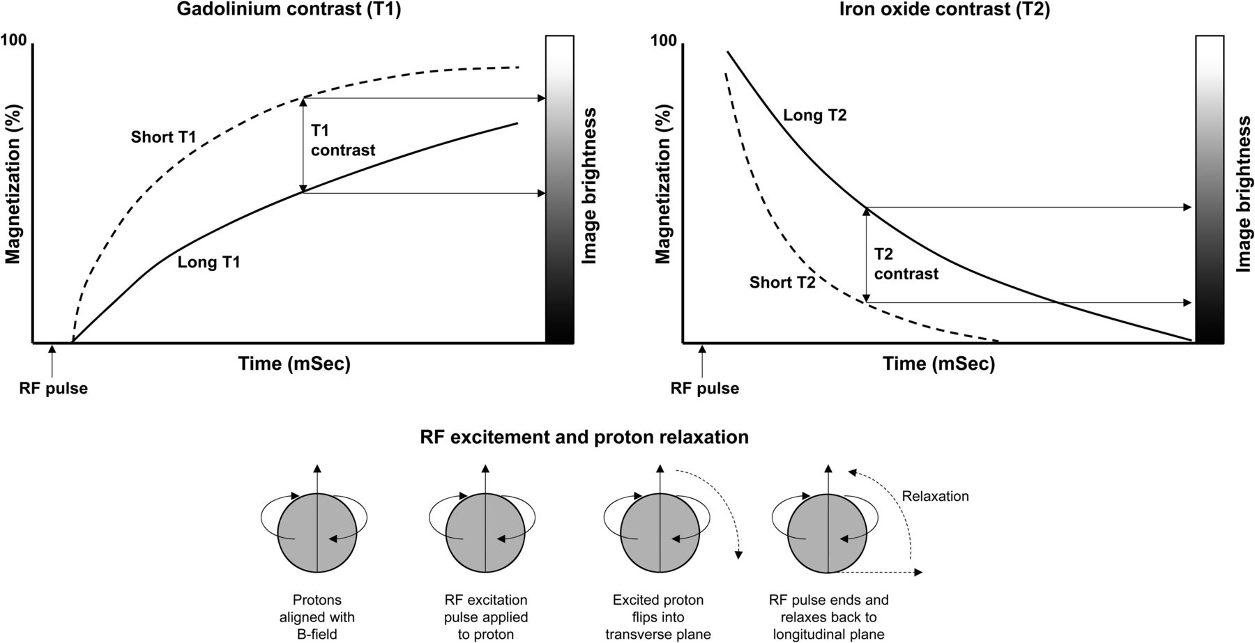
- FIGURE 7.
Schematic representation of principle of T1 and T2 contrast enhancement in MRI. As represented at bottom of figure, hydrogen (protons) initially aligns with magnetic field. Radiofrequency (RF) excitation pulse is applied to proton, which flips into transverse plane. RF pulse ends, allowing proton to relax back to longitudinal plane. T1 plot (top left) shows effect of shortening relaxation time with gadolinium contrast and resultant positive enhancement of contrast. Likewise, T2 plot (top right) shows effect of shortening relaxation time with iron oxide contrast and resultant negative enhancement of contrast.


Tables
- TABLE 1
Key Properties of Iodinated Contrast Agents That Influence Their Behavior, Efficiency, and Adverse Reaction Risk
Contrast agent Iodine concentration (mg/mL) Osmolality (mOsm/kg water) Viscosity (mPa/s) (37°C) Ionic monomer (HOCM) Up to 400 1,400–2,100 Ionic dimer (HOCM) 320 600 Nonionic monomer (LOCM) Up to 350 600–800 Nonionic dimer (IOCM) 320 290 Human serum 3.2–4 290 1.5–2.0 Ionic monomers Amidotrizoic acid 146 1,690 8.5 Amidotrizoate-meg 350 1,530 7.5 Ioxitalaminic acid 2,130 Nonionic monomers Iohexol 240, 300, 350 500, 690, 880 3.3, 6.1, 10.6 Iopamidol 200, 300, 370 413, 616, 796 2.0, 4.7, 8.6 Ioxilan 350 695 4.6 Iopromide 370 780 9.5 Ioversol 320 702 5.8 Iomeprol 350 618 7.5 Iobitridol 350 915 10.0 Ionic dimers ioxaglate 320 580 7.5 Nonionic dimers Iodixanol 320 290 11.4 Iotrolan 300 320 8.1 Optimizing key properties of intravenous CT contrast agents has resulted in evolution to those that are easier to use, with lower intravenous toxicity and fewer adverse effects (frequency and severity) (6,8,12).
- TABLE 2
Incidence of Adverse Reactions to Iodinated Intravenous Contrast Agents (10–13,16–18)
Reaction type Ionic Nonionic Ionic HOCM Nonionic LOCM/IOCM Mild 15% 3% Moderate 1%–2% 0.2%–0.4% Severe 0.2% 0.04% 0.22% 0.04% Fatal 0.0006% 0.0006% Overall 13% 3% Delayed 2%–4% for nonionic dimer 0.5%–1% for ionic and nonionic monomers 12.5% for CT with intravenous contrast 10% for CT without intravenous contrast Extravasation 0.04%–0.2% for mechanical power injectors 0.04%–0.2% for mechanical power injectors Contrast-induced nephropathy 1%–3% in normal renal function 12%–27% in renal impairment 50% in diabetic nephropathy Contrast agent Structure Ionicity Clearance T0.5 (h) Osmolality (mOsm/kg water) Viscosity at 37°C (cP) T1 relaxivity (L/mmol-s) Gadopentetate dimeglumine Linear Ionic 1.6 1,960 2.9 4.1 Gadoteridol Macrocyclic Nonionic 1.57 630 1.3 4.1 Gadodiamide Linear Nonionic 1.3 789 1.4 4.3 Gadoversetamide Linear Nonionic 1.73 1,110 2.0 5.2 Gadobenate dimeglumine Linear Ionic 1.2–2 1,970 5.3 6.3 Gadoterate Macrocyclic Ionic 1.6 1,350 2.4 3.6 Gadobutrol Macrocyclic Nonionic 1.81 1,603 5.0 5.2 Gadoxetate Linear Ionic 0.93 688 1.2 6.9 Gadofosveset Linear Ionic 1,110 3.0 19 Blood 290 1.5–2 With exception of gadoxetate, dose is 0.1 mmol/kg. Gadoxetate dose is 0.025 mmol/kg (27,29,31).


