


Article Figures & Data
Figures
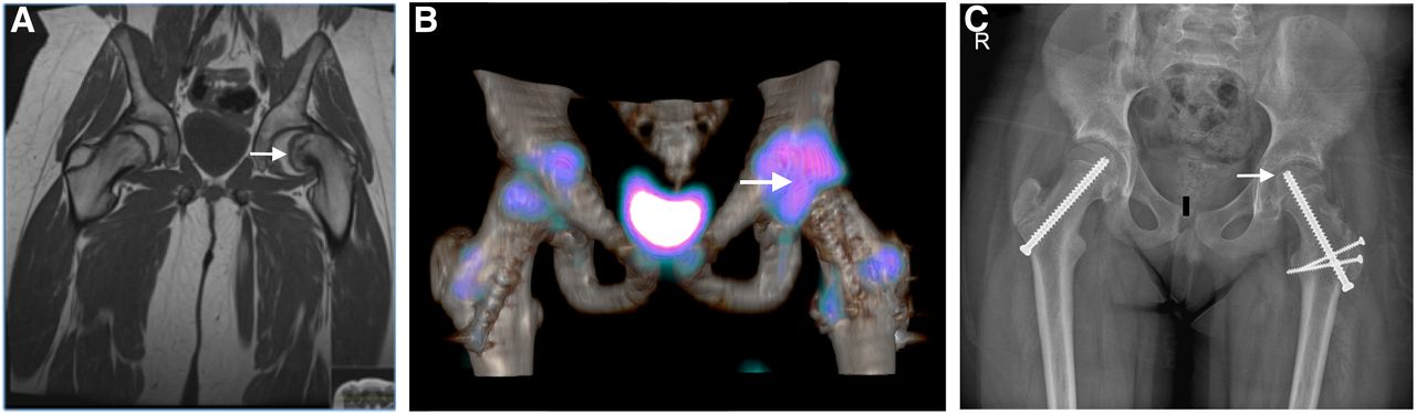
- FIGURE 1.
(A) Coronal T1-weighted MR image showing left slipped capital femoral epiphysis (arrow). (B) Axial fused 3-dimensional SPECT/CT image showing uptake in femoral head (arrow), with satisfactory appearance of articular surface. (C) Radiograph obtained at 2-y follow-up showing smooth outline of left femoral head (arrow).
- FIGURE 2.
(A) Coronal T1-weighted MR image showing left slipped capital epiphysis (arrow). (B) Postoperative axial fused 3-dimensional SPECT/CT image showing absence of uptake in left femoral head (arrow), in keeping with nonviable region. (C) Radiograph obtained at 2-y follow-up showing collapse of left femoral head (arrow) at same site where 3-dimensional SPECT/CT showed nonviable femoral head.


Jump to section
Related Articles
Cited By...
- No citing articles found.