
Main menu

  • Home
  • Content
    • Current
      • JNMT Supplement
    • Ahead of print
    • Past Issues
    • Continuing Education
    • JNMT Podcast
    • SNMMI Annual Meeting Abstracts
  • Subscriptions
    • Subscribers
    • Rates
    • Journal Claims
    • Institutional and Non-member
  • Authors
    • Submit to JNMT
    • Information for Authors
    • Assignment of Copyright
    • AQARA Requirements
  • Info
    • Reviewers
    • Permissions
    • Advertisers
    • Corporate & Special Sales
  • About
    • About Us
    • Editorial Board
    • Contact Information
  • More
    • Alerts
    • Feedback
    • Help
    • SNMMI Journals
  • SNMMI
    • JNMT
    • JNM
    • SNMMI Journals
    • SNMMI

User menu

  • Subscribe
  • My alerts
  • Log in
  • My Cart

Search

  • Advanced search
Journal of Nuclear Medicine Technology
  • SNMMI
    • JNMT
    • JNM
    • SNMMI Journals
    • SNMMI
  • Subscribe
  • My alerts
  • Log in
  • My Cart
Journal of Nuclear Medicine Technology

Advanced Search

  • Home
  • Content
    • Current
    • Ahead of print
    • Past Issues
    • Continuing Education
    • JNMT Podcast
    • SNMMI Annual Meeting Abstracts
  • Subscriptions
    • Subscribers
    • Rates
    • Journal Claims
    • Institutional and Non-member
  • Authors
    • Submit to JNMT
    • Information for Authors
    • Assignment of Copyright
    • AQARA Requirements
  • Info
    • Reviewers
    • Permissions
    • Advertisers
    • Corporate & Special Sales
  • About
    • About Us
    • Editorial Board
    • Contact Information
  • More
    • Alerts
    • Feedback
    • Help
    • SNMMI Journals
  • Watch or Listen to JNMT Podcast
  • Visit SNMMI on Facebook
  • Join SNMMI on LinkedIn
  • Follow SNMMI on Twitter
  • Subscribe to JNMT RSS feeds
Review ArticleTeaching Case Studies

Looking Beyond the Indication: Bone Scan for Trauma Providing Unsuspected Initial Diagnosis of Acute Critical Lower Limb Arterial Compromise

Fathima Fijula Palot Manzil, Samuel E. Almodovar and Pradeep G. Bhambhvani
Journal of Nuclear Medicine Technology March 2018, 46 (1) 67-68; DOI: https://doi.org/10.2967/jnmt.117.200618
Fathima Fijula Palot Manzil
1Clinical Radiology, Weill Cornell Medical College, Qatar, and Department of Radiology, Hamad General Hospital, Doha, Qatar
  • Find this author on Google Scholar
  • Find this author on PubMed
  • Search for this author on this site
Samuel E. Almodovar
2Department of Radiology, Orlando VA Medical Center, Orlando, Florida; and
  • Find this author on Google Scholar
  • Find this author on PubMed
  • Search for this author on this site
Pradeep G. Bhambhvani
3Division of Molecular Imaging and Therapeutics, Department of Radiology, University of Alabama at Birmingham, Birmingham, Alabama
  • Find this author on Google Scholar
  • Find this author on PubMed
  • Search for this author on this site
  • Article
  • Figures & Data
  • Info & Metrics
  • PDF
Loading

Abstract

We report a case of rare incidental initial detection of unsuspected arterial supply compromise to the right lower extremity on a bone scan obtained to rule out fracture in a patient with right hip pain after a fall. Follow-up CT angiogram confirmed near-complete occlusion of right lower extremity arteries for which patient underwent above-knee amputation.

  • three phase bone scan
  • ischemia
  • CT angiogram
  • amputation

Three-phase bone scintigraphy is a useful tool to assess arterial flow, relative soft-tissue perfusion, and variations in bone turnover. Systematic evaluation of all 3 phases is crucial to detect skeletal and unsuspected extraskeletal abnormalities.

CASE REPORT

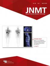
A 101-y-old patient with a history of atrial fibrillation presented to our hospital with right hip pain and difficulty walking after a fall. A plain CT scan of the bony pelvis and 3-phase 99mTc-methylene diphosphonate bone scintigraphy performed to rule out clinically suspected occult hip bone injury were negative for fracture. However, scintigraphy showed near-absent radiotracer activity within the right lower limb arteries on the flow phase (Fig. 1A), decreased soft-tissue uptake around the right knee and lower leg on the blood-pool phase (Fig. 1B), and decreased radiotracer uptake in the right femur, especially distally, and the lower leg bones on the delayed skeletal phase (Fig. 1C). Follow-up urgent CT angiogram on the same day demonstrated extensive thromboembolism of right lower limb arteries (Fig. 2). The right leg turned extremely painful and cold after a day. On physical examination, there was no palpable or audible right femoral, popliteal, and pedal flow. The patient had limited right knee flexion and limited sensation from the knee downward and could not move the right foot, and underwent right transfemoral above-knee amputation.

FIGURE 1.
  • Download figure
  • Open in new tab
  • Download powerpoint
FIGURE 1.

(A) Blood-flow images of 3-phase bone scintigraphy of thighs in anterior projection shows near-absent radiotracer uptake within right common iliac, external iliac (thick arrow), and proximal superficial femoral arteries (thin arrow). (B) Blood-pool images at knee/leg level in anterior and posterior projections show decreased soft-tissue uptake in right knee and leg (arrows). (C) Delayed whole-body bone scan in anterior and posterior projections show decreased radiotracer uptake in right femur, especially distally, and bones of right lower leg (thin transverse arrows). Increased linear uptake at medial right thigh (oblique arrows) possibly represents muscle injury. Note urinary activity in diaper (thick arrows).

FIGURE 2.
  • Download figure
  • Open in new tab
  • Download powerpoint
FIGURE 2.

CT angiogram of pelvis/lower limbs demonstrates extensive thromboembolism of right common iliac, external iliac, common femoral, popliteal, anterior, and posterior tibial arteries (white arrows).

DISCUSSION

Major factors determining radiotracer uptake in 3-phase bone scintigraphy are local blood flow, extraction efficiency, and osteoblastic activity. Impaired circulation, hydration level, renal function, and age also influence uptake (1,2). A bone scan is a useful adjunct to choose appropriate amputation level by aiding in distinguishing viable from nonviable tissue (3).

Assessing symmetry of radiotracer distribution is crucial. Venous or lymphatic obstruction and inflammation can cause asymmetrically increased tracer uptake. Unilateral paralysis can reduce ipsilateral uptake with increased contralateral activity from compensatory overuse. Decreased radiotracer activity can be seen in compromised arterial flow.

The functional status of the limbs could be improved with adequate collateral flow (4). However, because of minimal time available for collateral development in cases of acute thromboembolism, chances for above-knee amputation are doubled compared with chronic ischemia (5).

CONCLUSION

While interpreting bone scans, it is important not to confine our attention only to the detection of skeletal abnormalities. In our case, bone scan led to the initial diagnosis of critical ischemia of lower limb and apparent necrosis of thigh muscles, which led to further investigations, definitive diagnosis, and treatment. Any asymmetry in radiotracer uptake pattern in all phases on a 3-phase bone scan should not be overlooked.

DISCLOSURE

No potential conflict of interest relevant to this article was reported.

Footnotes

  • Published online Nov. 10, 2017.

REFERENCES

  1. 1.↵
    1. Gholamrezanezhad A
    1. Medina-Gálvez N,
    2. Pedraz T
    . Nuclear medicine in musculoskeletal disorders: clinical approach. In: Gholamrezanezhad A, ed. 12 Chapters on Nuclear Medicine. Rijeka, Croatia: InTech; 2011:97–135.
  2. 2.↵
    1. Love C,
    2. Din AS,
    3. Tomas MB,
    4. et al
    . Radionuclide bone imaging: an illustrative review. Radiographics. 2003;23:341–358.
    OpenUrlCrossRefPubMed
  3. 3.↵
    1. Masaoka S
    . Evaluation of arterial obstructive leg and foot disease by three-phase bone scintigraphy. Ann Nucl Med. 2001;15:281–287.
    OpenUrlPubMed
  4. 4.↵
    1. Weitz JI,
    2. Byrne J,
    3. Clagett GP,
    4. et al
    . Diagnosis and treatment of chronic arterial insufficiency of the lower extremities: a critical review. Circulation. 1996;94:3026–3049.
    OpenUrlFREE Full Text
  5. 5.↵
    1. Campbell WB,
    2. Marriott S,
    3. Eve R,
    4. et al
    . Amputation for acute ischaemia is associated with increased comorbidity and higher amputation level. Cardiovasc Surg. 2003;11:121–123.
    OpenUrlCrossRefPubMed
  • Received for publication August 10, 2017.
  • Accepted for publication August 28, 2017.
PreviousNext
Back to top

In this issue

Journal of Nuclear Medicine Technology: 46 (1)
Journal of Nuclear Medicine Technology
Vol. 46, Issue 1
March 1, 2018
  • Table of Contents
  • About the Cover
  • Index by author
Print
Download PDF
Article Alerts
Sign In to Email Alerts with your Email Address
Email Article

Thank you for your interest in spreading the word on Journal of Nuclear Medicine Technology.

NOTE: We only request your email address so that the person you are recommending the page to knows that you wanted them to see it, and that it is not junk mail. We do not capture any email address.

Enter multiple addresses on separate lines or separate them with commas.
Looking Beyond the Indication: Bone Scan for Trauma Providing Unsuspected Initial Diagnosis of Acute Critical Lower Limb Arterial Compromise
(Your Name) has sent you a message from Journal of Nuclear Medicine Technology
(Your Name) thought you would like to see the Journal of Nuclear Medicine Technology web site.
Citation Tools
Looking Beyond the Indication: Bone Scan for Trauma Providing Unsuspected Initial Diagnosis of Acute Critical Lower Limb Arterial Compromise
Fathima Fijula Palot Manzil, Samuel E. Almodovar, Pradeep G. Bhambhvani
Journal of Nuclear Medicine Technology Mar 2018, 46 (1) 67-68; DOI: 10.2967/jnmt.117.200618

Citation Manager Formats

  • BibTeX
  • Bookends
  • EasyBib
  • EndNote (tagged)
  • EndNote 8 (xml)
  • Medlars
  • Mendeley
  • Papers
  • RefWorks Tagged
  • Ref Manager
  • RIS
  • Zotero
Share
Looking Beyond the Indication: Bone Scan for Trauma Providing Unsuspected Initial Diagnosis of Acute Critical Lower Limb Arterial Compromise
Fathima Fijula Palot Manzil, Samuel E. Almodovar, Pradeep G. Bhambhvani
Journal of Nuclear Medicine Technology Mar 2018, 46 (1) 67-68; DOI: 10.2967/jnmt.117.200618
Twitter logo Facebook logo LinkedIn logo Mendeley logo
  • Tweet Widget
  • Facebook Like
  • Google Plus One
Bookmark this article

Jump to section

  • Article
    • Abstract
    • CASE REPORT
    • DISCUSSION
    • CONCLUSION
    • DISCLOSURE
    • Footnotes
    • REFERENCES
  • Figures & Data
  • Info & Metrics
  • PDF

Related Articles

  • No related articles found.
  • PubMed
  • Google Scholar

Cited By...

  • No citing articles found.
  • Google Scholar

More in this TOC Section

  • High-Sensitivity Troponin Elevation in a Young Woman with Typical Chest Pain: The Heart of the Matter
  • Pulmonary Adenocarcinoma Revealed by Parathyroid Scintigraphy: An Incidental Case to Remember
  • Prominent Right Ventricular Tracer Uptake: A Harbinger of Multivessel Coronary Artery Disease
Show more Teaching Case Studies

Similar Articles

Keywords

  • Three phase bone scan
  • Ischemia
  • CT angiogram
  • Amputation
SNMMI

© 2025 SNMMI

Powered by HighWire