
Main menu

  • Home
  • Content
    • Current
      • JNMT Supplement
    • Ahead of print
    • Past Issues
    • Continuing Education
    • JNMT Podcast
    • SNMMI Annual Meeting Abstracts
  • Subscriptions
    • Subscribers
    • Rates
    • Journal Claims
    • Institutional and Non-member
  • Authors
    • Submit to JNMT
    • Information for Authors
    • Assignment of Copyright
    • AQARA Requirements
  • Info
    • Reviewers
    • Permissions
    • Advertisers
    • Corporate & Special Sales
  • About
    • About Us
    • Editorial Board
    • Contact Information
  • More
    • Alerts
    • Feedback
    • Help
    • SNMMI Journals
  • SNMMI
    • JNMT
    • JNM
    • SNMMI Journals
    • SNMMI

User menu

  • Subscribe
  • My alerts
  • Log in
  • Log out
  • My Cart

Search

  • Advanced search
Journal of Nuclear Medicine Technology
  • SNMMI
    • JNMT
    • JNM
    • SNMMI Journals
    • SNMMI
  • Subscribe
  • My alerts
  • Log in
  • Log out
  • My Cart
Journal of Nuclear Medicine Technology

Advanced Search

  • Home
  • Content
    • Current
    • Ahead of print
    • Past Issues
    • Continuing Education
    • JNMT Podcast
    • SNMMI Annual Meeting Abstracts
  • Subscriptions
    • Subscribers
    • Rates
    • Journal Claims
    • Institutional and Non-member
  • Authors
    • Submit to JNMT
    • Information for Authors
    • Assignment of Copyright
    • AQARA Requirements
  • Info
    • Reviewers
    • Permissions
    • Advertisers
    • Corporate & Special Sales
  • About
    • About Us
    • Editorial Board
    • Contact Information
  • More
    • Alerts
    • Feedback
    • Help
    • SNMMI Journals
  • Watch or Listen to JNMT Podcast
  • Visit SNMMI on Facebook
  • Join SNMMI on LinkedIn
  • Follow SNMMI on Twitter
  • Subscribe to JNMT RSS feeds
Review ArticleTeaching Case Studies

18F-FDG PET/CT Imaging of Primary Gastric Lymphoma

Brady S. Davis, Trevor A. Thompson and Ely A. Wolin
Journal of Nuclear Medicine Technology December 2016, 44 (4) 263-264; DOI: https://doi.org/10.2967/jnmt.116.176842
Brady S. Davis
Department of Radiology, David Grant Medical Center, Travis AFB, California
  • Find this author on Google Scholar
  • Find this author on PubMed
  • Search for this author on this site
Trevor A. Thompson
Department of Radiology, David Grant Medical Center, Travis AFB, California
  • Find this author on Google Scholar
  • Find this author on PubMed
  • Search for this author on this site
Ely A. Wolin
Department of Radiology, David Grant Medical Center, Travis AFB, California
  • Find this author on Google Scholar
  • Find this author on PubMed
  • Search for this author on this site
  • Article
  • Figures & Data
  • Info & Metrics
  • PDF
Loading

Abstract

Primary gastric lymphoma (PGL) accounts for less than 4% of gastric neoplasms. 18F-FDG PET with simultaneously acquired CT (18F-FDG PET/CT) allows for staging and differentiation from other gastric cancers. Rapid diagnosis and staging are important because chemotherapeutic response is generally favorable. We describe a case of an 83-y-old woman with stage II1 PGL. 18F-FDG PET/CT can be helpful to differentiate various gastric masses and is an important factor in the staging of PGL.

  • gastrointestinal
  • oncology
  • GI
  • lymphoma
  • PET
  • PET/CT
  • gastric cancer
  • primary gastric lymphoma

PET/CT with 18F-FDG can be useful to differentiate PGL from other nonlymphomatous gastric cancers and provides more accurate staging information, as illustrated by this case report.

CASE REPORT

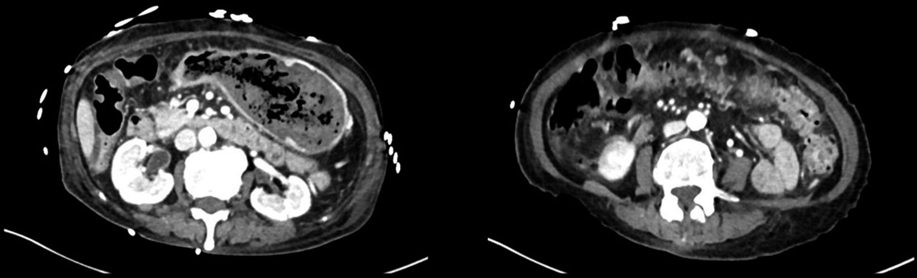
An 83-y-old woman presented with fatigue, weight loss, upper abdominal pain made worse by eating, and a palpable mid-epigastric mass. A CT scan noted wall thickening of the gastric pylorus and body, measuring up to 1.1 cm, and an enlarged 1.4-cm lymph node adjacent to the right gastroepiploic vessels. PET/CT obtained 70 min after the injection of 442.15 MBq (11.95 mCi) of 18F-FDG demonstrated diffuse gastric wall hypermetabolism and avidity of the enlarged lymph node and an SUVmax of 25 and 15.3, respectively (Fig. 1). Upper endoscopy revealed a large ulcerated distal gastric body and antrum mass extending across the pylorus. Subsequent biopsy was consistent with diffuse large B cell lymphoma and the patient was diagnosed with stage II1 gastrointestinal lymphoma. A CT scan 1 mo after chemotherapy showed improvement (Fig. 2).

FIGURE 1.
  • Download figure
  • Open in new tab
  • Download powerpoint
FIGURE 1.

Marked diffuse gastric wall 18F-FDG uptake with an enlarged and avid adjacent lymph node.

FIGURE 2.
  • Download figure
  • Open in new tab
  • Download powerpoint
FIGURE 2.

One-month follow-up CT shows significant improvement.

DISCUSSION

The stomach is the most common site for extranodal lymphoma, accounting for up to 50% of gastrointestinal lymphomas (1). Primary gastric lymphoma (PGL) is most commonly either low-grade mucosa-associated lymphoid tissue lymphoma or diffuse large B cell lymphoma, which vary in terms of histology, epidemiology, morbidity, and treatment yet appear similar on imaging.

18F-FDG PET/CT is valuable in both detecting and staging PGL. The sensitivity of 18F-FDG PET/CT for detecting the diffuse large B cell lymphoma subtype of PGL is estimated to be 97%–100% (2). The high sensitivity is helpful because submucosal disease can be missed on endoscopy.

The Lugano staging system defines stage I as confined to the source organ, stage II as abdominal lymph node spread (local II1, distant II2) or adjacent organ involvement (IIE), and stage IV as disseminated extranodal disease or supradiaphragmatic nodal involvement; no stage III is used. Given improved detection of unsuspected distant disease, particularly extranodal involvement, with 18F-FDG PET/CT over anatomic imaging alone, PET/CT has been shown to correctly upstage disease in 22% and downstage disease in 14% of cases (3). SUVmax has also been shown to correlate with aggressiveness, with higher uptake associated with advanced Lugano stage (2).

CT findings of focal wall thickening and secondary gastric outlet obstruction suggest gastric adenocarcinoma over PGL. 18F-FDG PET/CT, however, can quantitatively differentiate wall thickening associated with PGL and nonlymphomatous cancers because there is generally a linear relationship between SUVmax and maximal gastric wall thickness in nonlymphomatous cancers, whereas PGL does not show a correlation between the 2 measurements (4). Our patient had maximal gastric wall thickness of 1.1 cm with an SUVmax of 25.0. Wu et al. emphasize this point, showing that 18F-FDG metabolism was independent of wall thickness in PGL allowing for exceptionally avid gastric walls throughout a wide range of wall thickness, as is the case in our patient (4).

CONCLUSION

18F-FDG PET/CT can be useful to differentiate PGL from other nonlymphomatous gastric cancers and provides more accurate staging information.

DISCLOSURE

No potential conflict of interest relevant to this article was reported.

Footnotes

  • Published online Aug. 4, 2016.

REFERENCES

  1. 1.↵
    1. Paes FM,
    2. Kalkanis DG,
    3. Sideras PA,
    4. et al
    . FDG PET/CT of extranodal involvement in non-Hodgkin lymphoma and Hodgkin disease. Radiographics. 2010;30:269–291.
    OpenUrlCrossRefPubMed
  2. 2.↵
    1. Yi JH,
    2. Kim SJ,
    3. Choi JY,
    4. et al
    . 18F-FDG uptake and its clinical relevance in primary gastric lymphoma. Hematol Oncol. 2010;28:57–61.
    OpenUrlPubMed
  3. 3.↵
    1. Wu CX,
    2. Zhu ZH
    . Diagnosis and evaluation of gastric cancer by positron emission tomography. World J Gastroenterol. 2014;20:4574–4585.
    OpenUrl
  4. 4.↵
    1. Wu J,
    2. Zhu H,
    3. Li K,
    4. et al
    . 18F-fluorodeoxyglucose positron emission tomography/computed tomography findings of gastric lymphoma: comparisons with gastric cancer. Oncol Lett. 2014;8:1757–1764.
    OpenUrlPubMed
  • Received for publication April 13, 2016.
  • Accepted for publication June 18, 2016.
PreviousNext
Back to top

In this issue

Journal of Nuclear Medicine Technology: 44 (4)
Journal of Nuclear Medicine Technology
Vol. 44, Issue 4
December 1, 2016
  • Table of Contents
  • About the Cover
  • Index by author
Print
Download PDF
Article Alerts
Sign In to Email Alerts with your Email Address
Email Article

Thank you for your interest in spreading the word on Journal of Nuclear Medicine Technology.

NOTE: We only request your email address so that the person you are recommending the page to knows that you wanted them to see it, and that it is not junk mail. We do not capture any email address.

Enter multiple addresses on separate lines or separate them with commas.
18F-FDG PET/CT Imaging of Primary Gastric Lymphoma
(Your Name) has sent you a message from Journal of Nuclear Medicine Technology
(Your Name) thought you would like to see the Journal of Nuclear Medicine Technology web site.
Citation Tools
18F-FDG PET/CT Imaging of Primary Gastric Lymphoma
Brady S. Davis, Trevor A. Thompson, Ely A. Wolin
Journal of Nuclear Medicine Technology Dec 2016, 44 (4) 263-264; DOI: 10.2967/jnmt.116.176842

Citation Manager Formats

  • BibTeX
  • Bookends
  • EasyBib
  • EndNote (tagged)
  • EndNote 8 (xml)
  • Medlars
  • Mendeley
  • Papers
  • RefWorks Tagged
  • Ref Manager
  • RIS
  • Zotero
Share
18F-FDG PET/CT Imaging of Primary Gastric Lymphoma
Brady S. Davis, Trevor A. Thompson, Ely A. Wolin
Journal of Nuclear Medicine Technology Dec 2016, 44 (4) 263-264; DOI: 10.2967/jnmt.116.176842
Twitter logo Facebook logo LinkedIn logo Mendeley logo
  • Tweet Widget
  • Facebook Like
  • Google Plus One
Bookmark this article

Jump to section

  • Article
    • Abstract
    • CASE REPORT
    • DISCUSSION
    • CONCLUSION
    • DISCLOSURE
    • Footnotes
    • REFERENCES
  • Figures & Data
  • Info & Metrics
  • PDF

Related Articles

  • No related articles found.
  • PubMed
  • Google Scholar

Cited By...

  • No citing articles found.
  • Google Scholar

More in this TOC Section

  • High-Sensitivity Troponin Elevation in a Young Woman with Typical Chest Pain: The Heart of the Matter
  • Pulmonary Adenocarcinoma Revealed by Parathyroid Scintigraphy: An Incidental Case to Remember
  • Prominent Right Ventricular Tracer Uptake: A Harbinger of Multivessel Coronary Artery Disease
Show more Teaching Case Studies

Similar Articles

Keywords

  • gastrointestinal
  • oncology
  • GI
  • lymphoma
  • PET
  • PET/CT
  • Gastric Cancer
  • Primary Gastric Lymphoma
SNMMI

© 2025 SNMMI

Powered by HighWire