
Main menu

  • Home
  • Content
    • Current
      • JNMT Supplement
    • Ahead of print
    • Past Issues
    • Continuing Education
    • JNMT Podcast
    • SNMMI Annual Meeting Abstracts
  • Subscriptions
    • Subscribers
    • Rates
    • Journal Claims
    • Institutional and Non-member
  • Authors
    • Submit to JNMT
    • Information for Authors
    • Assignment of Copyright
    • AQARA Requirements
  • Info
    • Reviewers
    • Permissions
    • Advertisers
    • Corporate & Special Sales
  • About
    • About Us
    • Editorial Board
    • Contact Information
  • More
    • Alerts
    • Feedback
    • Help
    • SNMMI Journals
  • SNMMI
    • JNMT
    • JNM
    • SNMMI Journals
    • SNMMI

User menu

  • Subscribe
  • My alerts
  • Log in
  • My Cart

Search

  • Advanced search
Journal of Nuclear Medicine Technology
  • SNMMI
    • JNMT
    • JNM
    • SNMMI Journals
    • SNMMI
  • Subscribe
  • My alerts
  • Log in
  • My Cart
Journal of Nuclear Medicine Technology

Advanced Search

  • Home
  • Content
    • Current
    • Ahead of print
    • Past Issues
    • Continuing Education
    • JNMT Podcast
    • SNMMI Annual Meeting Abstracts
  • Subscriptions
    • Subscribers
    • Rates
    • Journal Claims
    • Institutional and Non-member
  • Authors
    • Submit to JNMT
    • Information for Authors
    • Assignment of Copyright
    • AQARA Requirements
  • Info
    • Reviewers
    • Permissions
    • Advertisers
    • Corporate & Special Sales
  • About
    • About Us
    • Editorial Board
    • Contact Information
  • More
    • Alerts
    • Feedback
    • Help
    • SNMMI Journals
  • Watch or Listen to JNMT Podcast
  • Visit SNMMI on Facebook
  • Join SNMMI on LinkedIn
  • Follow SNMMI on Twitter
  • Subscribe to JNMT RSS feeds
Review ArticleTeaching Case Studies

18F-FDG PET/CT Imaging of Hodgkin Lymphoma in a Child with Common Variable Immunodeficiency

Ebru Tatci, Inci Uslu Biner, Hikmet Gulsah Tanyildiz, Ozlem Ozmen, Atila Gokcek, Gurses Sahin and Zuhal Tazeler
Journal of Nuclear Medicine Technology December 2016, 44 (4) 259-260; DOI: https://doi.org/10.2967/jnmt.116.175745
Ebru Tatci
1Department of Nuclear Medicine, Ataturk Chest Diseases and Thoracic Surgery Training and Research Hospital, Ankara, Turkey
  • Find this author on Google Scholar
  • Find this author on PubMed
  • Search for this author on this site
Inci Uslu Biner
1Department of Nuclear Medicine, Ataturk Chest Diseases and Thoracic Surgery Training and Research Hospital, Ankara, Turkey
  • Find this author on Google Scholar
  • Find this author on PubMed
  • Search for this author on this site
Hikmet Gulsah Tanyildiz
2Department of Pediatric Oncology and Haematology, Dr. Sami Ulus Maternity and Children’s Health and Diseases Training and Research Hospital, Ankara, Turkey; and
  • Find this author on Google Scholar
  • Find this author on PubMed
  • Search for this author on this site
Ozlem Ozmen
1Department of Nuclear Medicine, Ataturk Chest Diseases and Thoracic Surgery Training and Research Hospital, Ankara, Turkey
  • Find this author on Google Scholar
  • Find this author on PubMed
  • Search for this author on this site
Atila Gokcek
3Department of Radiology, Ataturk Chest Diseases and Thoracic Surgery Training and Research Hospital, Ankara, Turkey
  • Find this author on Google Scholar
  • Find this author on PubMed
  • Search for this author on this site
Gurses Sahin
2Department of Pediatric Oncology and Haematology, Dr. Sami Ulus Maternity and Children’s Health and Diseases Training and Research Hospital, Ankara, Turkey; and
  • Find this author on Google Scholar
  • Find this author on PubMed
  • Search for this author on this site
Zuhal Tazeler
1Department of Nuclear Medicine, Ataturk Chest Diseases and Thoracic Surgery Training and Research Hospital, Ankara, Turkey
  • Find this author on Google Scholar
  • Find this author on PubMed
  • Search for this author on this site
  • Article
  • Figures & Data
  • Info & Metrics
  • PDF
Loading

Abstract

Common variable immunodeficiency is characterized by low levels of serum immunoglobulins and antibodies, recurrent infections, and a predisposition to malignancy. Here, we present the 18F-FDG PET/CT findings of a 7-y-old boy with common variable immunodeficiency and Hodgkin lymphoma.

  • immunodeficiency
  • PET/CT
  • lymphoma

Common variable immunodeficiency is one of the most common primary immune deficiencies, affecting approximately 1 in 10,000–50,000 people. Recurrent infections and granulomatous diseases are main features of this disorder. The risk of malignancy, especially gastric carcinoma and lymphoma, also increases in patients with common variable immunodeficiency (1). Here, we report the 18F-FDG PET/CT findings of a 7-y-old boy with common variable immunodeficiency and Hodgkin lymphoma.

CASE REPORT

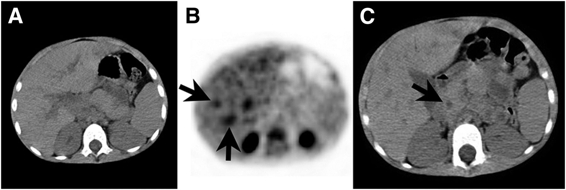
Hodgkin lymphoma was detected in a 7-y-old boy with common variable immunodeficiency. The patient had experienced recurrent lower respiratory tract infections, growth retardation, chronic diarrhea, and hypogammaglobulinemia since he was 4 y old. PET/CT for staging of Hodgkin lymphoma demonstrated 18F-FDG–avid supradiaphragmatic lymphadenopathies (Fig. 1). Abdominal lymphadenopathies seen on the CT images of the PET/CT examination did not show distinctive 18F-FDG uptake (Fig. 2). Widespread bronchiectasis, accompanying infiltration, and peribronchovascular thickening with intense 18F-FDG uptake in the lung fields were demonstrated bilaterally but predominantly in the left lung (Fig. 1). Dilatation of the ductus choledochus and 18F-FDG accumulation in the biliary tract, suggesting cholangitis, were also seen on the PET/CT images (Fig. 3). MR cholangiopancreatography revealed intra- and extrahepatic duct dilatation and lymph nodes in the hepatic hilus. High alanine aminotransferase and aspartate aminotransferase levels were noted. It was thought that an obstructive suppurative cholangitis had resulted from biliary obstruction provoked by enlarged lymph nodes. The patient received 6 cycles of doxorubicin, vinblastine, and dacarbazine and 3,000 cGy of total mediastinal radiotherapy. Bleomycin was not administered. Unfortunately, the patient died of sepsis 15 mo after the initial diagnosis. Informed consent for publication of the case was obtained from the patient’s parents.

FIGURE 1.
  • Download figure
  • Open in new tab
  • Download powerpoint
FIGURE 1.

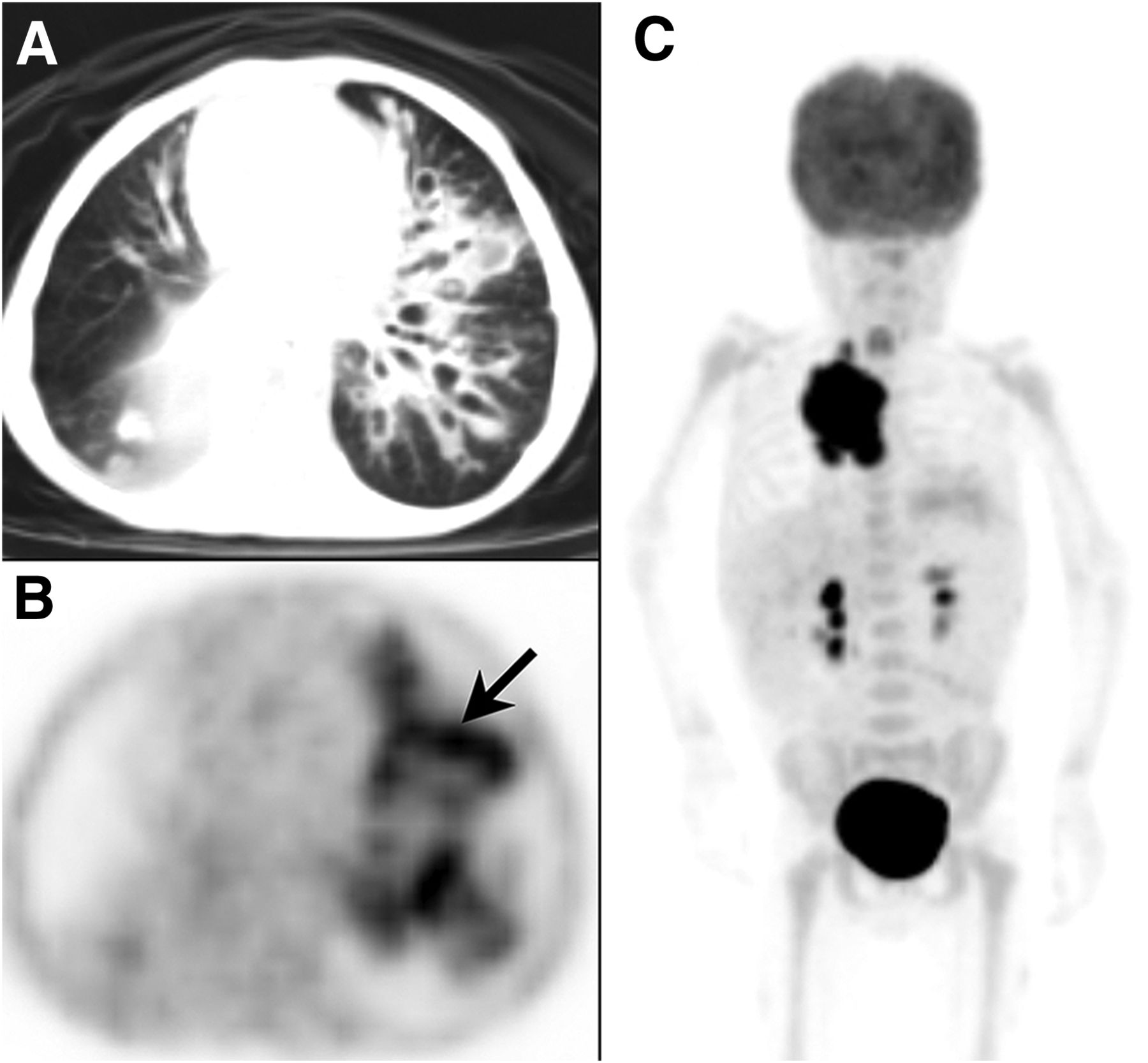
PET/CT demonstrated widespread bronchiectasis, accompanying infiltration, and peribronchovascular thickening in left lung (A and B, arrow) and 18F-FDG–avid supradiaphragmatic lymph nodes (C).

FIGURE 2.
  • Download figure
  • Open in new tab
  • Download powerpoint
FIGURE 2.

Abdominal lymph nodes were seen on the CT images (arrows), but they showed no distinctive uptake on the PET images.

FIGURE 3.
  • Download figure
  • Open in new tab
  • Download powerpoint
FIGURE 3.

18F-FDG PET/CT demonstrated 18F-FDG accumulation in biliary tract (A and B, arrows) and dilatation of ductus choledochus (C, arrow).

DISCUSSION

In our patient, infectious foci in the lung and biliary tract were detected on the 18F-FDG PET/CT scan. Recognition of coincidental infections is important in cancer patients under immunosuppression because they have an increased risk of sepsis secondary to the toxic effect of chemotherapy. 18F-FDG PET/CT may therefore be valuable for monitoring patients with common variable immunodeficiency. However, patients with this disorder are hypersensitive to x-irradiation and radiomimetic drugs such as bleomycin, and increased radiosensitivity is one of the risk factors for malignancy. It has been shown that lymphocytes derived from patients with common variable immunodeficiency are significantly more radiosensitive than those from healthy individuals and that chromosomal aberrations (chromatid breaks and gaps) in such patients increase after x-ray exposure (2). Significant radiation exposure occurs from PET/CT, even with a child-adapted low-dose regimen. In one study, the average radiation dose from 18F-FDG PET/CT was estimated to be 12.15 mSv: 5.89 mSv from 18F-FDG PET and 6.26 mSv from CT (3). 18F-FDG PET/MRI provides a lower radiation exposure, and the sensitivity and specificity of 18F-FDG PET/MR have been shown to be similar to those of 18F-FDG PET/CT for the staging of lymphoma patients (4).

CONCLUSION

18F-FDG PET/CT may be useful in monitoring malignancies and detecting infectious foci in pediatric patients. However, because of the risk of hypersensitivity to x-irradiation, PET/MR may be a better choice for staging lymphomas in patients with primary immune deficiencies.

DISCLOSURE

No potential conflict of interest relevant to this article was reported.

Footnotes

  • Published online Jun. 30, 2016.

REFERENCES

  1. 1.↵
    1. Salzer U,
    2. Warnatz K,
    3. Peter HH
    . Common variable immunodeficiency: an update. Arthritis Res Ther. 2012;14:223.
    OpenUrlCrossRefPubMed
  2. 2.↵
    1. Aghamohammadi A,
    2. Moin M,
    3. Kouhi A,
    4. et al
    . Chromosomal radiosensitivity in patients with common variable immunodeficiency. Immunobiology. 2008;213:447–454.
    OpenUrlPubMed
  3. 3.↵
    1. Kwon HW,
    2. Kim JP,
    3. Lee HJ,
    4. et al
    . Radiation dose from whole-body F-18 fluorodeoxyglucose positron emission tomography/computed tomography: nationwide survey in Korea. J Korean Med Sci. 2016;31(suppl):S69–S74.
    OpenUrl
  4. 4.↵
    1. Herrmann K,
    2. Queiroz M,
    3. Huellner MW,
    4. et al
    . Diagnostic performance of FDG-PET/MRI and WB-DW-MRI in the evaluation of lymphoma: a prospective comparison to standard FDG-PET/CT. BMC Cancer. 2015;15:1002.
    OpenUrl
  • Received for publication March 16, 2016.
  • Accepted for publication June 1, 2016.
PreviousNext
Back to top

In this issue

Journal of Nuclear Medicine Technology: 44 (4)
Journal of Nuclear Medicine Technology
Vol. 44, Issue 4
December 1, 2016
  • Table of Contents
  • About the Cover
  • Index by author
Print
Download PDF
Article Alerts
Sign In to Email Alerts with your Email Address
Email Article

Thank you for your interest in spreading the word on Journal of Nuclear Medicine Technology.

NOTE: We only request your email address so that the person you are recommending the page to knows that you wanted them to see it, and that it is not junk mail. We do not capture any email address.

Enter multiple addresses on separate lines or separate them with commas.
18F-FDG PET/CT Imaging of Hodgkin Lymphoma in a Child with Common Variable Immunodeficiency
(Your Name) has sent you a message from Journal of Nuclear Medicine Technology
(Your Name) thought you would like to see the Journal of Nuclear Medicine Technology web site.
Citation Tools
18F-FDG PET/CT Imaging of Hodgkin Lymphoma in a Child with Common Variable Immunodeficiency
Ebru Tatci, Inci Uslu Biner, Hikmet Gulsah Tanyildiz, Ozlem Ozmen, Atila Gokcek, Gurses Sahin, Zuhal Tazeler
Journal of Nuclear Medicine Technology Dec 2016, 44 (4) 259-260; DOI: 10.2967/jnmt.116.175745

Citation Manager Formats

  • BibTeX
  • Bookends
  • EasyBib
  • EndNote (tagged)
  • EndNote 8 (xml)
  • Medlars
  • Mendeley
  • Papers
  • RefWorks Tagged
  • Ref Manager
  • RIS
  • Zotero
Share
18F-FDG PET/CT Imaging of Hodgkin Lymphoma in a Child with Common Variable Immunodeficiency
Ebru Tatci, Inci Uslu Biner, Hikmet Gulsah Tanyildiz, Ozlem Ozmen, Atila Gokcek, Gurses Sahin, Zuhal Tazeler
Journal of Nuclear Medicine Technology Dec 2016, 44 (4) 259-260; DOI: 10.2967/jnmt.116.175745
Twitter logo Facebook logo LinkedIn logo Mendeley logo
  • Tweet Widget
  • Facebook Like
  • Google Plus One
Bookmark this article

Jump to section

  • Article
    • Abstract
    • CASE REPORT
    • DISCUSSION
    • CONCLUSION
    • DISCLOSURE
    • Footnotes
    • REFERENCES
  • Figures & Data
  • Info & Metrics
  • PDF

Related Articles

  • No related articles found.
  • PubMed
  • Google Scholar

Cited By...

  • No citing articles found.
  • Google Scholar

More in this TOC Section

  • High-Sensitivity Troponin Elevation in a Young Woman with Typical Chest Pain: The Heart of the Matter
  • Pulmonary Adenocarcinoma Revealed by Parathyroid Scintigraphy: An Incidental Case to Remember
  • Prominent Right Ventricular Tracer Uptake: A Harbinger of Multivessel Coronary Artery Disease
Show more Teaching Case Studies

Similar Articles

Keywords

  • Immunodeficiency
  • PET/CT
  • lymphoma
SNMMI

© 2025 SNMMI

Powered by HighWire