


Article Figures & Data
Figures
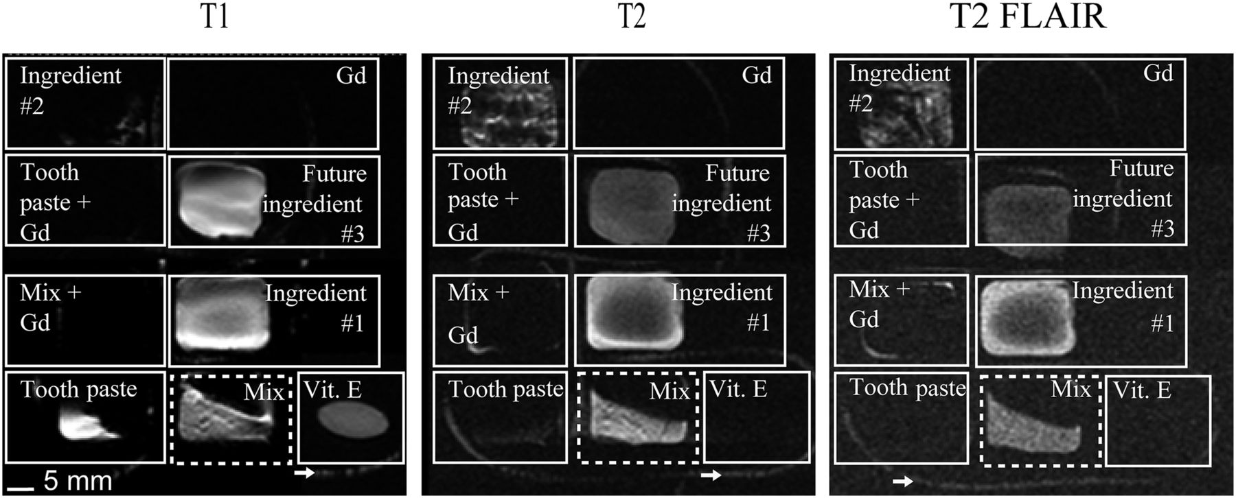
- FIGURE 1.
Initial test of imaging compound (mix) and other ingredients on T1-weighted, T2-weighted, and T2-weighted FLAIR MRI sequences. Arrows indicate compound inside 2-mm-diameter surgical tubing. Commonly used vitamin E tablet disappears in T2-weighted sequences. Ingredient 1 = olive oil; ingredient 2 = purified butter; future ingredient 3 = water.
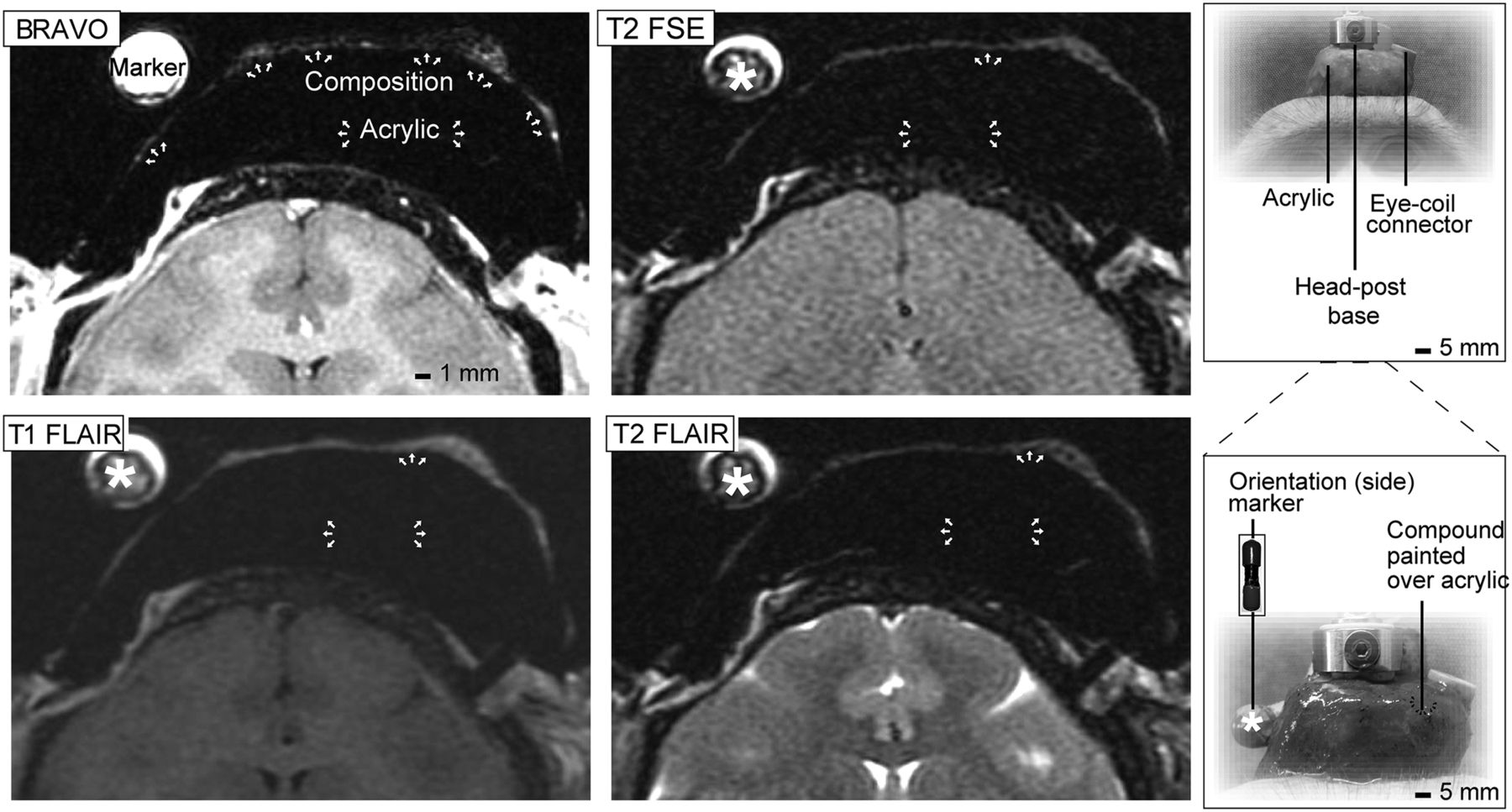
- FIGURE 2.
MRI test of compound in various monkey brain sequences. Thin, white layer of compound (top arrows) is seen on top of dental acrylic (opaque mound indicated by lower white arrows) in all scans. Photographs at right show dental acrylic implant in monkey, orientation marker (8-mm-diameter plastic tube filled with compound), and compound placement. FSE = fast spin echo.
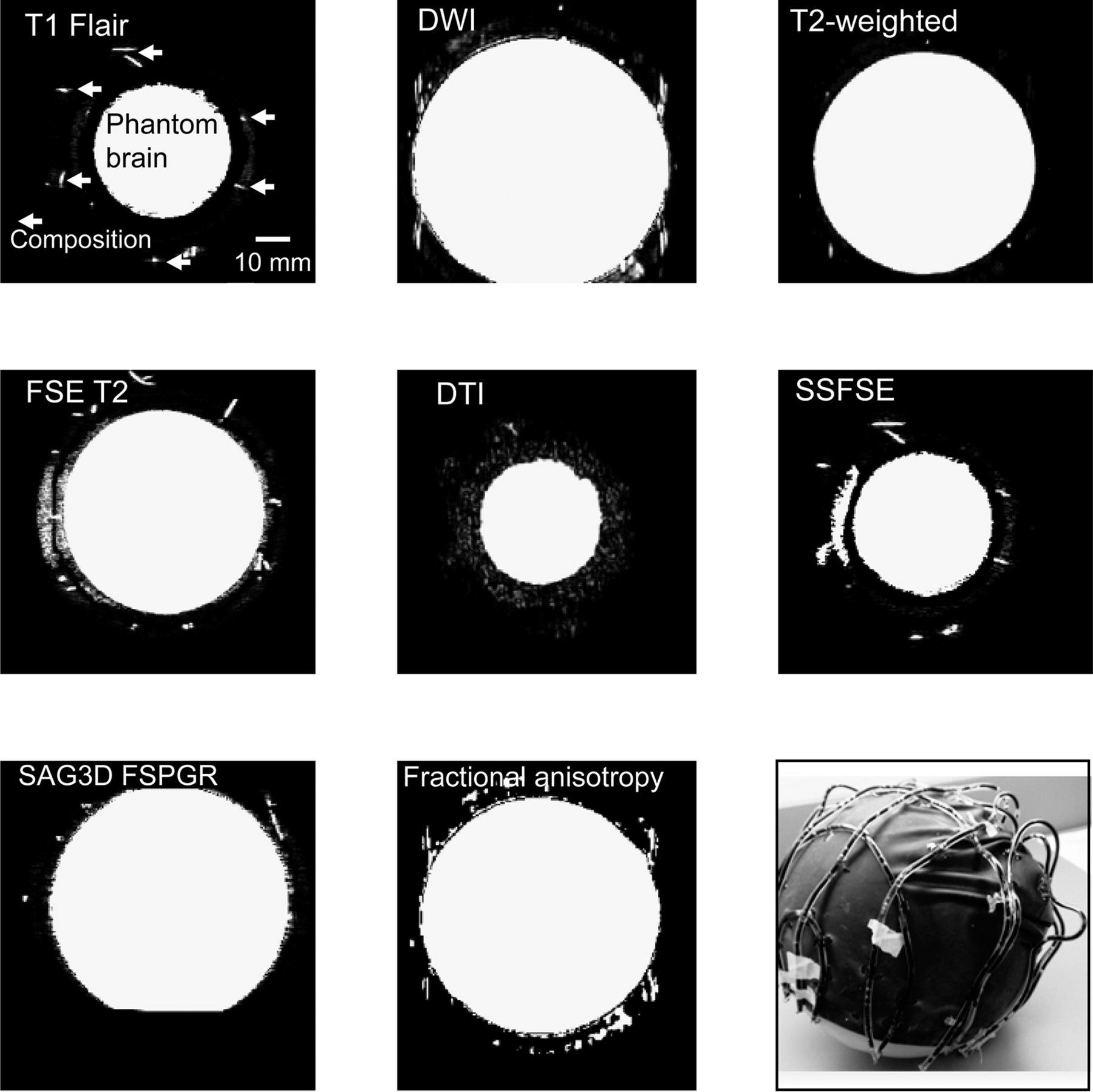
- FIGURE 3.
MRI test of compound in helmet-over-phantom scans. Arrows mark example locations in cross-sectional view of “brain,” where compound is highly MR-visible. Almost all sequences show the compound. Photograph at bottom right shows phantom brain wearing plastic helmet with network of vinyl tubes containing compound. DTI = diffusion tensor imaging; DWI = diffusion-weighted imaging; FSE = fast spin echo; FSPGR = fast spoiled gradient-recalled echo; SAG3D = sagittal 3D; SSFSE = single-shot fast spin echo.
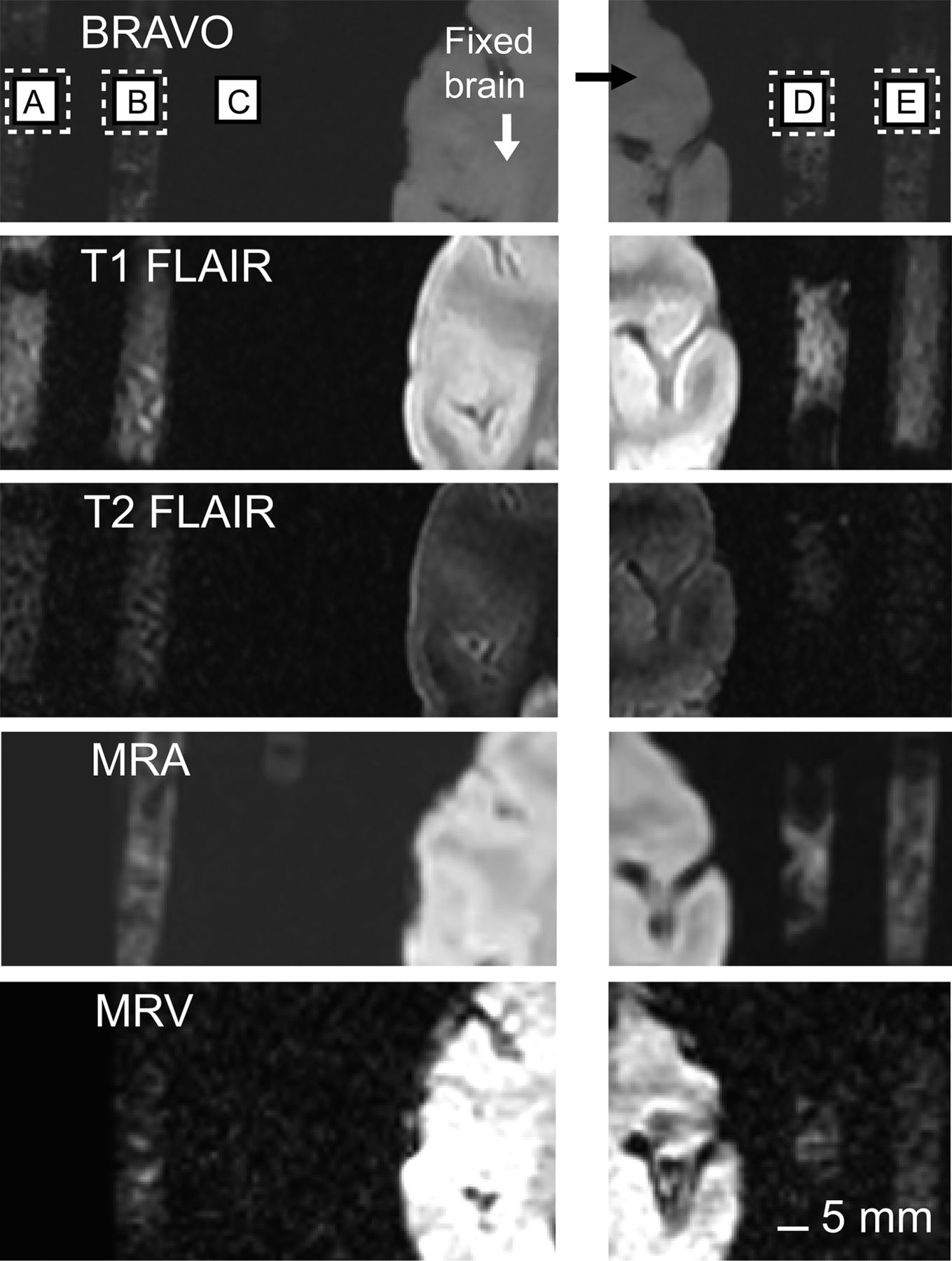
- FIGURE 4.
MRI test of various formulations of compound: formulation 1 mixed with dental acrylic (A), formulation 1 mixed with acrylic (B), dental acrylic alone (C), formulation 1 without acrylic (D), and formulation 2 without acrylic (E). All were put in 1-mL surgical syringes for stability during scans and ease in visual comparisons. Dental acrylic alone is MR-invisible in all sequences. MRA = MR angiography; MRV = MR venography.
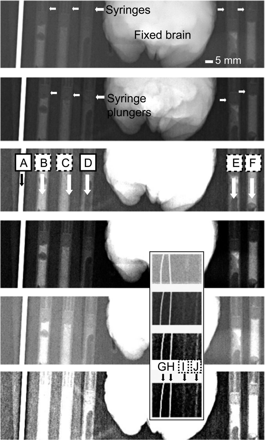
- FIGURE 5.
Radiography test of various formulations of compound: stainless steel guide tube (2-mm-diameter rod [A]), formulation 1 mixed with dental acrylic in 1-mL surgical syringe (B), formulation 2 mixed with acrylic in 1-mL surgical syringe (C), acrylic alone in 1-mL surgical syringe (D), formulation 1 without acrylic in 1-mL surgical syringe (E), and formulation 2 without acrylic in 1-mL surgical syringe (F). Inset: 75-μm-thick microelectrode (G), 250-μm-thick microelectrode (H), formulation 1 without acrylic in 0.5-mm-wide plastic guide tube (I), and formulation 2 without acrylic in 0.5-mm-wide plastic guide tube (J). Same scan is presented with increasing contrast from top to bottom panels.
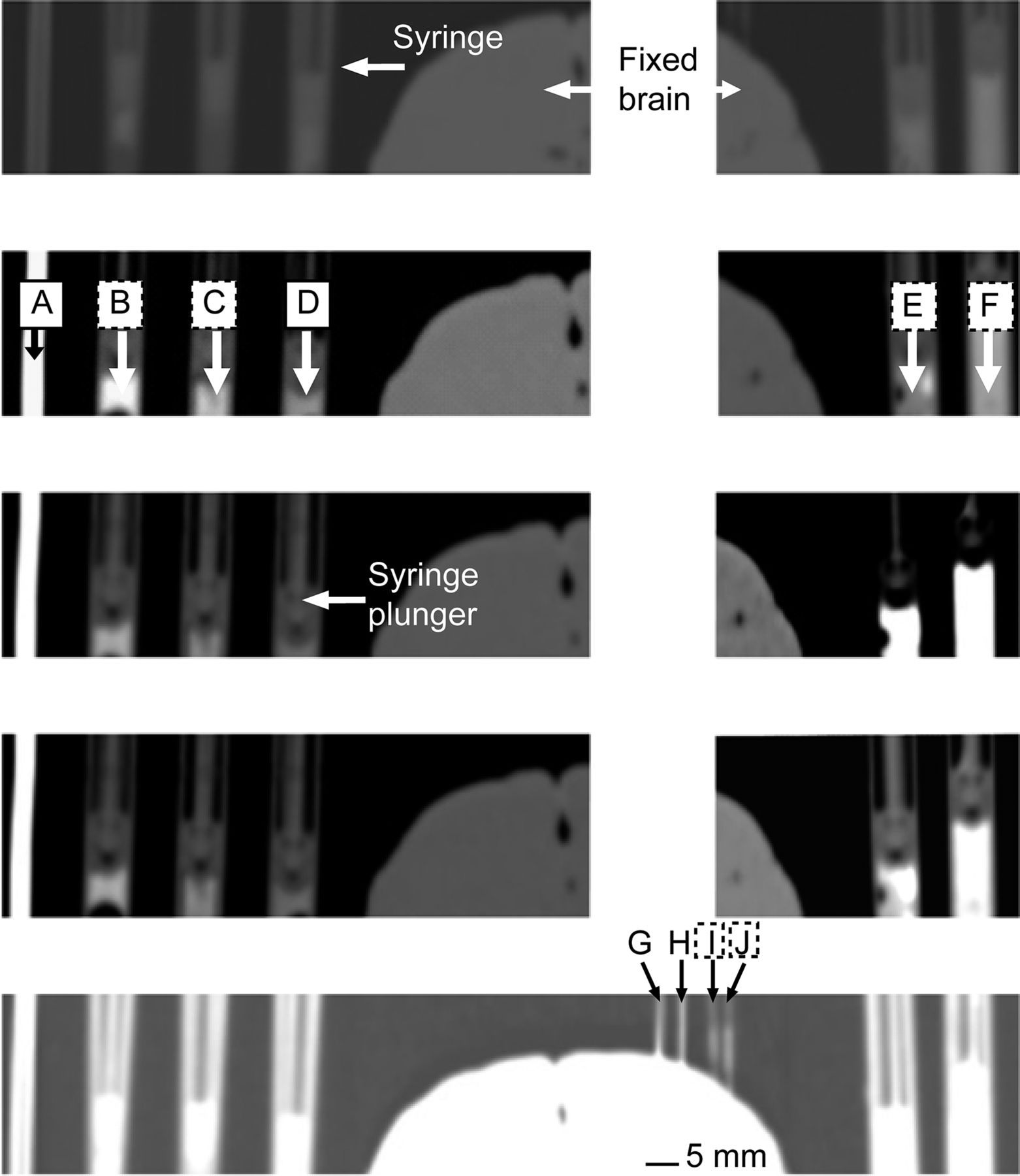
- FIGURE 6.
CT test of various formulations of compound: stainless steel guide tube (2-mm-diameter rod [A]), formulation 1 mixed with dental acrylic in 1-mL surgical syringe (B), formulation 2 mixed with acrylic in 1-mL surgical syringe (C), acrylic alone in 1-mL surgical syringe (D), formulation 1 without acrylic in 1-mL surgical syringe (E), formulation 2 without acrylic in 1-mL surgical syringe (F), 75-μm-thick microelectrode (G), 250-μm-thick microelectrode (H), formulation 1 without acrylic in 0.5-mm-wide plastic guide tube (I), and formulation 2 without acrylic in 0.5-mm-wide plastic guide tube (J). Same scan is presented with increasing contrast from top to bottom panels.
- FIGURE 7.
High- and low-dose 3D CT test of compound: stainless steel guide tube (2-mm-diameter rod [A]), formulation 1 mixed with dental acrylic in 1-mL surgical syringe (B), formulation 2 mixed with acrylic in 1-mL surgical syringe (C), acrylic alone in 1-mL surgical syringe (D), 75-μm-thick microelectrode (E), 250-μm-thick microelectrode (F), formulation 1 without acrylic in 0.5-mm-wide plastic guide tube (G), and formulation 2 without acrylic in 0.5-mm-wide plastic guide tube (H).


Tables
Figure Series Type Echo time (ms) Repetition time (ms) Inversion time (ms) Thickness (mm) Matrix Resolution Pixel spacing (mm) Scanner 1 T1 3D 4 34 0 1 120 × 120 256 × 256 0.47 × 0.47 Signa T2 2D 97.27 5,200 0 2 120 × 120 256 × 256 0.47 × 0.47 Signa FLAIR 2D 120.64 10,002 2,200 2 120 × 120 224 × 256 0.47 × 0.47 Signa 2 T2 fast spin echo 2D 105.76 10,818 2 384 × 256 512 × 512 0.27 × 0.27 Discovery T1 FLAIR 2D 27.68 2,300.06 970.54 2 320 × 224 512 × 512 0.27 × 0.27 Discovery T2 FLAIR 2D 125.60 8,000 2,250 2 320 × 224 512 × 512 0.27 × 0.27 Discovery BRAVO 3D 4.10 9.36 450 2 384 × 256 512 × 512 0.27 × 0.27 Discovery 3 T1 FLAIR 2D 16.81 3,000.01 1,000 5 288 × 192 512 × 512 0.49 × 0.49 Discovery Diffusion-weighted 2D 80.8 8,000 0 5 128 × 128 224 × 256 0.94 × 0.94 Discovery T2 fast spin echo 2D 80.4 8,000 0 4 128 × 128 256 × 256 0.94 × 0.94 Discovery Diffusion tensor 2D 80.4 8,000 0 4 128 × 128 256 × 256 0.94 × 0.94 Discovery Single-shot fast spin echo 2D 40.10 953.78 0 5 288 × 192 512 × 512 0.43 × 0.43 Discovery SAG 3D FSPGR 3D 2.55 6.32 500 1 128 × 128 256 × 256 0.98 × 0.98 Discovery Fractional anisotropy 2D 80.4 8,000 0 4 128 × 128 256 × 256 0.94 × 0.94 Discovery 4 BRAVO 3D 2.55 6.32 500 1 128 × 128 256 × 256 0.98 × 0.98 Discovery T1 FLAIR 2D 24 2,300 2 320 × 244 140 × 140 0.44 × 0.57 Discovery T2 FLAIR 2D 120 8,000 2,250 2 320 × 244 140 × 140 0.44 × 0.57 Discovery MR angiography 3D Out of phase 23 1.2 256 × 192 140 × 105 0.55 × 0.55 Discovery MR venography 2D Minimum Minimum 1.5 256 × 192 140 × 105 0.55 × 0.55 Discovery *1.5-T Signa Excite (GE Healthcare).
†3-T Discovery MR750 (GE Healthcare).
SAG FSPGR = sagittal fast spoiled gradient-recalled echo.
kVp Tube current (mA) Modality Filter Area–dose product (dGy ⋅ cm ⋅ cm) Resolution Field of view Exposure (mAs) Pixel spacing (mm) Exposure time (ms) 62 101 Bone densitometry Multiple/copper 0.09 865 × 1,136 173 × 227 1.61 0.19 × 0.19 16 Scan type Total delivered (mAs) Total DLP (mGy ⋅ cm) Type Tube potential (kV) mAs Reference quality milliamperage CT dose index volume (mGy) DLP mGy ⋅ cm Rotation time (s) Section collimation (mm) Figure High dose 1,628 190 Topogram 120 30 mA 5.3 0.6 6/7 HR 120 9.09 190 0.5 Low dose 854 89 Topogram 120 35 mA 5.3 0.62 7 HR 50 3.80 89 0.5 DLP = dose–length product; total DLP = DLP of entire examination (estimated radiation exposure).


Jump to section
Related Articles
Cited By...
- No citing articles found.