
Main menu

  • Home
  • Content
    • Current
      • JNMT Supplement
    • Ahead of print
    • Past Issues
    • Continuing Education
    • JNMT Podcast
    • SNMMI Annual Meeting Abstracts
  • Subscriptions
    • Subscribers
    • Rates
    • Journal Claims
    • Institutional and Non-member
  • Authors
    • Submit to JNMT
    • Information for Authors
    • Assignment of Copyright
    • AQARA Requirements
  • Info
    • Reviewers
    • Permissions
    • Advertisers
    • Corporate & Special Sales
  • About
    • About Us
    • Editorial Board
    • Contact Information
  • More
    • Alerts
    • Feedback
    • Help
    • SNMMI Journals
  • SNMMI
    • JNMT
    • JNM
    • SNMMI Journals
    • SNMMI

User menu

  • Subscribe
  • My alerts
  • Log in
  • My Cart

Search

  • Advanced search
Journal of Nuclear Medicine Technology
  • SNMMI
    • JNMT
    • JNM
    • SNMMI Journals
    • SNMMI
  • Subscribe
  • My alerts
  • Log in
  • My Cart
Journal of Nuclear Medicine Technology

Advanced Search

  • Home
  • Content
    • Current
    • Ahead of print
    • Past Issues
    • Continuing Education
    • JNMT Podcast
    • SNMMI Annual Meeting Abstracts
  • Subscriptions
    • Subscribers
    • Rates
    • Journal Claims
    • Institutional and Non-member
  • Authors
    • Submit to JNMT
    • Information for Authors
    • Assignment of Copyright
    • AQARA Requirements
  • Info
    • Reviewers
    • Permissions
    • Advertisers
    • Corporate & Special Sales
  • About
    • About Us
    • Editorial Board
    • Contact Information
  • More
    • Alerts
    • Feedback
    • Help
    • SNMMI Journals
  • Watch or Listen to JNMT Podcast
  • Visit SNMMI on Facebook
  • Join SNMMI on LinkedIn
  • Follow SNMMI on Twitter
  • Subscribe to JNMT RSS feeds
Research ArticleImaging

PET/CT for Pancreatic Malignancy: Potential and Pitfalls

Priyanka Jha and Bijan Bijan
Journal of Nuclear Medicine Technology June 2015, 43 (2) 92-97; DOI: https://doi.org/10.2967/jnmt.114.145458
Priyanka Jha
Department of Radiology and Nuclear Medicine, University of California Davis Medical Center, Sacramento, California
  • Find this author on Google Scholar
  • Find this author on PubMed
  • Search for this author on this site
Bijan Bijan
Department of Radiology and Nuclear Medicine, University of California Davis Medical Center, Sacramento, California
  • Find this author on Google Scholar
  • Find this author on PubMed
  • Search for this author on this site
  • Article
  • Figures & Data
  • Supplemental
  • Info & Metrics
  • PDF
Loading

Article Figures & Data

Figures

  • Tables
  • Additional Files
  • FIGURE 1.
    • Download figure
    • Open in new tab
    • Download powerpoint
    FIGURE 1.

    Initial staging PET/CT in a 59-y-old man with pancreatic mass of unknown etiology. (A) Axial fusion PET/CT fusion images demonstrate centrally necrotic mass with hypermetabolic peripheral rim. (B) Axial procedural CT image demonstrates targeted biopsy in progress. PET/CT was used to target hypermetabolic rim to increase diagnostic confidence. Arrows point to peripheral rim.
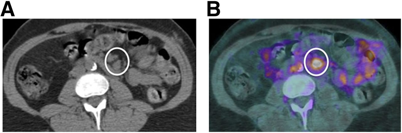
  • FIGURE 2.
    • Download figure
    • Open in new tab
    • Download powerpoint
    FIGURE 2.

    Initial staging PET/CT in a 65-y-old man with known pancreatic adenocarcinoma. (A) Axial noncontrast CT demonstrates enlarged mesenteric lymph nodes (circle). (B) Corresponding PET/CT fusion image demonstrates intense 18F-FDG uptake within nodes suggestive of metastatic involvement. Physiologic uptake is present within small bowel.
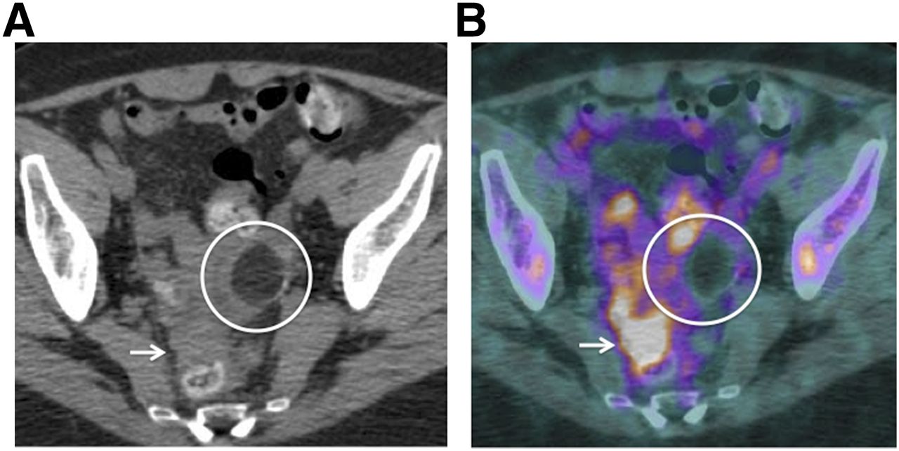
  • FIGURE 3.
    • Download figure
    • Open in new tab
    • Download powerpoint
    FIGURE 3.

    Initial staging PET/CT in a 63-y-old woman with pancreatic adenocarcinoma. (A) Noncontrast axial CT images demonstrates ill-defined peritoneal masses within pelvic cul-de-sac (arrow). This is another difficult location to evaluate with CT alone. (B) Corresponding PET/CT image demonstrates these masses to be intensely hypermetabolic (arrow), helpful in diagnosing peritoneal/mesenteric metastases. Incidental note is made of fat density mass (circle) within left adnexa, which does not demonstrate any 18F-FDG uptake consistent with dermoid/mature cystic teratoma.
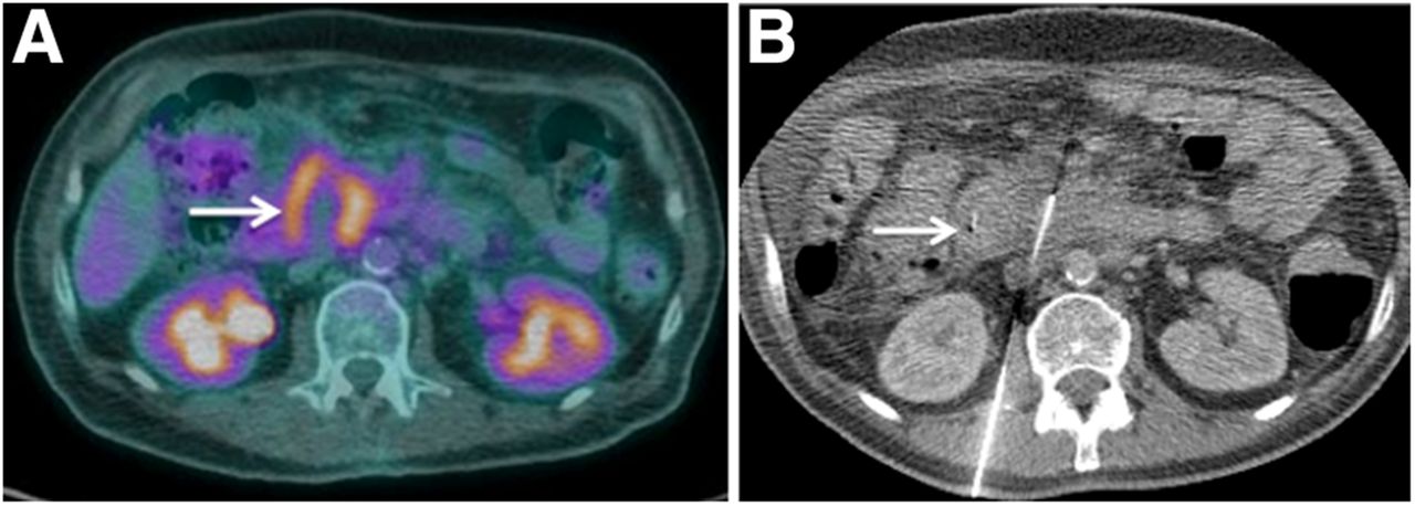
  • FIGURE 4.
    • Download figure
    • Open in new tab
    • Download powerpoint
    FIGURE 4.

    PET/CT performed for evaluating response to chemotherapy in a 69-y-old man with pancreatic adenocarcinoma. (A) Noncontrast CT shows small pleural-based mass adjacent to left lung apex (arrow). (B) Corresponding PET/CT demonstrates its hypermetabolic nature, suggestive of pleural metastasis. This was previously not identified on CECT, and presence of 18F-FDG uptake led to improved identification and, hence, more accurate staging assessment with PET/CT.
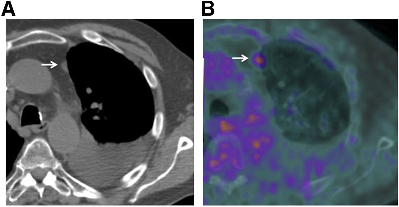
  • FIGURE 5.
    • Download figure
    • Open in new tab
    • Download powerpoint
    FIGURE 5.

    Restaging PET/CT in a 71-y-old woman with pancreatic adenocarcinoma. Hypermetabolic focus at right lung base (arrow) (A) without corresponding nodule on CT component of examination (B) represents misregistration of hepatic activity mimicking a lung nodule.
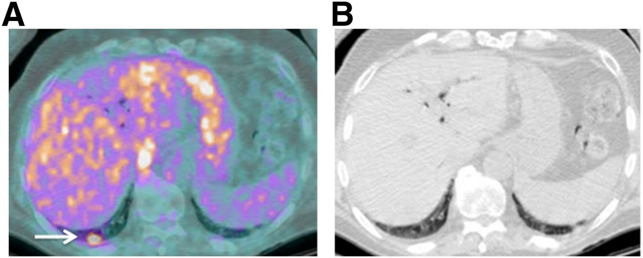
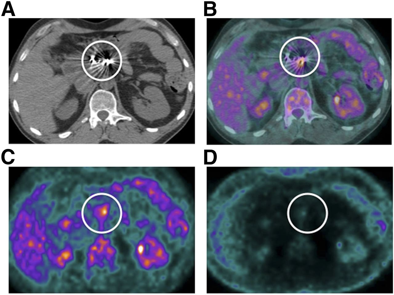
  • FIGURE 6.
    • Download figure
    • Open in new tab
    • Download powerpoint
    FIGURE 6.

    Restaging PET/CT in a 68-y-old man post Whipple’s procedure with multiple surgical clips at surgical site. (A) Dense beam hardening limits evaluation for anatomic details (circle). Fused PET/CT (B) and attenuation-corrected PET (C) demonstrate hypermetabolic focus (circle) corresponding to surgical clips. (D) However, no uptake is present on non–attenuation-corrected image, suggesting that this is attenuation-overcorrection artifact mimicking a lesion.

Tables

  • Figures
  • Additional Files
    • View popup
    TABLE 1

    Imaging Modalities to Assess for Pancreatic Malignancy in Order of Preference

    Disease featuresImaging modalities
    Primary tumor1. CECT
    2. Contrast-enhanced MR imaging
    3. PET/CT
    Locoregional Spread1. CECT
    2. PET/CT
    Vascular invasion1. CECT
    2. Contrast-enhanced MR imaging
    Distant metastases1. PET/CT
    2. CECT

Additional Files

  • Figures
  • Tables
  • Supplemental Data

    Files in this Data Supplement:

    • Supplemental Data
PreviousNext
Back to top

In this issue

Journal of Nuclear Medicine Technology: 43 (2)
Journal of Nuclear Medicine Technology
Vol. 43, Issue 2
June 1, 2015
  • Table of Contents
  • About the Cover
  • Index by author
Print
Download PDF
Article Alerts
Sign In to Email Alerts with your Email Address
Email Article

Thank you for your interest in spreading the word on Journal of Nuclear Medicine Technology.

NOTE: We only request your email address so that the person you are recommending the page to knows that you wanted them to see it, and that it is not junk mail. We do not capture any email address.

Enter multiple addresses on separate lines or separate them with commas.
PET/CT for Pancreatic Malignancy: Potential and Pitfalls
(Your Name) has sent you a message from Journal of Nuclear Medicine Technology
(Your Name) thought you would like to see the Journal of Nuclear Medicine Technology web site.
Citation Tools
PET/CT for Pancreatic Malignancy: Potential and Pitfalls
Priyanka Jha, Bijan Bijan
Journal of Nuclear Medicine Technology Jun 2015, 43 (2) 92-97; DOI: 10.2967/jnmt.114.145458

Citation Manager Formats

  • BibTeX
  • Bookends
  • EasyBib
  • EndNote (tagged)
  • EndNote 8 (xml)
  • Medlars
  • Mendeley
  • Papers
  • RefWorks Tagged
  • Ref Manager
  • RIS
  • Zotero
Share
PET/CT for Pancreatic Malignancy: Potential and Pitfalls
Priyanka Jha, Bijan Bijan
Journal of Nuclear Medicine Technology Jun 2015, 43 (2) 92-97; DOI: 10.2967/jnmt.114.145458
Twitter logo Facebook logo LinkedIn logo Mendeley logo
  • Tweet Widget
  • Facebook Like
  • Google Plus One
Bookmark this article

Jump to section

  • Article
    • Abstract
    • PET/CT APPLICATION IN INITIAL MANAGEMENT OF PANCREATIC MALIGNANCY
    • PET/CT APPLICATION IN STAGING AND PRESURGICAL PLANNING
    • PET/CT APPLICATIONS IN PRERADIOTHERAPY PLANNING
    • PET/CT APPLICATIONS TOWARD PROGNOSIS, RESPONSE TO THERAPY, RECURRENCE, AND SUBSEQUENT MANAGEMENT
    • LIMITATIONS OF PET/CT
    • FUTURE DIRECTIONS
    • CONCLUSION
    • DISCLOSURE
    • Footnotes
    • REFERENCES
  • Figures & Data
  • Supplemental
  • Info & Metrics
  • PDF

Related Articles

  • PubMed
  • Google Scholar

Cited By...

  • Clinical Predictors of Unresectable Disease at Laparotomy in Patients With Pancreatic Ductal Adenocarcinoma Planning to Undergo Surgical Resection
  • Dual-Modality Immuno-PET and Near-Infrared Fluorescence Imaging of Pancreatic Cancer Using an Anti-Prostate Stem Cell Antigen Cys-Diabody
  • Google Scholar

More in this TOC Section

  • Improvement of Anatomic Alignment and Image Quality Using a Respiratory Motion Reduction Block in Oncologic PET/CT
  • Single- Versus Dual-Time-Point Imaging for Transthyretin Cardiac Amyloid Using 99mTc-Pyrophosphate
  • Early 10-Minute Postinjection [18F]F-FAPI-42 uEXPLORER Total-Body PET/CT Scanning Protocol for Staging Lung Cancer Using HYPER Iterative Reconstruction
Show more Imaging

Similar Articles

Keywords

  • Pancreatic malignancy
  • pancreas adenocarcinoma
  • PET/CT
  • PET/MR
  • pancreatic adenocarcinoma
SNMMI

© 2025 SNMMI

Powered by HighWire