


Article Figures & Data
Figures
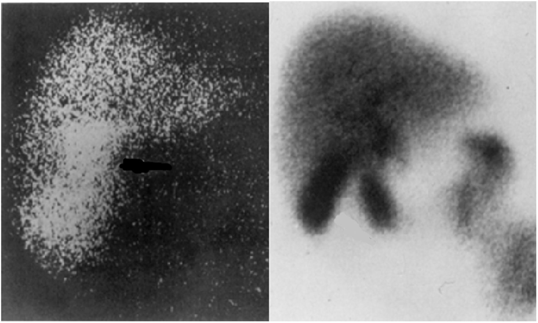
- FIGURE 1.
123I-rose bengal cholescintiscan (left) and 99mTc-HIDA cholescintiscan (right).
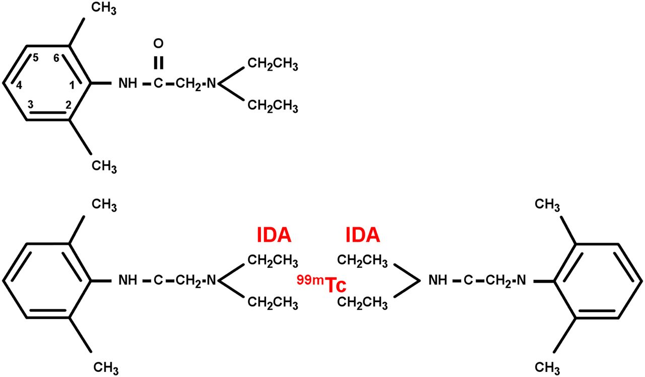
- FIGURE 2.
Chemistry of 99mTc-HIDA radiopharmaceuticals: lidocaine (A) and 99mTc-mebrofenin (B). Two lidocaine analogs are bichelated to 99mTc by IDA.
- FIGURE 3.
Cystic duct sign. (A) 99mTc-HIDA scan ordered to rule out acute cholecystitis. Images show focal accumulation of activity medial to usual position of gallbladder, which remains mostly unchanged over time. (B) Ten-minute SPECT/CT image in same patient shows that focal activity is in cystic duct just distal to occluding cystic duct stone. (Reprinted with permission of (27).)
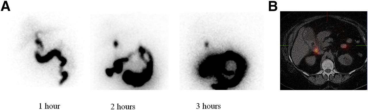
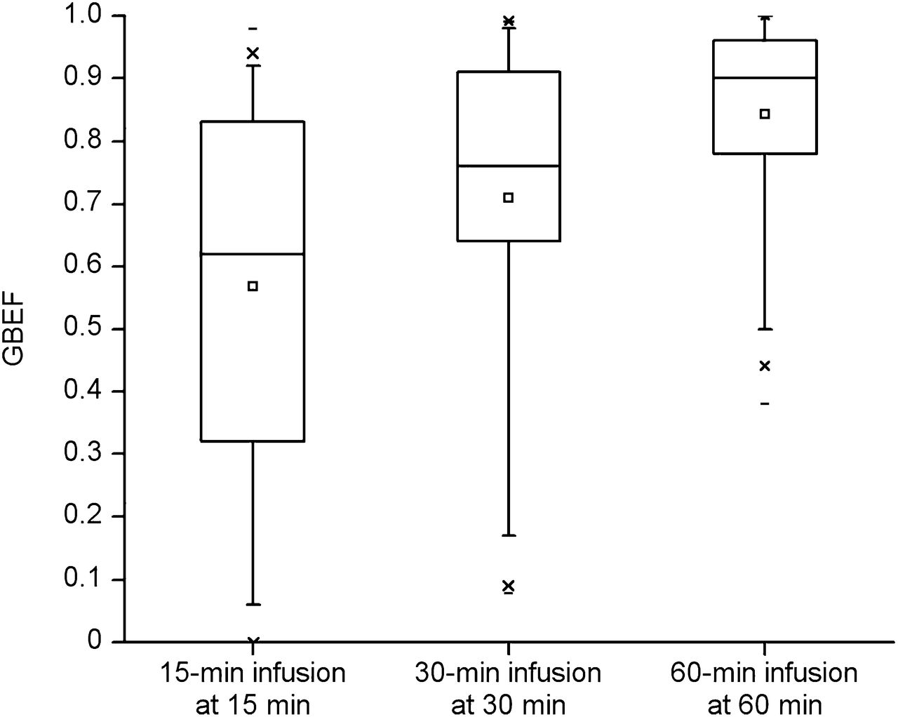
- FIGURE 4.
Comparison trial of multicenter sincalide infusion method. Box plots show distribution of GBEF values for 3 different infusion groups for different lengths of infusion time. The 60-min infusion had the highest GBEFs and the lowest variability. Boxes represent interquartile range (25th−75th percentiles, median line in center, mean is square), bars represent fifth and 95th percentiles, ×s represent first and 95th percentiles, and dash is minimum and maximum. (Reprinted with permission of (48).)
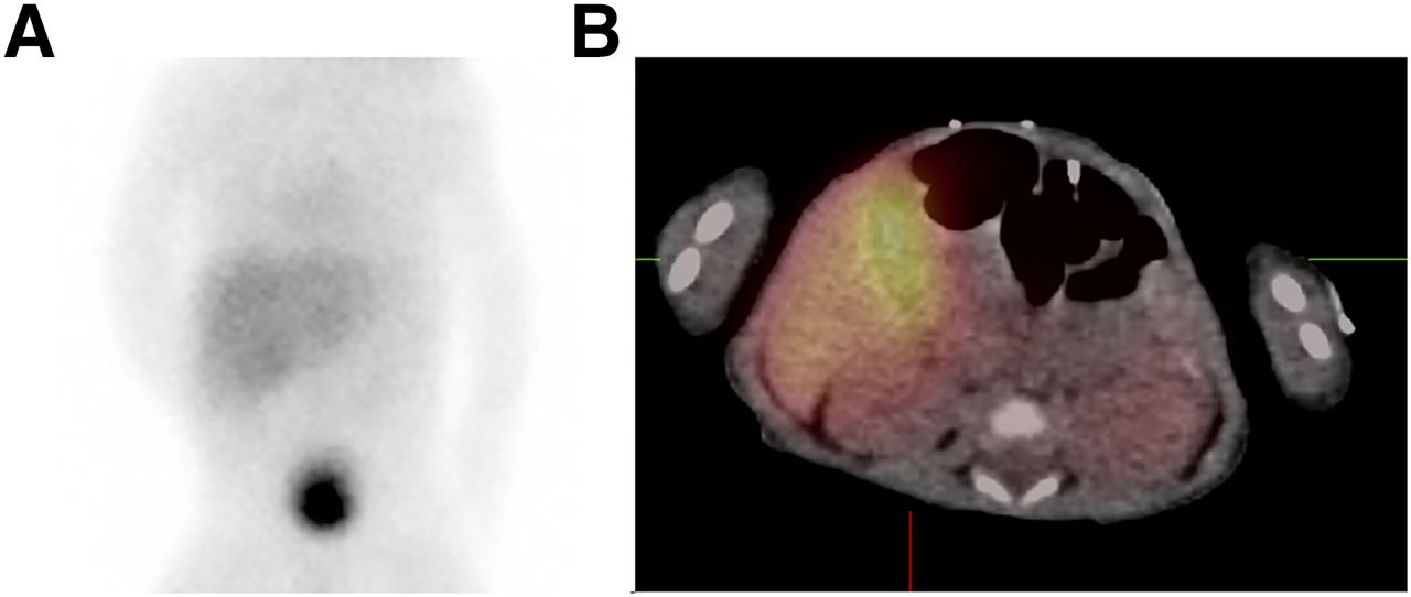
- FIGURE 5.
Study ordered to confirm or exclude biliary atresia. (A) Six-hour 99mTc-HIDA planar static image shows equivocal gallbladder filling and biliary-to-bowel transit. (B) Ten-minute SPECT/CT acquisition confirms activity in gallbladder, excluding biliary atresia. (Reprinted with permission of (27).)


Tables
- TABLE 1
Accuracy of Ultrasonography vs. Cholescintigraphy for Acute Cholecystitis: Direct-Comparison Investigation
Cholescintigraphy Ultrasonography Publication Year No. of patients Sensitivity Specificity Sensitivity Specificity Stadalnik et al. (5) 1978 120 87 100 70 93 Zeman et al. (11) 1981 200 98 81 67 82 Worthen et al. (8) 1981 113 95 100 76 100 Ralls et al. (10) 1982 59 86 84 86 90 Freitas et al. (6) 1982 195 98 90 81 60 Samuels et al. (7) 1983 194 97 93 97 64 Chatziioannou et al. (9) 2000 107 92 89 40 89


