
Main menu

  • Home
  • Content
    • Current
      • JNMT Supplement
    • Ahead of print
    • Past Issues
    • Continuing Education
    • JNMT Podcast
    • SNMMI Annual Meeting Abstracts
  • Subscriptions
    • Subscribers
    • Rates
    • Journal Claims
    • Institutional and Non-member
  • Authors
    • Submit to JNMT
    • Information for Authors
    • Assignment of Copyright
    • AQARA Requirements
  • Info
    • Reviewers
    • Permissions
    • Advertisers
    • Corporate & Special Sales
  • About
    • About Us
    • Editorial Board
    • Contact Information
  • More
    • Alerts
    • Feedback
    • Help
    • SNMMI Journals
  • SNMMI
    • JNMT
    • JNM
    • SNMMI Journals
    • SNMMI

User menu

  • Subscribe
  • My alerts
  • Log in
  • My Cart

Search

  • Advanced search
Journal of Nuclear Medicine Technology
  • SNMMI
    • JNMT
    • JNM
    • SNMMI Journals
    • SNMMI
  • Subscribe
  • My alerts
  • Log in
  • My Cart
Journal of Nuclear Medicine Technology

Advanced Search

  • Home
  • Content
    • Current
    • Ahead of print
    • Past Issues
    • Continuing Education
    • JNMT Podcast
    • SNMMI Annual Meeting Abstracts
  • Subscriptions
    • Subscribers
    • Rates
    • Journal Claims
    • Institutional and Non-member
  • Authors
    • Submit to JNMT
    • Information for Authors
    • Assignment of Copyright
    • AQARA Requirements
  • Info
    • Reviewers
    • Permissions
    • Advertisers
    • Corporate & Special Sales
  • About
    • About Us
    • Editorial Board
    • Contact Information
  • More
    • Alerts
    • Feedback
    • Help
    • SNMMI Journals
  • Watch or Listen to JNMT Podcast
  • Visit SNMMI on Facebook
  • Join SNMMI on LinkedIn
  • Follow SNMMI on Twitter
  • Subscribe to JNMT RSS feeds
Review ArticleTeaching Case Studies

Painful Knee Arthroplasty

Katie Traylor, Prashant Jolepalem and Paresh Mahajan
Journal of Nuclear Medicine Technology June 2014, 42 (2) 122-123; DOI: https://doi.org/10.2967/jnmt.113.132753
Katie Traylor
Department of Diagnostic Radiology and Molecular Imaging, William Beaumont Health System, Royal Oak, Michigan
  • Find this author on Google Scholar
  • Find this author on PubMed
  • Search for this author on this site
Prashant Jolepalem
Department of Diagnostic Radiology and Molecular Imaging, William Beaumont Health System, Royal Oak, Michigan
  • Find this author on Google Scholar
  • Find this author on PubMed
  • Search for this author on this site
Paresh Mahajan
Department of Diagnostic Radiology and Molecular Imaging, William Beaumont Health System, Royal Oak, Michigan
  • Find this author on Google Scholar
  • Find this author on PubMed
  • Search for this author on this site
  • Article
  • Figures & Data
  • Info & Metrics
  • PDF
Loading

Abstract

A triple-tracer nuclear medicine study that incorporates 99mTc-sulfur colloid, 111In-labeled leukocytes, and 99mTc-methylene diphosphonate can be useful for the diagnosis of aseptic loosening in a patient after total-knee arthroplasty, as demonstrated in this case study. The triple-tracer technique takes less time and is more accurate than a bone scan alone.

  • bone
  • instrumentation
  • joint/muscle
  • 111In-labeled autologous leukocytes
  • 99mTc-MDP
  • 99mTc-sulfur colloid

This article discusses the importance of the use of a triple-tracer study in the postoperative knee to diagnose an aseptic knee. The components of the triple tracer will be discussed, as well as how the results of this study can aid the orthopedic surgeon in designing a treatment plan that will be successful for the patient.

CASE REPORT

A 59-y-old woman with a history of rheumatoid arthritis and osteoarthritis presented with 6 mo of progressive right knee pain, which did not improve with conservative management. She had undergone cemented total-right-knee arthroplasty 2 y previously for osteoarthritis. She was previously pain-free and had not undergone recent trauma. She was afebrile and had a mildly elevated white blood cell count of 10.7 billion/L. A triple-tracer study was requested to evaluate for septic prosthesis loosening.

The patient was first injected with 373.7 MBq (10.1 mCi) of 99mTc-sulfur colloid, and spot views of the knees were obtained. The patient was then injected with 20.72 MBq (0.56 mCi) of 111In-labeled leukocytes, and similar-projection 24-h delayed images of the knees were obtained. Lastly, the patient was injected with 862.1 MBq (23.3 mCi) of 99mTc-methylene diphosphonate, and acquisition of dynamic flow and static blood-pool images followed immediately afterward. After 3–4 h of tracer localization, spot views of the knees were obtained.

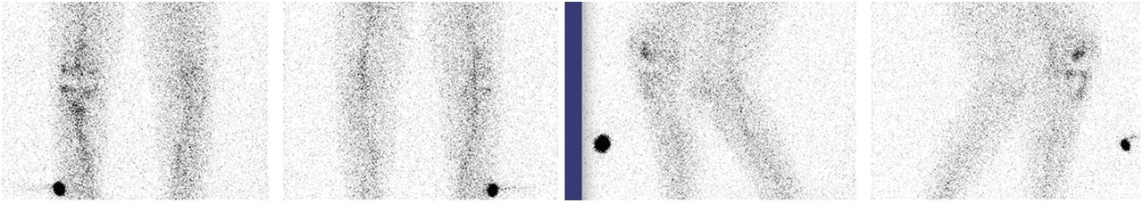
Comparison of the 99mTc-sulfur colloid and 111In-labeled leukocyte images (Fig. 1) did not demonstrate discordant uptake of the latter to suggest infection. On the 3-phase 99mTc-methylene diphosphonate bone scan, there was periprosthetic hyperemia near the tibial component on the early images (Fig. 2). On the delayed images, there was increased uptake at the interface between the tibial plateau native bone and the prosthesis, worse medially (Fig. 3).

FIGURE 1.
  • Download figure
  • Open in new tab
  • Download powerpoint
FIGURE 1.

Anterior/posterior (left) and medial/lateral (right) projection images of knees after injection with 99mTc-sulfur colloid.

FIGURE 2.
  • Download figure
  • Open in new tab
  • Download powerpoint
FIGURE 2.

Anterior/posterior (left) and medial/lateral (right) projection images of knees after injection with 111In-labeled autologous leukocytes.

FIGURE 3.
  • Download figure
  • Open in new tab
  • Download powerpoint
FIGURE 3.

(A) Anterior projection blood-flow images with right-sided marker. (B) Blood-pool 99mTc-methylene diphosphonate images of knees in anterior/posterior projection. (C) Delayed static images of knees in anterior/posterior and medial/lateral projections.

The triple-tracer findings were consistent with aseptic failure of the prosthesis. Surgical exploration did not reveal any signs of infection; however, the tibial component had subsided into the medial tibial plateau and was easily dislodged during removal, concordant with the imaging findings. A new prosthesis was inserted, and after a short convalescent period, the patient was discharged home.

DISCUSSION

More than 700,000 total-knee arthroplasties are performed in the United States annually, of which more than one quarter will eventually need revision. The most frequent cause, aseptic loosening, is an inflammatory reaction to the prosthetic components (1). Subsequently, the particulate debris activates tissue phagocytes but cannot be broken down by cellular enzymes. The continued secretion of proinflammatory cytokines and proteolytic enzymes damages the bone and cartilage and further activates immune cells. The heightened inflammatory response leads to osteolysis and, eventually, loosening of the prosthesis (2).

The infection rate after primary total-knee arthroplasty is approximately 1%, but the treatments of aseptic loosening and infected hardware are very different. Therefore, it is extremely important to differentiate them. Often the initial study is a bone scan; however, it has a variable sensitivity of 67%–100% for diagnosing an infected knee replacement (3,4).

CONCLUSION

In this patient, the orthopedic surgeon was clinically concerned about infection and wanted the patient to expeditiously undergo a 1-stop-shop scan to exclude it. We performed the 99mTc-sulfur colloid scan followed by the 111In-labeled leukocyte scan and then the 3-phase bone scan to complete the triple-tracer protocol. This sequence takes the shortest amount of time and has higher accuracy than the bone scan alone (4).

DISCLOSURE

No potential conflict of interest relevant to this article was reported.

Footnotes

  • Published online Mar. 13, 2014.

REFERENCES

  1. 1.↵
    1. Rini JN,
    2. Bhargava KK,
    3. Tronco GG,
    4. et al
    . PET with FDG-labeled leukocytes versus scintigraphy with In-111 oxine-labeled leukocytes for detection of infection. Radiology. 2006;238:978–987.
    OpenUrlCrossRefPubMed
  2. 2.↵
    1. Henkin RE,
    2. Bova D,
    3. Dillehay GL,
    4. et al
    . Nuclear Medicine. 2nd ed. St. Louis, MO: Mosby; 2006:343–346.
  3. 3.↵
    1. Love C,
    2. Marwin SE,
    3. Palestro CJ
    . Nuclear medicine and the infected joint replacement. Semin Nucl Med. 2009;39:66–78.
    OpenUrlCrossRefPubMed
  4. 4.↵
    1. Love C,
    2. Marwin SE,
    3. Tomas MB,
    4. et al
    . Diagnosing infection in the failed joint replacement: a comparison of coincidence detection fluorine-18-FDG and indium-111-labeled leukocyte/technetium-99m-sulfur colloid marrow imaging. J Nucl Med. 2004;45:1864–1871.
    OpenUrlAbstract/FREE Full Text
  • Received for publication September 17, 2013.
  • Accepted for publication November 24, 2013.
PreviousNext
Back to top

In this issue

Journal of Nuclear Medicine Technology: 42 (2)
Journal of Nuclear Medicine Technology
Vol. 42, Issue 2
June 1, 2014
  • Table of Contents
  • About the Cover
  • Index by author
Print
Download PDF
Article Alerts
Sign In to Email Alerts with your Email Address
Email Article

Thank you for your interest in spreading the word on Journal of Nuclear Medicine Technology.

NOTE: We only request your email address so that the person you are recommending the page to knows that you wanted them to see it, and that it is not junk mail. We do not capture any email address.

Enter multiple addresses on separate lines or separate them with commas.
Painful Knee Arthroplasty
(Your Name) has sent you a message from Journal of Nuclear Medicine Technology
(Your Name) thought you would like to see the Journal of Nuclear Medicine Technology web site.
Citation Tools
Painful Knee Arthroplasty
Katie Traylor, Prashant Jolepalem, Paresh Mahajan
Journal of Nuclear Medicine Technology Jun 2014, 42 (2) 122-123; DOI: 10.2967/jnmt.113.132753

Citation Manager Formats

  • BibTeX
  • Bookends
  • EasyBib
  • EndNote (tagged)
  • EndNote 8 (xml)
  • Medlars
  • Mendeley
  • Papers
  • RefWorks Tagged
  • Ref Manager
  • RIS
  • Zotero
Share
Painful Knee Arthroplasty
Katie Traylor, Prashant Jolepalem, Paresh Mahajan
Journal of Nuclear Medicine Technology Jun 2014, 42 (2) 122-123; DOI: 10.2967/jnmt.113.132753
Twitter logo Facebook logo LinkedIn logo Mendeley logo
  • Tweet Widget
  • Facebook Like
  • Google Plus One
Bookmark this article

Jump to section

  • Article
    • Abstract
    • CASE REPORT
    • DISCUSSION
    • CONCLUSION
    • DISCLOSURE
    • Footnotes
    • REFERENCES
  • Figures & Data
  • Info & Metrics
  • PDF

Related Articles

  • No related articles found.
  • PubMed
  • Google Scholar

Cited By...

  • No citing articles found.
  • Google Scholar

More in this TOC Section

  • High-Sensitivity Troponin Elevation in a Young Woman with Typical Chest Pain: The Heart of the Matter
  • Pulmonary Adenocarcinoma Revealed by Parathyroid Scintigraphy: An Incidental Case to Remember
  • Prominent Right Ventricular Tracer Uptake: A Harbinger of Multivessel Coronary Artery Disease
Show more Teaching Case Studies

Similar Articles

Keywords

  • bone
  • instrumentation
  • Joint/Muscle
  • 111In-labeled autologous leukocytes
  • 99mTc-MDP
  • 99mTc-sulfur colloid
SNMMI

© 2025 SNMMI

Powered by HighWire