  • Main menu
  • User menu
  • Search
  • English ▼
    • English
    • Afrikaans
    • Albanian
    • Amharic
    • Arabic
    • Armenian
    • Azerbaijani
    • Basque
    • Belarusian
    • Bengali
    • Bosnian
    • Bulgarian
    • Catalan
    • Cebuano
    • Chichewa
    • Chinese (Simplified)
    • Chinese (Traditional)
    • Corsican
    • Croatian
    • Czech
    • Danish
    • Dutch
    • Esperanto
    • Estonian
    • Filipino
    • Finnish
    • French
    • Frisian
    • Galician
    • Georgian
    • German
    • Greek
    • Gujarati
    • Haitian Creole
    • Hausa
    • Hawaiian
    • Hebrew
    • Hindi
    • Hmong
    • Hungarian
    • Icelandic
    • Igbo
    • Indonesian
    • Irish
    • Italian
    • Japanese
    • Javanese
    • Kannada
    • Kazakh
    • Khmer
    • Korean
    • Kurdish (Kurmanji)
    • Kyrgyz
    • Lao
    • Latin
    • Latvian
    • Lithuanian
    • Luxembourgish
    • Macedonian
    • Malagasy
    • Malay
    • Malayalam
    • Maltese
    • Maori
    • Marathi
    • Mongolian
    • Myanmar (Burmese)
    • Nepali
    • Norwegian
    • Pashto
    • Persian
    • Polish
    • Portuguese
    • Punjabi
    • Romanian
    • Russian
    • Samoan
    • Scottish Gaelic
    • Serbian
    • Sesotho
    • Shona
    • Sindhi
    • Sinhala
    • Slovak
    • Slovenian
    • Somali
    • Spanish
    • Sudanese
    • Swahili
    • Swedish
    • Tajik
    • Tamil
    • Telugu
    • Thai
    • Turkish
    • Ukrainian
    • Urdu
    • Uzbek
    • Vietnamese
    • Welsh
    • Xhosa
    • Yiddish
    • Yoruba
    • Zulu

Main menu

  • Home
  • Content
    • Current
      • JNMT Supplement
    • Ahead of print
    • Past Issues
    • Continuing Education
    • JNMT Podcast
    • SNMMI Annual Meeting Abstracts
  • Subscriptions
    • Subscribers
    • Rates
    • Journal Claims
    • Institutional and Non-member
  • Authors
    • Submit to JNMT
    • Information for Authors
    • Assignment of Copyright
    • AQARA Requirements
  • Info
    • Reviewers
    • Permissions
    • Advertisers
    • Corporate & Special Sales
  • About
    • About Us
    • Editorial Board
    • Contact Information
  • More
    • Alerts
    • Feedback
    • Help
    • SNMMI Journals
  • SNMMI
    • JNMT
    • JNM
    • SNMMI Journals
    • SNMMI

User menu

  • Subscribe
  • My alerts
  • Log in
  • My Cart

Search

  • Advanced search
Journal of Nuclear Medicine Technology
  • SNMMI
    • JNMT
    • JNM
    • SNMMI Journals
    • SNMMI
  • Subscribe
  • My alerts
  • Log in
  • My Cart
Journal of Nuclear Medicine Technology

Advanced Search

English ▼
  • English
  • Afrikaans
  • Albanian
  • Amharic
  • Arabic
  • Armenian
  • Azerbaijani
  • Basque
  • Belarusian
  • Bengali
  • Bosnian
  • Bulgarian
  • Catalan
  • Cebuano
  • Chichewa
  • Chinese (Simplified)
  • Chinese (Traditional)
  • Corsican
  • Croatian
  • Czech
  • Danish
  • Dutch
  • Esperanto
  • Estonian
  • Filipino
  • Finnish
  • French
  • Frisian
  • Galician
  • Georgian
  • German
  • Greek
  • Gujarati
  • Haitian Creole
  • Hausa
  • Hawaiian
  • Hebrew
  • Hindi
  • Hmong
  • Hungarian
  • Icelandic
  • Igbo
  • Indonesian
  • Irish
  • Italian
  • Japanese
  • Javanese
  • Kannada
  • Kazakh
  • Khmer
  • Korean
  • Kurdish (Kurmanji)
  • Kyrgyz
  • Lao
  • Latin
  • Latvian
  • Lithuanian
  • Luxembourgish
  • Macedonian
  • Malagasy
  • Malay
  • Malayalam
  • Maltese
  • Maori
  • Marathi
  • Mongolian
  • Myanmar (Burmese)
  • Nepali
  • Norwegian
  • Pashto
  • Persian
  • Polish
  • Portuguese
  • Punjabi
  • Romanian
  • Russian
  • Samoan
  • Scottish Gaelic
  • Serbian
  • Sesotho
  • Shona
  • Sindhi
  • Sinhala
  • Slovak
  • Slovenian
  • Somali
  • Spanish
  • Sudanese
  • Swahili
  • Swedish
  • Tajik
  • Tamil
  • Telugu
  • Thai
  • Turkish
  • Ukrainian
  • Urdu
  • Uzbek
  • Vietnamese
  • Welsh
  • Xhosa
  • Yiddish
  • Yoruba
  • Zulu
  • Home
  • Content
    • Current
    • Ahead of print
    • Past Issues
    • Continuing Education
    • JNMT Podcast
    • SNMMI Annual Meeting Abstracts
  • Subscriptions
    • Subscribers
    • Rates
    • Journal Claims
    • Institutional and Non-member
  • Authors
    • Submit to JNMT
    • Information for Authors
    • Assignment of Copyright
    • AQARA Requirements
  • Info
    • Reviewers
    • Permissions
    • Advertisers
    • Corporate & Special Sales
  • About
    • About Us
    • Editorial Board
    • Contact Information
  • More
    • Alerts
    • Feedback
    • Help
    • SNMMI Journals
  • Watch or Listen to JNMT Podcast
  • Visit SNMMI on Facebook
  • Join SNMMI on LinkedIn
  • Follow SNMMI on Twitter
  • Subscribe to JNMT RSS feeds
Review ArticleTeaching Case Studies

Atypical Presentation of Sarcoidosis on 18F-FDG PET/CT

Prashant Jolepalem, Katie Zanyk McLean and Ching-Yee Oliver Wong
Journal of Nuclear Medicine Technology June 2014, 42 (2) 114-115; DOI: https://doi.org/10.2967/jnmt.113.132654
Prashant Jolepalem
1Department of Diagnostic Radiology and Molecular Imaging, Oakland University William Beaumont School of Medicine and Beaumont Health System, Royal Oak, Michigan; and
  • Find this author on Google Scholar
  • Find this author on PubMed
  • Search for this author on this site
Katie Zanyk McLean
2Oakland University William Beaumont School of Medicine, Rochester Hills, Michigan
  • Find this author on Google Scholar
  • Find this author on PubMed
  • Search for this author on this site
Ching-Yee Oliver Wong
1Department of Diagnostic Radiology and Molecular Imaging, Oakland University William Beaumont School of Medicine and Beaumont Health System, Royal Oak, Michigan; and
  • Find this author on Google Scholar
  • Find this author on PubMed
  • Search for this author on this site
  • Article
  • Figures & Data
  • Info & Metrics
  • PDF
Loading

Abstract

On 18F-FDG PET, inflammatory processes can be one of the most confounding factors for interpretation, particularly when the presentation of the process is atypical, as it was in the case of sarcoidosis reported here. Clinicians should be aware that sarcoidosis may mimic lung malignancies and lymphomas, regardless of typical or atypical appearance on 18F-FDG PET.

  • oncology
  • lymphoma
  • PET/CT
  • FDG
  • atypical
  • sarcoidosis

The presence of inflammatory processes on 18F-FDG PET can be one of the most confounding factors for interpretation. Sarcoidosis and other granulomatous processes in the lungs are one of the more common examples of this problem. Quantitative criteria as well as familiarity with the typical appearance of granulomatous disease has somewhat obviated this problem. This case demonstrates an example of how an atypical presentation of sarcoidosis can easily be mistaken for malignancy in a patient being evaluated with PET scanning for diagnostic purposes.

CASE REPORT

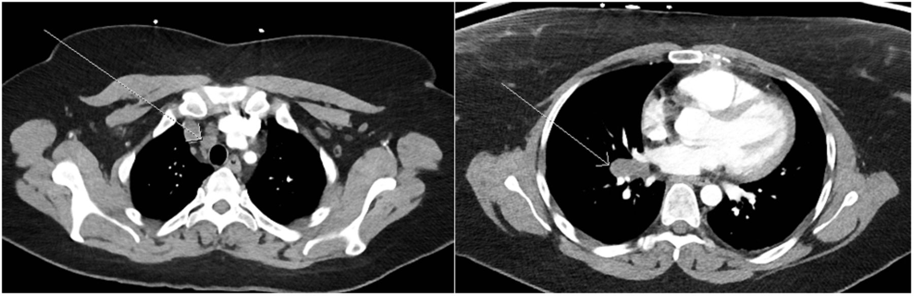
A 35-y-old woman with a history of multiple pulmonary emboli presented with dyspnea and chest pain. A subsequent CT study using a pulmonary embolism protocol did not demonstrate a pulmonary embolism but did detect several prominent mediastinal lymph nodes (Fig. 1). The size and appearance of the lymph nodes raised suspicion for lymphoma.

FIGURE 1.
  • Download figure
  • Open in new tab
  • Download powerpoint
FIGURE 1.

Contrast-enhanced CT scan demonstrating lymphadenopathy in right high paratracheal (left side) and right hilar (right side) lymph nodes (arrows).

An 18F-FDG PET/CT scan was ordered for diagnostic and staging purposes (Fig. 2). The study was performed after injection with 555 MBq of the radiotracer and a 90-min uptake time. The whole-body PET images were obtained for 3 min per bed position, with 6 positions to cover from the base of the skull to the middle of the thighs, using a 3-slice overlap. The images were acquired using the 3-dimensional high-sensitivity mode with an axial field of view in a 256 × 256 matrix. Then, iterative image reconstruction was performed on a 128 × 128 matrix using an ordered-subset expectation maximization algorithm for 30 subsets and 2 iterations, with a 7.0-mm postreconstruction filter. Concomitant CT data were used for attenuation correction using 140 kVp, 120–200 mA, and a 3-mm slice thickness.

FIGURE 2.
  • Download figure
  • Open in new tab
  • Download powerpoint
FIGURE 2.

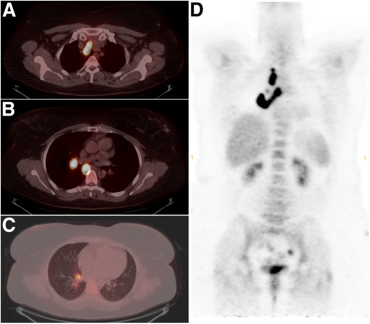
(A–C) Cross sections of PET/CT images showing intensely 18F-FDG–avid lymph nodes in right high paratracheal (A), right hilar and subcarinal (B), and interlobar and lobar (C) regions. (D) Coronal PET image demonstrating distribution pattern of metabolically active lymph nodes.

The images demonstrated abnormal multifocal 18F-FDG uptake only in the right suprahilar (maximum standardized uptake value [SUVmax], 16), right paratracheal (SUVmax, 23), and subcarinal (SUVmax, 21) lymph nodes, with likely station 11 and 12 lymph nodes (SUVmax, 11). This uptake followed a lymphatic drainage pattern suggestive of a pulmonary neoplasm in the right upper lobe. However, there was no evidence of an 18F-FDG–avid parenchymal lesion, leaving open the possibility of lymphoma despite the lack of lymph conglomeration that is typical of such high-grade 18F-FDG activity. Nonetheless, tissue sampling was indicated, and subsequent biopsy of the subcarinal lymph node was consistent with sarcoidosis.

DISCUSSION

18F-FDG uptake can be nonspecific and is associated with a variety of conditions, including infectious processes (pneumonia), chronic inflammatory processes (granulomatous disease), or acute inflammatory processes (postradiation changes) (1). These inflammatory conditions have in common the increased 18F-FDG uptake by neutrophils, macrophages, and lymphocytes that may mimic neoplastic 18F-FDG uptake (2). These conditions may be indistinguishable from malignancy on the basis of PET alone. The 18F-FDG PET study in this case demonstrated several discrete high-grade 18F-FDG–avid lymph nodes in the subcarinal, right paratracheal, and right suprahilar lymph nodes, with likely station 11 and 12 lymph nodes.

The appearance of several discrete, intensely avid lymph nodes is not typical of lymphoma, because cases with such high-grade activity (SUVmax > 10) typically present with lymph node conglomerations. Additionally, other clusters of lymph nodes are typically 18F-FDG–avid, such as the axillary and cervical nodes (3).

The anatomic distribution of the 18F-FDG–avid lymph nodes was consistent with the drainage pattern of a right-upper-lobe neoplasm, as the malignant cells typically deposit into the right paratracheal lymph nodes and may skip stations 10–14 (4). Nevertheless, an 18F-FDG–avid neoplasm was not identified on the imaging, suggesting another etiology.

Activated granulomatous disease (histoplasmosis or sarcoidosis) or pneumonia is often a consideration in cases with a similar appearance. Although pneumonia will also follow anatomic lymph node drainage, the activity is often higher peripherally and decreases in the central lymph drainage sites, as is the case with lung cancer (1,4). Additionally, there was no anatomic evidence of a consolidative or infiltrative process. Conversely, sarcoidosis will typically demonstrate higher activity in the central mediastinal nodes than in the hilar and lobar nodes, will affect both sides of the chest symmetrically, will not follow any specific lymph node drainage, and often does not present with a parenchymal lung lesion. Chundru et al. (1) proposed criteria based on case reviews to distinguish sarcoid from malignancy, including symmetric uptake in the hila; calcifications in the lymph nodes, liver, or spleen; and unexpected lymphatic drainage. However, the presentation of the lesions in this case did not meet any of these criteria. Therefore, the histopathologic diagnosis of sarcoidosis was unexpected.

18F-FDG PET/CT has many proven applications beyond cancer imaging; its superior anatomic localization of increased metabolic activity has been found to be more sensitive for inflammatory or infectious processes such as sarcoidosis than are other nuclear imaging techniques such as 67Ga imaging (2). However, the presence of such metabolically active benign processes can present a challenge when one interprets PET in the setting of cancer. Despite many studies using various criteria and semiquantitative methods to predict tumor phenotype or distinguish benign from malignant findings, atypical presentations (such as sarcoid in this case) are always possibilities that should be considered. Even as the accuracy of 18F-FDG PET imaging improves, its findings should always be considered in conjunction with the clinical setting and confirmed with tissue sampling when possible.

CONCLUSION

Interpretation of 18F-FDG PET scans can be complex because of the multiple etiologies of increased 18F-FDG uptake, including neoplasms, infectious conditions, and noninfectious inflammatory conditions. Clinicians should be aware that sarcoidosis may mimic lung malignancies and lymphomas, regardless of typical or atypical appearance on 18F-FDG PET.

DISCLOSURE

No potential conflict of interest relevant to this article was reported.

Footnotes

  • Published online Mar. 13, 2014.

REFERENCES

  1. 1.↵
    1. Chundru S,
    2. Wong CY,
    3. Wu D,
    4. et al
    . Granulomatous disease: is it a nuisance or an asset during PET/computed tomography evaluation of lung cancers? Nucl Med Commun 2008;29:623–627.
    OpenUrlCrossRefPubMedGoogle Scholar
  2. 2.↵
    1. Braun JJ,
    2. Kessler R,
    3. Constantinesco A,
    4. Imperiale A
    . 18F-FDG PET/CT in sarcoidosis management: review and report of 20 cases. Eur J Nucl Med Mol Imaging 2008;35:1537–1543.
    OpenUrlCrossRefPubMedGoogle Scholar
  3. 3.↵
    1. Ngeow JYY,
    2. Quek RHH,
    3. Ng DCE,
    4. et al
    . High SUV uptake on FDG–PET/CT predicts for an aggressive B-cell lymphoma in a prospective study of primary FDG–PET/CT staging in lymphoma. Ann Oncol 2009;20:1543–1547.
    OpenUrlAbstract/FREE Full TextGoogle Scholar
  4. 4.↵
    1. UyBico SJ,
    2. Wu CC,
    3. Suh RD,
    4. Le NH,
    5. Brown K,
    6. Krishnam MS
    . Lung cancer staging essentials: the new TNM staging system and potential imaging pitfalls. Radiographics 2010;30:1163–1181.
    OpenUrlCrossRefPubMedGoogle Scholar
  • Received for publication September 16, 2013.
  • Accepted for publication October 22, 2013.
PreviousNext
Back to top

In this issue

Journal of Nuclear Medicine Technology: 42 (2)
Journal of Nuclear Medicine Technology
Vol. 42, Issue 2
June 1, 2014
  • Table of Contents
  • About the Cover
  • Index by author
Print
Download PDF
Article Alerts
Email Article
Citation Tools
Share
Atypical Presentation of Sarcoidosis on 18F-FDG PET/CT
Prashant Jolepalem, Katie Zanyk McLean, Ching-Yee Oliver Wong
Journal of Nuclear Medicine Technology Jun 2014, 42 (2) 114-115; DOI: 10.2967/jnmt.113.132654
Twitter logo Facebook logo LinkedIn logo Mendeley logo
  • Tweet Widget
Bookmark this article

Jump to section

  • Article
    • Abstract
    • CASE REPORT
    • DISCUSSION
    • CONCLUSION
    • DISCLOSURE
    • Footnotes
    • REFERENCES
  • Figures & Data
  • Info & Metrics
  • PDF

Related Articles

  • No related articles found.
  • PubMed
  • Google Scholar

Cited By...

  • No citing articles found.
  • Google Scholar

More in this TOC Section

  • High-Sensitivity Troponin Elevation in a Young Woman with Typical Chest Pain: The Heart of the Matter
  • Prominent Right Ventricular Tracer Uptake: A Harbinger of Multivessel Coronary Artery Disease
  • SPECT/CT for Discrimination Between Active and Inactive Os Trigonum in Posterior Ankle Syndrome
Show more Teaching Case Studies

Similar Articles

  • Placement
  • Placement
  • Placement
  • Placement
  • Placement
See more

Keywords

  • oncology
  • lymphoma
  • PET/CT
  • FDG
  • atypical
  • sarcoidosis
SNMMI

© 2025 SNMMI

Powered by HighWire
Alerts for this Article
Sign In to Email Alerts with your Email Address
Email this Article

Thank you for your interest in spreading the word on Journal of Nuclear Medicine Technology.

NOTE: We only request your email address so that the person you are recommending the page to knows that you wanted them to see it, and that it is not junk mail. We do not capture any email address.

Enter multiple addresses on separate lines or separate them with commas.
Atypical Presentation of Sarcoidosis on 18F-FDG PET/CT
(Your Name) has sent you a message from Journal of Nuclear Medicine Technology
(Your Name) thought you would like to see the Journal of Nuclear Medicine Technology web site.
Citation Tools
Atypical Presentation of Sarcoidosis on 18F-FDG PET/CT
Prashant Jolepalem, Katie Zanyk McLean, Ching-Yee Oliver Wong
Journal of Nuclear Medicine Technology Jun 2014, 42 (2) 114-115; DOI: 10.2967/jnmt.113.132654

Citation Manager Formats

  • BibTeX
  • Bookends
  • EasyBib
  • EndNote (tagged)
  • EndNote 8 (xml)
  • Medlars
  • Mendeley
  • Papers
  • RefWorks Tagged
  • Ref Manager
  • RIS
  • Zotero

We use cookies on this site to enhance your user experience

By clicking any link on this page you are giving your consent for us to set cookies.