


Article Figures & Data
Figures
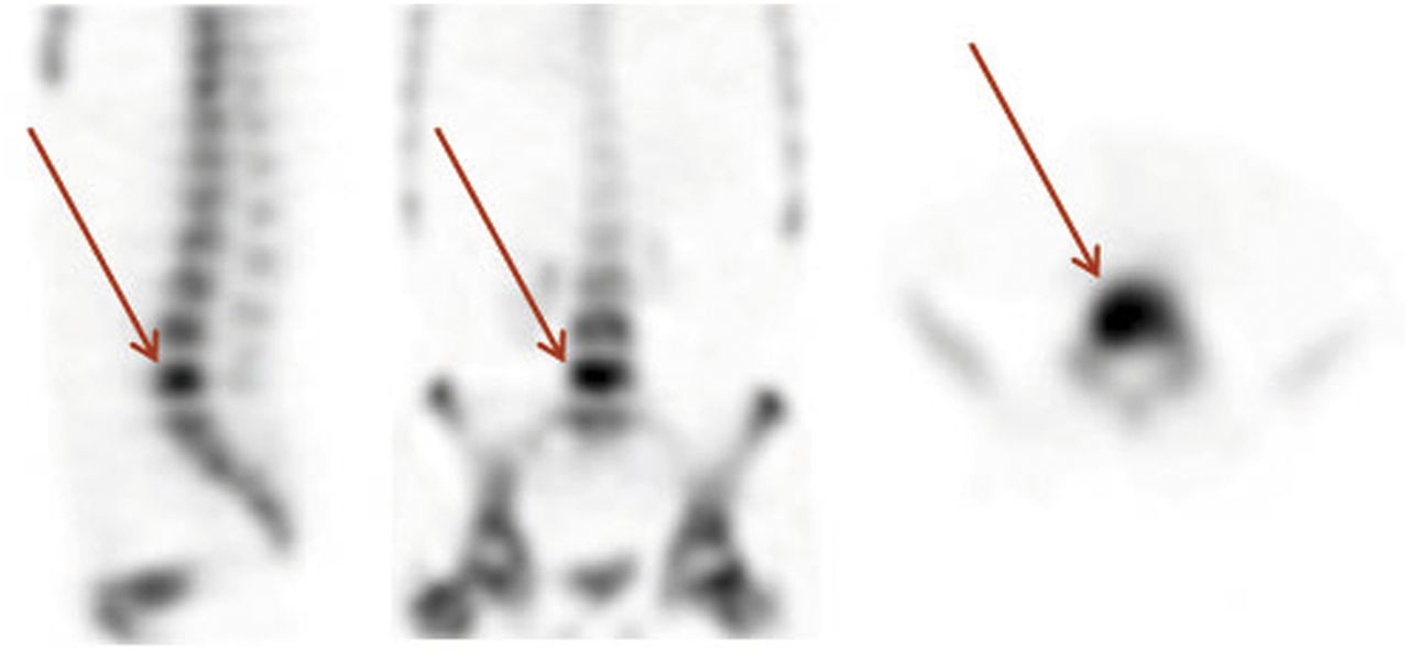
- FIGURE 1.
99mTc-hydroxymethylene diphosphonate bone SPECT scan of patient with osteomyelitis of fifth lumbar vertebra (arrows).
- FIGURE 2.
18F-FDG PET/CT scan of patient with proven Escherichia coli infection of vascular graft. Focal, intense uptake of 18F-FDG (arrows) is sign of infection, in comparison with more diffusely increased physiologic uptake along graft.
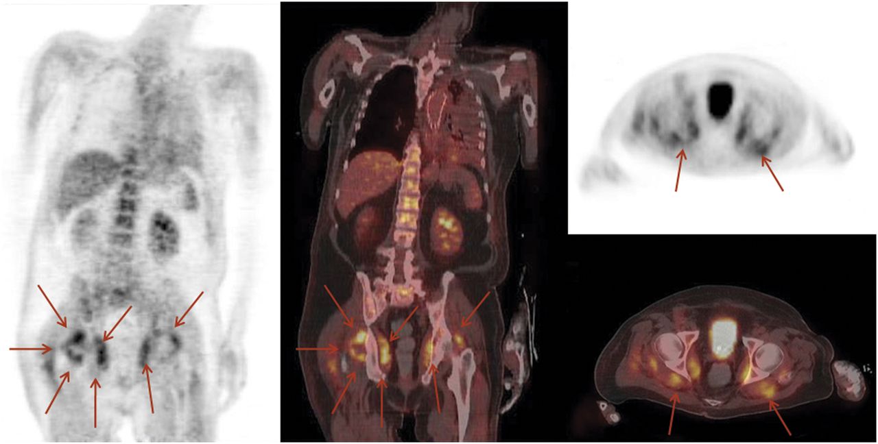
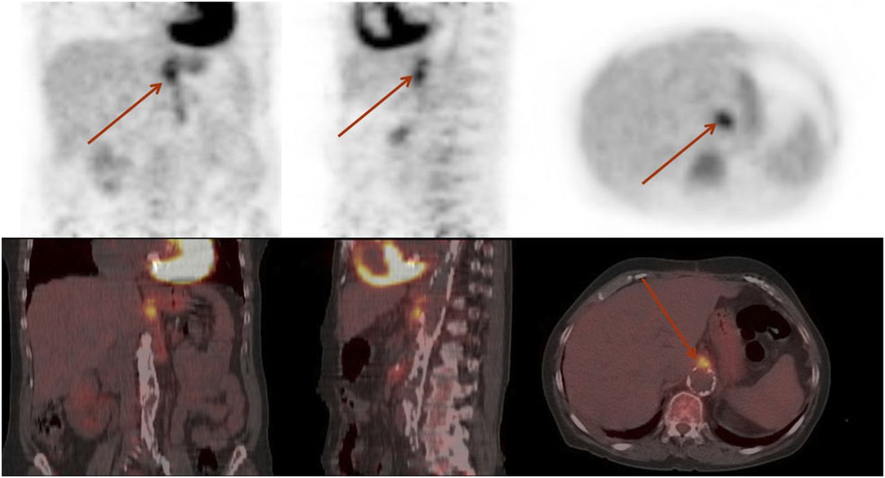
- FIGURE 3.
18F-FDG PET/CT scan of patient with FUO after placement of aortic stent for treatment of aneurysm of thoracic aorta. Stent infection was considered most likely cause of FUO, but 18F-FDG PET showed bilaterally increased uptake in region of dorsal hip muscles related to abscess formation (arrows). This scan demonstrates value of whole-body PET in patients with FUO.
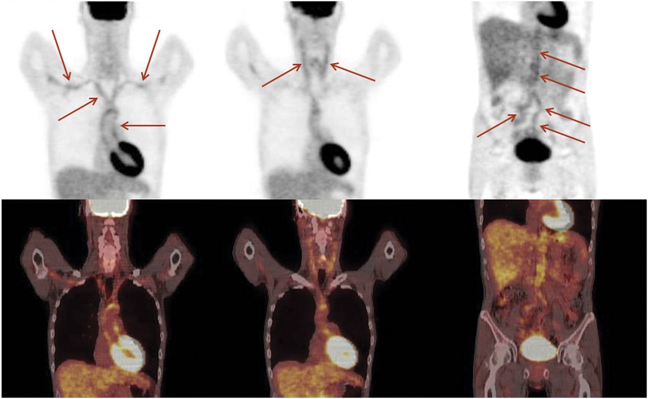
- FIGURE 4.
Patient with a history of FUO. Physical examination and ultrasonography of temporal artery were negative. Erythrocyte sedimentation and C-reactive protein were elevated; otherwise, laboratory parameters did not indicate vasculitis. 18F-FDG PET scan shows increased uptake in thoracic aorta and subclavian arteries (left), carotid arteries (middle), and abdominal aorta together with iliac arteries (right) (arrows). Vasculitis was diagnosed. This example supports role of PET for detection of vasculitis as underlying cause of FUO.
- FIGURE 5.
18F-FDG PET/CT scan of patient with sarcoidosis. Maximal-intensity projection on left side gives overview of sites of disease, including cervical area, axillae (green arrows), mediastinum (blue arrows), and inguinal lymph nodes (red arrows).
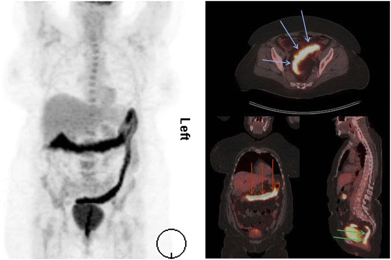
- FIGURE 6.
18F-FDG PET/CT scan of patient with ulcerative colitis. Maximal-intensity projection on left side gives overview of extent of disease, which includes transverse colon (red arrows on coronal slice), sigmoid (blue arrows on transversal slice), and rectum (green arrows on sagittal slice).


Tables
Radiopharmaceutical Mechanism 18F-FDG Macrophages (metabolically active cells), leukocytes 99mTc/111In-labeled autologous WBCs (leukocytes) Active migration into sites of inflammation 99mTc-labeled bisphosphonates Uptake in sites of increased perfusion and extravasation (early phase) and increased bone formation (late phase) 67Ga-citrate Increased perfusion, extravasation due to increased vessel permeability, locally binding to lactoferrin 99mTc-labeled nanocolloids Uptake in macrophages (inflammation, bone marrow, liver, spleen) 99mTc/111In-labeled proteins (IgG, albumin) Extravasation (increased perfusion and vessel permeability)


Jump to section
Related Articles
Cited By...
- No citing articles found.