


Article Figures & Data
Figures
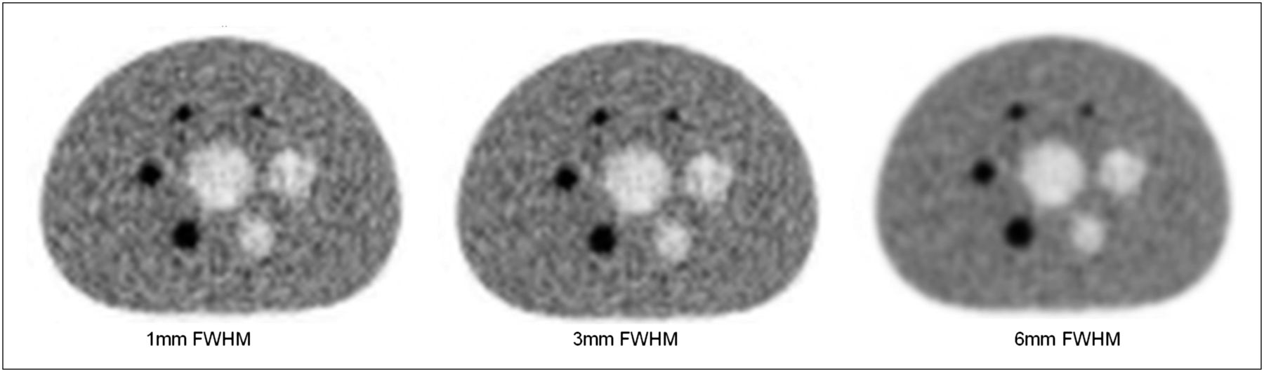
- FIGURE 1.
Transversal image of phantom of International Electrotechnical Commission for image quality test following NEMA protocol NU 2-2007 for different postfilters. Reconstructions were done with OSEM, 3 iterations, 24 subsets, matrix size of 200 × 200, and sphere-to-background ratio of 4:1.
- FIGURE 2.
A 67-y-old woman with HNSCC of oral cavity and intensive 18F-FDG uptake of primary tumor on PET. In left neck level II, 1 lymph node with intensive 18F-FDG uptake (thick arrow) was histologically confirmed as lymph node metastasis anterior to left jugular vein on corresponding contrast-enhanced CT (thick arrow). Second lymph node in left neck level II with moderate 18F-FDG uptake on PET (thin arrow) was confirmed benign lesion posterior and lateral to left jugular vein on CT (thin arrow).
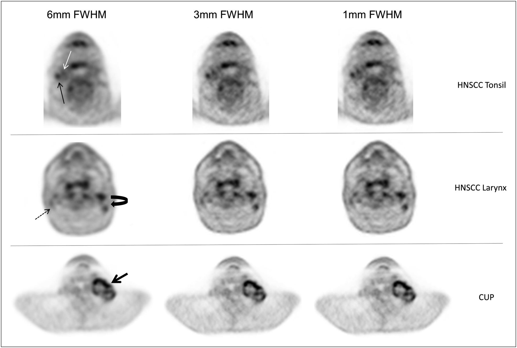
- FIGURE 3.
Postfiltered images at 1, 3, and 6 mm in FWHM and influence of postfiltering on image quality and accurate detection of lymph node metastases. In patient with HNSCC of right tonsil, first lymph node metastasis was diagnosed correctly on all postfiltered images (thin solid black arrow), whereas second malignant node (white arrow) was misdiagnosed as false-negative finding in all postfiltered images, most probably due to small lesion size and SUV. In patient with HNSCC of larynx, benign lymph nodes could hardly be delineated on 6-mm images (dashed arrow). One- and 3-mm images enabled correct assessment of benign lymph nodes in right neck level II (dashed arrow) and metastases in left neck levels II and III (curved arrow). Diagnostic visualization of large necrotic lymph node metastases at level II on left side of neck patient with cancer of unknown primary (CUP; thick straight arrow) did not differ in any postfiltered images despite increased image blurring with 6 mm in FWHM.

Tables
Gaussian filter (mm in FWHM) Sphere size (mm) 1 3 6 10 32.0 ± 4.4 29.8 ± 3.5 21.1 ± 1.6 13 56.8 ± 2.6 54.5 ± 2.1 42.6 ± 0.6 17 63.0 ± 4.0 61.7 ± 3.6 53.2 ± 2.1 22 75.0 ± 4.9 73.6 ± 5.1 64.9 ± 4.4 28 64.7 ± 1.6 64.3 ± 1.4 61.3 ± 1.4 37 66.8 ± 1.4 66.5 ± 1.2 64.0 ± 1.2 Data are image contrast (Q) (%).
SNR All-pass* 1 mm in FWHM 3 mm in FWHM 6 mm in FWHM Sphere size (mm) 200 MBq 350 MBq 200 MBq 350 MBq 200 MBq 350 MBq 200 MBq 350 MBq 19.9 16.9 ± 0.9 29.5 ± 0.4 16.0 ± 1.0 27.2 ± 2.2 18.7 ± 0.01 33.9 ± 1.2 21.8 ± 0.2 59.1 ± 0.8 15.5 14.7 ± 0.5 24.9 ± 0.4 14.2 ± 0.6 24.2 ± 1.4 16.8 ± 0.3 29.0 ± 0.5 18.9 ± 0.3 48.5 ± 1.1 12.2 11.3 ± 0.7 17.0 ± 0.1 10.6 ± 0.8 16.4 ± 0.9 13.1 ± 0.2 20.0 ± 0.4 14.2 ± 0.2 33.9 ± 0.7 8.7 7.7 ± 0.4 10.8 ± 0.2 7.3 ± 0.5 10.6 ± 0.6 8.4 ± 0.1 12.6 ± 0.2 9.3 ± 0.2 20.5 ± 0.6 4.4 2.0 ± 0.1 3.0 ± 0.2 1.9 ± 0.1 2.6 ± 0.2 2.3 ± 0.1 3.5 ± 0.2 2.6 ± 0.1 6.1 ± 0.1 ↵* Reconstructed images without any postprocessing filter method.
- TABLE 3
Filter-Dependant Differences in Maximum SUV Levels in Benign and Malignant Lymph Nodes
Gaussian filter (mm) Histology Lymph nodes Mean maximum SUV SD Minimum Maximum 6 Benign 41 2.89 0.59 2.2 5.2 Malignant 82 4.20 1.94 1.8 11.0 3 Benign 41 3.23 0.73 2.4 6.4 Malignant 82 4.77 2.39 1.4 13.6 1 Benign 41 3.25 0.81 1.7 6.8 Malignant 82 4.85 2.43 2.5 14.6 - TABLE 4
Lymph Node Detection Sensitivity Analysis of Consensus Reading for 1, 3, and 6 mm in FWHM
Gaussian filter (mm) Histology No. of lymph nodes True-positive finding False-positive finding True-negative finding False-negative finding 6 Malignant 95 73 22 19 9 Benign 28 3 Malignant 112 79 33 8 3 Benign 11 1 Malignant 114 78 36 5 4 Benign 9 No. of total lymph nodes: 82, malignant; 41, benign.
- TABLE 5
Receiver-Operating-Characteristic Analysis of Maximum SUV Levels for Benign and Malignant Lymph Nodes for 1, 3, and 6 mm in FWHM
Gaussian filter (mm) Histology Mean maximum SUV Total area under curve, κ-values P > χ2 6 Benign 2.89 0.76 P < 0.0001 Malignant 4.2 0.76 P < 0.0001 3 Benign 3.23 0.77 P < 0.0001 Malignant 4.77 0.77 P < 0.0001 1 Benign 3.25 0.78 P < 0.0001 Malignant 4.85 0.78 P < 0.0001


