


Article Figures & Data
Figures
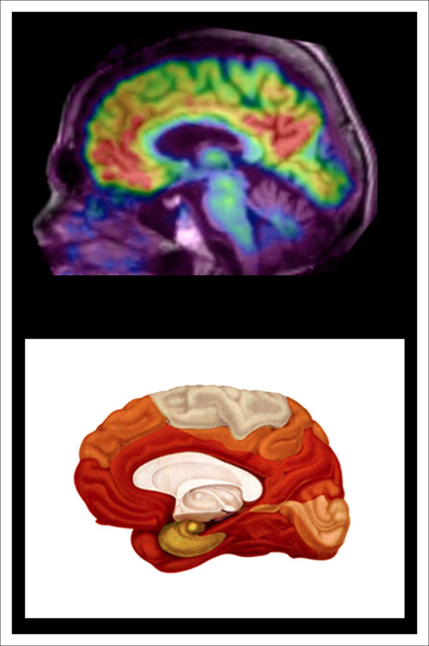
- FIGURE 1.
Comparison of 11C-PiB PET with postmortem distribution of amyloid plaques. (Top) Representative midsagittal PET image shows regional uptake of 11C-PiB overlaid on MRI, reflecting Aβ plaque burden in brain of participant with AD. (Bottom) Schematic drawing demonstrates stage C of Aβ deposition in human brain as described by Braak and Braak based on postmortem study of 2,700 brains (16). There is excellent concordance, with marked 11C-PiB binding in frontal and anterior cortex, posterior cingulate gyrus, and precuneus (posterior medial parietal), with relative sparing of sensorimotor and occipital regions.
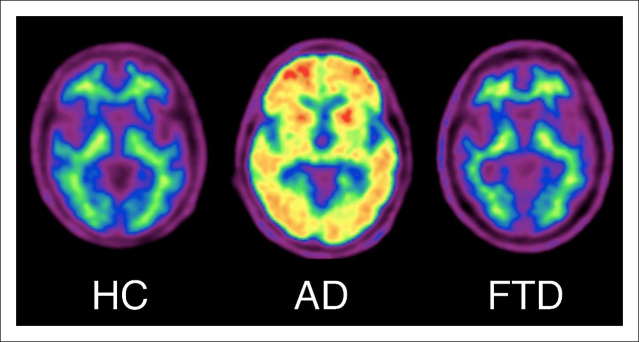
- FIGURE 2.
18F-florbetaben PET images of healthy aging (HC), AD, and frontotemporal dementia (FTD). 18F-florbetaben images show nonspecific white matter retention in healthy elderly individual, cortical and striatal binding in patient with AD, but no cortical binding in patient with frontotemporal dementia.
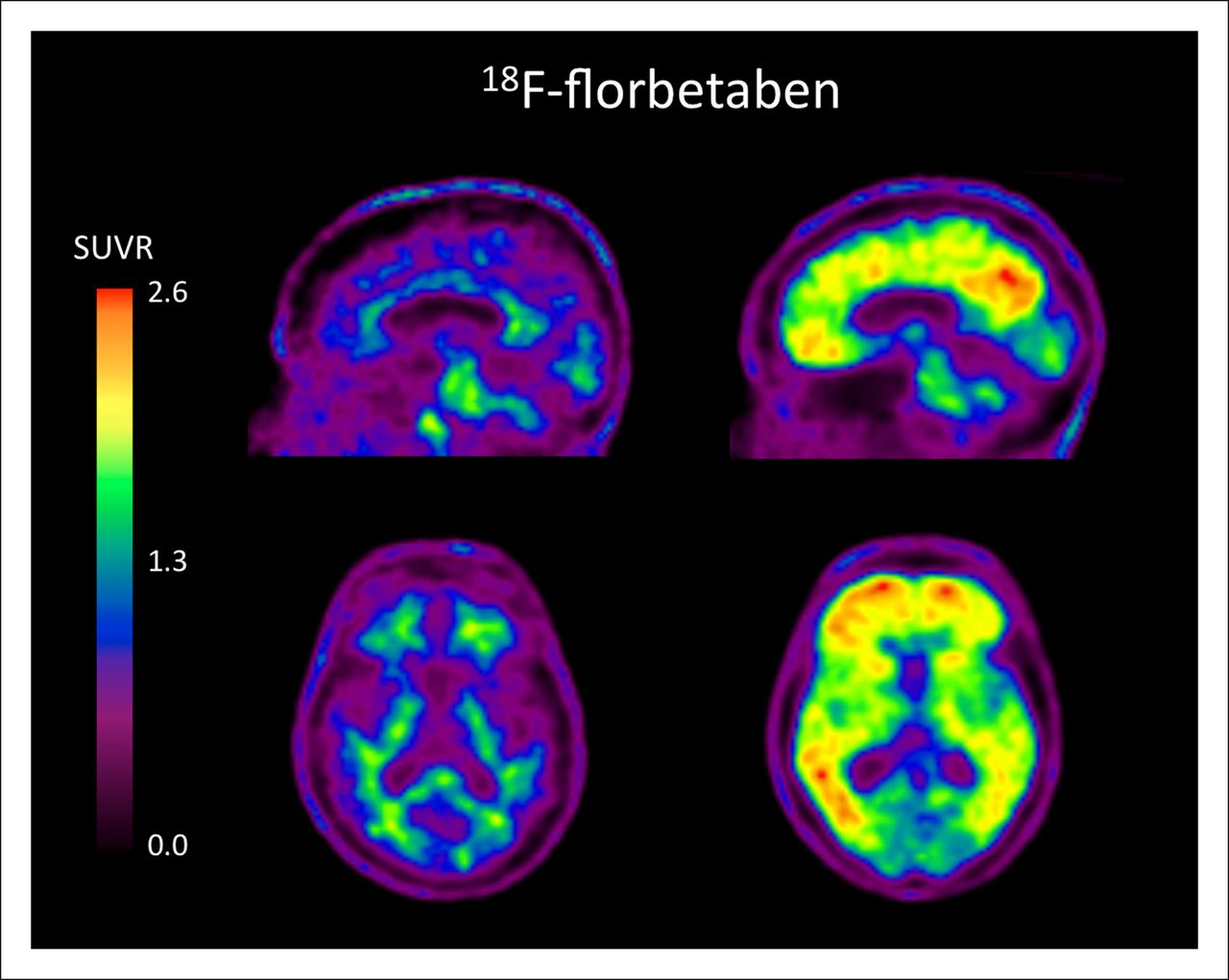
- FIGURE 3.
18F-florbetaben images of 2 elderly subjects with MCI. Scan on left is negative for brain amyloid. Uptake in corpus callosum and pons is clearly seen on midsagittal image and only in white matter on transverse slice. This subject remained cognitively stable over 2 y of follow-up. Scan on right is positive for brain amyloid, with binding in frontal cortex, posterior cingulate gyrus, precuneus, and lateral temporal cortex. This subject progressed to AD over 2 y of follow-up. SUVR = SUV ratio.
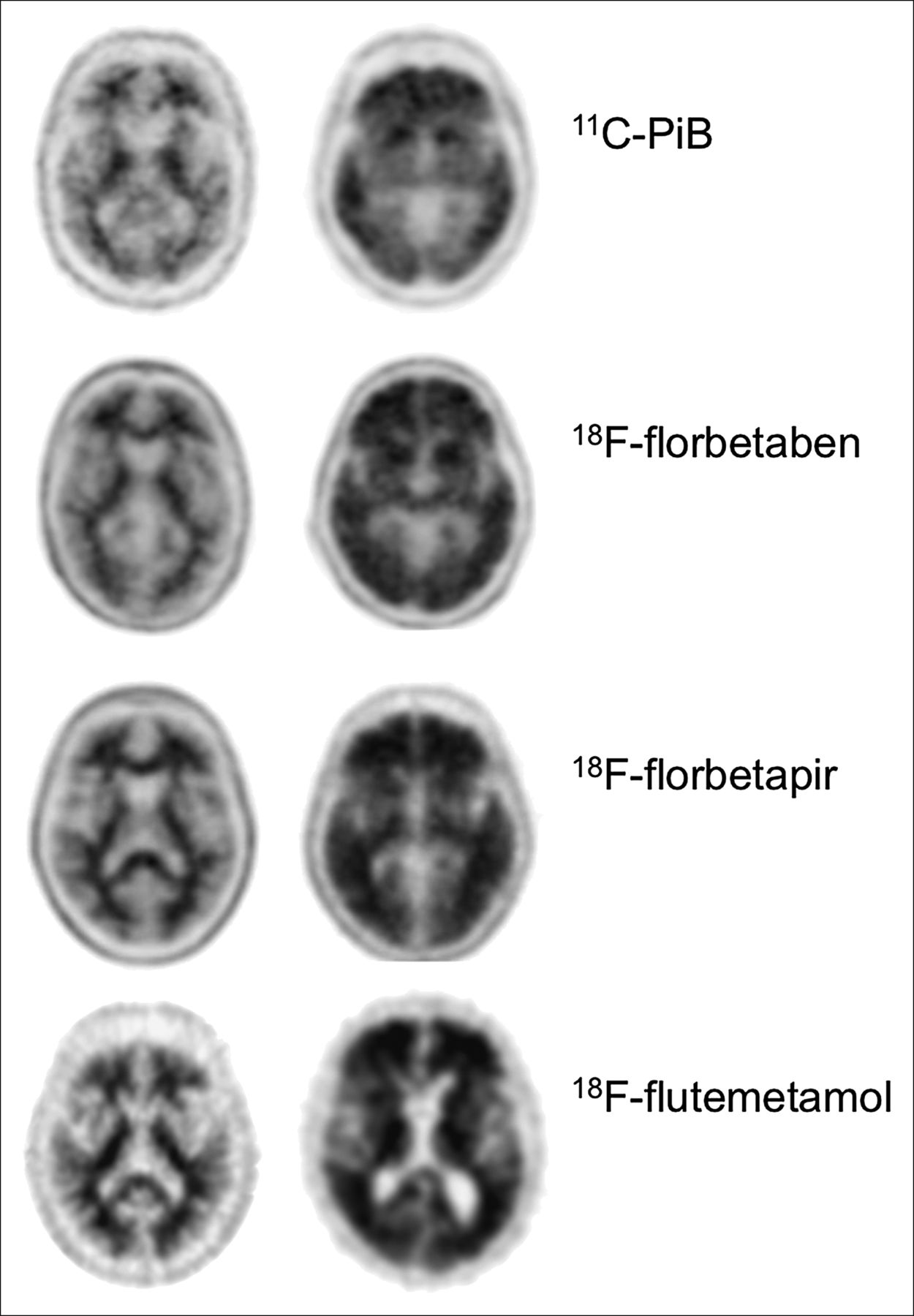
- FIGURE 4.
11C-PiB, 18F-florbetaben, 18F-florbetapir, and 18F-flutemetamol images of healthy subjects and AD patients. Images on left are negative for brain amyloid and show distinctive pattern of retention in white matter. In contrast, positive scans on right illustrate that uptake in cortical gray matter obscures normal white matter pattern and that binding extends to outer edge of brain. Images were obtained from different centers (Austin Health, Avid Radiopharmaceuticals, and University of Pittsburgh) and were acquired using different cameras and reconstruction algorithms.


Jump to section
Related Articles
Cited By...
- Atypical Parkinsonian Syndromes: Structural, Functional, and Molecular Imaging Features
- Impact of PET Reconstruction on Amyloid-{beta} Quantitation in Cross-Sectional and Longitudinal Analyses
- Inter-scanner A{beta}-amyloid PET harmonization using barrel phantom spatial resolution matching
- Impact of PET reconstruction on A{beta}-amyloid quantitation in cross-sectional and longitudinal analyses
- Tau, {beta}-amyloid, and glucose metabolism following service-related Traumatic Brain Injury in Vietnam war veterans: The AIBL-VETS study
- Impact of Reference and Target Region Selection on Amyloid PET SUV Ratios in the Phase 1b PRIME Study of Aducanumab
- 18F-FDG PET Improves Diagnosis in Patients with Focal-Onset Dementias
- Technical Considerations in Brain Amyloid PET Imaging with 18F-Florbetapir