  • Main menu
  • User menu
  • Search
  • English ▼
    • English
    • Afrikaans
    • Albanian
    • Amharic
    • Arabic
    • Armenian
    • Azerbaijani
    • Basque
    • Belarusian
    • Bengali
    • Bosnian
    • Bulgarian
    • Catalan
    • Cebuano
    • Chichewa
    • Chinese (Simplified)
    • Chinese (Traditional)
    • Corsican
    • Croatian
    • Czech
    • Danish
    • Dutch
    • Esperanto
    • Estonian
    • Filipino
    • Finnish
    • French
    • Frisian
    • Galician
    • Georgian
    • German
    • Greek
    • Gujarati
    • Haitian Creole
    • Hausa
    • Hawaiian
    • Hebrew
    • Hindi
    • Hmong
    • Hungarian
    • Icelandic
    • Igbo
    • Indonesian
    • Irish
    • Italian
    • Japanese
    • Javanese
    • Kannada
    • Kazakh
    • Khmer
    • Korean
    • Kurdish (Kurmanji)
    • Kyrgyz
    • Lao
    • Latin
    • Latvian
    • Lithuanian
    • Luxembourgish
    • Macedonian
    • Malagasy
    • Malay
    • Malayalam
    • Maltese
    • Maori
    • Marathi
    • Mongolian
    • Myanmar (Burmese)
    • Nepali
    • Norwegian
    • Pashto
    • Persian
    • Polish
    • Portuguese
    • Punjabi
    • Romanian
    • Russian
    • Samoan
    • Scottish Gaelic
    • Serbian
    • Sesotho
    • Shona
    • Sindhi
    • Sinhala
    • Slovak
    • Slovenian
    • Somali
    • Spanish
    • Sudanese
    • Swahili
    • Swedish
    • Tajik
    • Tamil
    • Telugu
    • Thai
    • Turkish
    • Ukrainian
    • Urdu
    • Uzbek
    • Vietnamese
    • Welsh
    • Xhosa
    • Yiddish
    • Yoruba
    • Zulu

Main menu

  • Home
  • Content
    • Current
      • JNMT Supplement
    • Ahead of print
    • Past Issues
    • Continuing Education
    • JNMT Podcast
    • SNMMI Annual Meeting Abstracts
  • Subscriptions
    • Subscribers
    • Rates
    • Journal Claims
    • Institutional and Non-member
  • Authors
    • Submit to JNMT
    • Information for Authors
    • Assignment of Copyright
    • AQARA Requirements
  • Info
    • Reviewers
    • Permissions
    • Advertisers
    • Corporate & Special Sales
  • About
    • About Us
    • Editorial Board
    • Contact Information
  • More
    • Alerts
    • Feedback
    • Help
    • SNMMI Journals
  • SNMMI
    • JNMT
    • JNM
    • SNMMI Journals
    • SNMMI

User menu

  • Subscribe
  • My alerts
  • Log in
  • My Cart

Search

  • Advanced search
Journal of Nuclear Medicine Technology
  • SNMMI
    • JNMT
    • JNM
    • SNMMI Journals
    • SNMMI
  • Subscribe
  • My alerts
  • Log in
  • My Cart
Journal of Nuclear Medicine Technology

Advanced Search

English ▼
  • English
  • Afrikaans
  • Albanian
  • Amharic
  • Arabic
  • Armenian
  • Azerbaijani
  • Basque
  • Belarusian
  • Bengali
  • Bosnian
  • Bulgarian
  • Catalan
  • Cebuano
  • Chichewa
  • Chinese (Simplified)
  • Chinese (Traditional)
  • Corsican
  • Croatian
  • Czech
  • Danish
  • Dutch
  • Esperanto
  • Estonian
  • Filipino
  • Finnish
  • French
  • Frisian
  • Galician
  • Georgian
  • German
  • Greek
  • Gujarati
  • Haitian Creole
  • Hausa
  • Hawaiian
  • Hebrew
  • Hindi
  • Hmong
  • Hungarian
  • Icelandic
  • Igbo
  • Indonesian
  • Irish
  • Italian
  • Japanese
  • Javanese
  • Kannada
  • Kazakh
  • Khmer
  • Korean
  • Kurdish (Kurmanji)
  • Kyrgyz
  • Lao
  • Latin
  • Latvian
  • Lithuanian
  • Luxembourgish
  • Macedonian
  • Malagasy
  • Malay
  • Malayalam
  • Maltese
  • Maori
  • Marathi
  • Mongolian
  • Myanmar (Burmese)
  • Nepali
  • Norwegian
  • Pashto
  • Persian
  • Polish
  • Portuguese
  • Punjabi
  • Romanian
  • Russian
  • Samoan
  • Scottish Gaelic
  • Serbian
  • Sesotho
  • Shona
  • Sindhi
  • Sinhala
  • Slovak
  • Slovenian
  • Somali
  • Spanish
  • Sudanese
  • Swahili
  • Swedish
  • Tajik
  • Tamil
  • Telugu
  • Thai
  • Turkish
  • Ukrainian
  • Urdu
  • Uzbek
  • Vietnamese
  • Welsh
  • Xhosa
  • Yiddish
  • Yoruba
  • Zulu
  • Home
  • Content
    • Current
    • Ahead of print
    • Past Issues
    • Continuing Education
    • JNMT Podcast
    • SNMMI Annual Meeting Abstracts
  • Subscriptions
    • Subscribers
    • Rates
    • Journal Claims
    • Institutional and Non-member
  • Authors
    • Submit to JNMT
    • Information for Authors
    • Assignment of Copyright
    • AQARA Requirements
  • Info
    • Reviewers
    • Permissions
    • Advertisers
    • Corporate & Special Sales
  • About
    • About Us
    • Editorial Board
    • Contact Information
  • More
    • Alerts
    • Feedback
    • Help
    • SNMMI Journals
  • Watch or Listen to JNMT Podcast
  • Visit SNMMI on Facebook
  • Join SNMMI on LinkedIn
  • Follow SNMMI on Twitter
  • Subscribe to JNMT RSS feeds
Research ArticleCONTINUING EDUCATION

A New Era of Clinical Dopamine Transporter Imaging Using 123I-FP-CIT

Eunkyung Park
Journal of Nuclear Medicine Technology December 2012, 40 (4) 222-228; DOI: https://doi.org/10.2967/jnmt.112.111617
Eunkyung Park
Department of Nuclear Medicine, School of Medicine and Biomedical Sciences, University at Buffalo, State University of New York, Buffalo, New York
  • Find this author on Google Scholar
  • Find this author on PubMed
  • Search for this author on this site
  • Article
  • Figures & Data
  • Info & Metrics
  • PDF
Loading

Article Figures & Data

Figures

  • Tables
  • FIGURE 1.
    • Download figure
    • Open in new tab
    • Download powerpoint
    FIGURE 1.

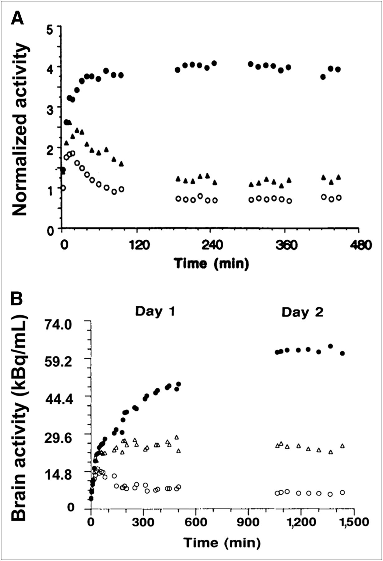
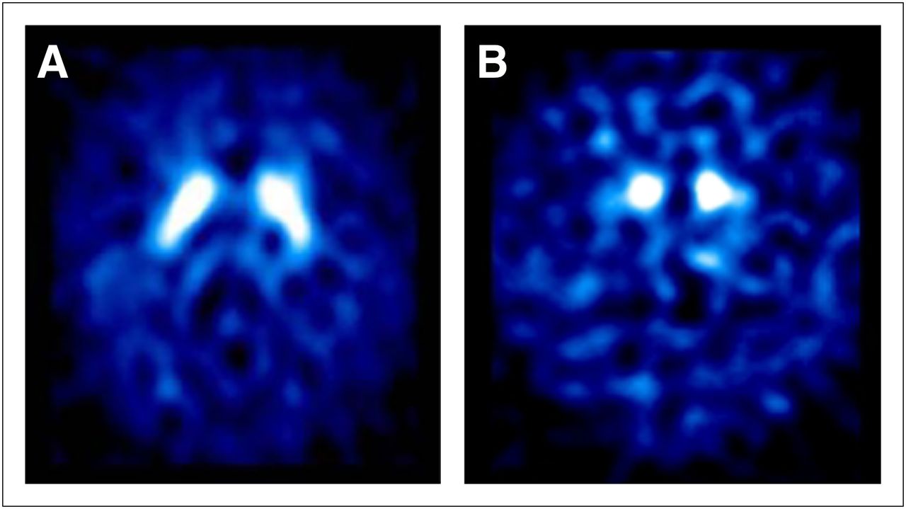
    Faster kinetics of 123I-FP-CIT. In human striatum, 123I-FP-CIT (A) is taken up more rapidly than 123I-β-CIT (B). Practical clinical advantage of fast kinetics of 123I-FP-CIT is that patients can be scanned on same day 3–6 h after injection. (Panel B reprinted with permission of (26).)

  • FIGURE 2.
    • Download figure
    • Open in new tab
    • Download powerpoint
    FIGURE 2.

    Brain 123I-FP-CIT SPECT images. Transaxial slices at striatal level. (A) Normal 123I-FP-CIT uptake, which is often described as comma shaped, is seen in healthy subject. (B) Striatal uptake is decreased in patient with PD. Reduction is more prominent in putamen than in caudate nucleus.

  • FIGURE 3.
    • Download figure
    • Open in new tab
    • Download powerpoint
    FIGURE 3.

    Superior spatial resolution of brain 18F-FP-CIT PET images. Superior spatial resolution of brain PET enables us to see caudate nucleus and putamen separately, and to analyze small brain structures such as in brain stem area. Another advantage of 18F-FP-CIT PET is that it can be acquired 2–3 h after injection.

  • FIGURE 4.
    • Download figure
    • Open in new tab
    • Download powerpoint
    FIGURE 4.

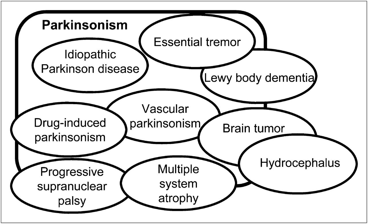
    Variety of diseases mimicking idiopathic PD.

  • FIGURE 5.
    • Download figure
    • Open in new tab
    • Download powerpoint
    FIGURE 5.

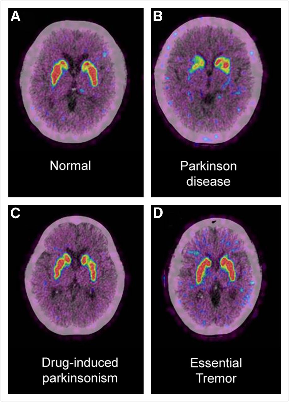
    Differential diagnosis of parkinsonism using brain 18F-FP-CIT PET. Brain PET/CT images of 18F-FP-CIT uptake at level of striatum demonstrate different DAT density in different conditions. DAT density is decreased in PD patient (B), whereas DAT density is normal in healthy subject (A) and in patients with drug-induced parkinsonism (C) and essential tremor (D). DAT imaging is useful for differential diagnosis of various causes of parkinsonism. Clinical diagnosis of parkinsonism is quite often straightforward, obviating additional tests, but when overlap and incomplete syndromes are present, improvements in diagnostic accuracy may be possible using DAT imaging.

  • FIGURE 6.
    • Download figure
    • Open in new tab
    • Download powerpoint
    FIGURE 6.

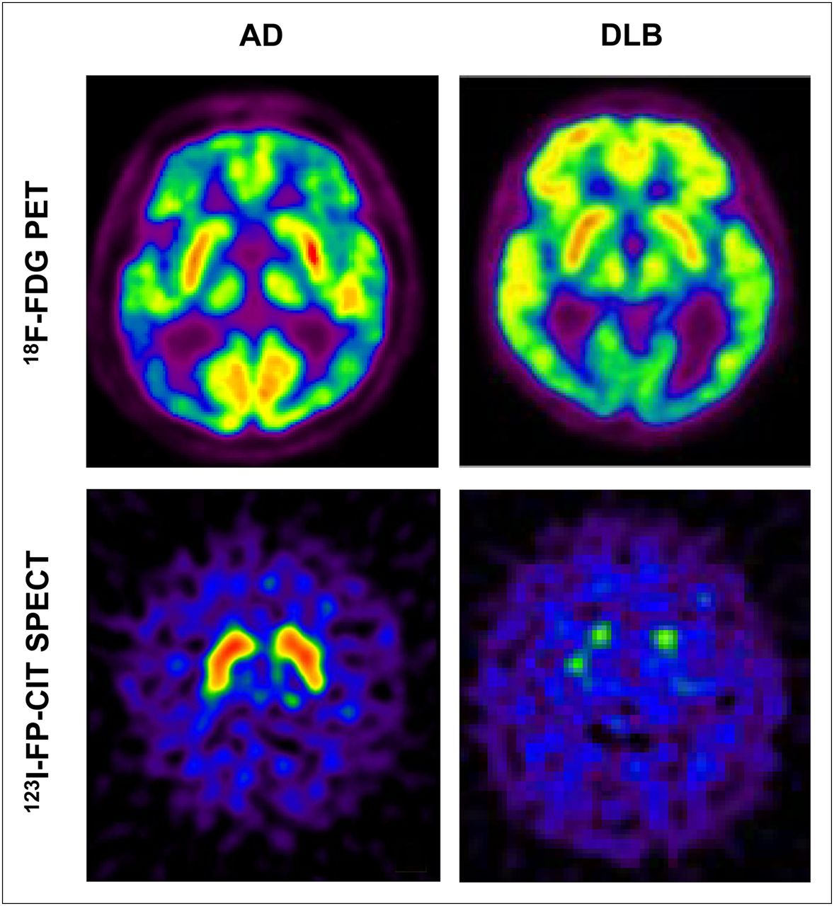
    Differential diagnosis of dementia using brain 123F-FP-CIT SPECT. The 2 most common forms of dementia, Alzheimer disease (AD) and DLB, share clinical features and are often difficult to differentiate. 18F-FDG PET is useful when typical bilateral temporoparietal hypometabolism is seen in AD and occipital hypometabolism is seen in DLB, as demonstrated in these images; however, that is not always case. With DAT imaging, for example, using 123I-FP-CIT, these 2 disorders can be more easily distinguished. Striatal dopaminergic deficit occurs in DLB but not in AD.

  • FIGURE 7.
    • Download figure
    • Open in new tab
    • Download powerpoint
    FIGURE 7.

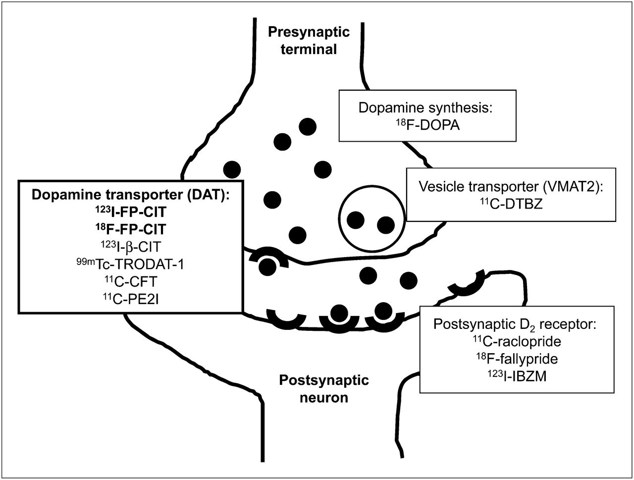
    Molecular targets for imaging dopamine system. Illustration is of dopamine nerve terminal and examples of several radioligands that enable in vivo demonstration of dopamine synthesis, rerelease into synaptic cleft, uptake by postsynaptic receptors, and active reuptake by DAT, respectively. Each step of dopamine synthesis, release, and reuptake can be imaged and measured with nuclear medicine molecular imaging techniques.

Tables

  • Figures
    • View popup
    TABLE 1

    Approval Year for Clinical Use of Radioligands for DAT Imaging

    RadioligandUnited StatesElsewhere
    123I-β-CIT (DOPASCAN; MAP Medical Technologies Oy)—Europe*, Japan*
    123I-FP-CIT (DaTSCAN)20112000 in Europe, 2011 in Korea
    18F-FP-CIT—2008 in Korea
    99mTc-TRODAT-1—2005 in Taiwan
    • ↵* Before 123I-FP-CIT, but exact date unknown.

PreviousNext
Back to top

In this issue

Journal of Nuclear Medicine Technology: 40 (4)
Journal of Nuclear Medicine Technology
Vol. 40, Issue 4
December 1, 2012
  • Table of Contents
  • About the Cover
  • Index by author
Print
Download PDF
Article Alerts
Email Article
Citation Tools
Share
A New Era of Clinical Dopamine Transporter Imaging Using 123I-FP-CIT
Eunkyung Park
Journal of Nuclear Medicine Technology Dec 2012, 40 (4) 222-228; DOI: 10.2967/jnmt.112.111617
Twitter logo Facebook logo LinkedIn logo Mendeley logo
  • Tweet Widget
Bookmark this article

Jump to section

  • Article
    • Abstract
    • A NEW ERA OF CLINICAL DOPAMINE TRANSPORTER IMAGING USING 123I-LABELED 2ς-CARBOMETHOXY-3ς-(4-IODOPHENYL)-N-(3-FLUOROPROPYL)NORTROPANE (123I-FP-CIT)
    • DAT DENSITY AND 123I-FP-CIT UPTAKE
    • WHY DO WE IMAGE STRIATAL DAT DENSITY?
    • RADIOLIGANDS FOR DAT IMAGING AND THEIR CLINICAL AVAILABILITY
    • CHARACTERISTICS OF 123I-FP-CIT
    • DAT IMAGING PROTOCOL USING 123I-FP-CIT
    • VISUAL INTERPRETATION AND SEMIQUANTITATIVE ANALYSIS
    • CLINICAL APPLICATION OF DAT IMAGING
    • DIFFERENTIAL DIAGNOSIS OF PARKINSONISM
    • DIAGNOSTIC VALUE OF DAT IMAGING
    • DAT DENSITY AND DISEASE SEVERITY
    • COST EFFECTIVENESS OF DAT IMAGING
    • CONCLUSION
    • Acknowledgments
    • Footnotes
    • REFERENCES
  • Figures & Data
  • Info & Metrics
  • PDF

Related Articles

  • Errata
  • PubMed
  • Google Scholar

Cited By...

  • No citing articles found.
  • Google Scholar

More in this TOC Section

  • Illuminating the Hidden: Standardizing Cardiac MIBG Imaging for Sympathetic Dysfunction
  • PET/CT Case Series: Unmasking the Mystery of Cardiac Sarcoidosis
  • Delivery Methods of Radiopharmaceuticals: Exploring Global Strategies to Minimize Occupational Radiation Exposure
Show more Continuing Education

Similar Articles

Keywords

  • dopamine transporter
  • Parkinson disease
  • FP-CIT
  • ioflupane
  • DaTscan
SNMMI

© 2025 SNMMI

Powered by HighWire
Alerts for this Article
Sign In to Email Alerts with your Email Address
Email this Article

Thank you for your interest in spreading the word on Journal of Nuclear Medicine Technology.

NOTE: We only request your email address so that the person you are recommending the page to knows that you wanted them to see it, and that it is not junk mail. We do not capture any email address.

Enter multiple addresses on separate lines or separate them with commas.
A New Era of Clinical Dopamine Transporter Imaging Using 123I-FP-CIT
(Your Name) has sent you a message from Journal of Nuclear Medicine Technology
(Your Name) thought you would like to see the Journal of Nuclear Medicine Technology web site.
Citation Tools
A New Era of Clinical Dopamine Transporter Imaging Using 123I-FP-CIT
Eunkyung Park
Journal of Nuclear Medicine Technology Dec 2012, 40 (4) 222-228; DOI: 10.2967/jnmt.112.111617

Citation Manager Formats

  • BibTeX
  • Bookends
  • EasyBib
  • EndNote (tagged)
  • EndNote 8 (xml)
  • Medlars
  • Mendeley
  • Papers
  • RefWorks Tagged
  • Ref Manager
  • RIS
  • Zotero

We use cookies on this site to enhance your user experience

By clicking any link on this page you are giving your consent for us to set cookies.